Artist Inspiration: Plant Cure (Part 3)

Plant Cure evite test11+SarahThe New York Academy of Medicine Library and CENTRAL BOOKING collaborated on the exhibition Plant Cure.  For this exhibition, five artists were selected to do research at the Academy Library over six months to produce work with their own unique take on medicinal plants. The project will culminate with an exhibition at CENTRAL BOOKING on the Lower East Side from September 6-October 29, 2017. Part 1 and 2 can be read here and here.

Maddy Rosenberg

Plant Cure, the collaborative project between CENTRAL BOOKING and The New York Academy of Medicine Library, percolating for over a year, is about to open to the public on September 6. The project includes a two month-long exhibition featuring the work of 19 international artists, vitrines documenting the inspiration and process of the five Artists in Residence, a catalog, and a program of events at both venues.

I feel fortunate to have had the opportunity to assemble the work of these artists who bring their own unique interpretations of plants and their medicinal qualities. From meticulously rendered drawings and water colors to an installation of sculptural free-standing collage works that seem to multiply from pedestal to pedestal, to a medicine cabinet that is beyond the expected, these works offer the viewer science from an artistic slant. In addition, interspersed within the CENTRAL BOOKING exhibition is a video-projected panorama of the Drs. Barry and Bobbi Coller Rare Book Reading Room and a floor to ceiling cabinet of curiosities.

But Plant Cure was more than a curatorial project for me. I enjoyed being the “honorary” sixth artist in the Academy Library. It was an opportunity for me to go beyond researching ideas for shaping the exhibition at CENTRAL BOOKING, mindful, as well, of pursuing material for my own studio work. The personal one-on-one access to exquisitely designed and illustrated books dating back hundreds of years was like hitting the mother lode for me, with the aid of the fountain of information and helpful direction of Arlene Shaner steering every visit through the vast possibilities.

My aim was to take my interest in medical museums and historic medical texts that are chock full of hand drawn images, add the science of the medicinal usefulness of plants combined with their aesthetic qualities, and make art out of it. The artists’ orientation at the library got the thought processes churning when I saw the collection of Burdock Blood Bitters advertising cards as a way of linking the plants with the cure. Always one to add an element to the flat page, whether it be built in pop-ups or interactive movable components, I requested in my next visit to see several of the anatomical flapbooks, and drew considerably from ones such as The BodyScope (1948) by Ralph H. Segal and Anatomicum Vivum (1720) by Christoph von Hellwig. The plant references I used came heavily from The Herball; or Generall Historie of Plantes (1597) by John Gerarde and published in London in 1597.

01.M. Rosenberg_reference Herball_watermark

Inspiration: The Herball; or Generall Historie of Plantes (1597) by John Gerarde.

From there emerged an artist’s book of my own medicinal cards with composite images on one side and texts of the cures on the reverse, tucked into pockets of an accordion book to hold them, framed by the drawings of the plants themselves, much like a traditional book of hours. A second artist’s book that is even more a two-dimensional object that becomes a three-dimensional structure through pop-ups and its own flaps, is in process. I am certain with all the material I was able to accumulate and am still digesting, pieces of the Academy Library collection will wind up in many more of my works to come.

Medicinal cards. Maddy Rosenberg (2017).

I am happy to introduce Mary Ting, the last of our five official CENTRAL BOOKING artists at the New York Academy of Medicine Library, who comes with a long interest in medicinal plants through family heritage and her own love of gardening.

Mary Ting

Secluded away in the library of the New York Academy of Medicine, surrounded by bookcases of historical medical texts, I have been intoxicated by the books containing magical illustrations of astonishing beauty and text that entice unanswerable questions. I have been looking and re-looking at the Hortus Sanitatus, various medical botany books, and anatomical flap books. Of particular interest for me are common medicinal plants, (such as ginseng, valerian, mandrake, snakeweed, dandelion, foxglove) ones that have figured in my garden, family life, and are also of cultural interest.

1.Tingreferences_watermark

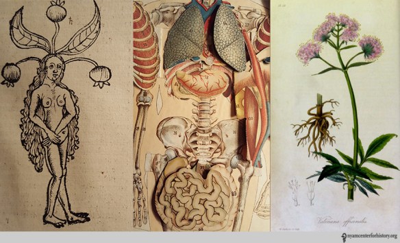
Academy Library references: Pages from Hortus Sanitatis (1517); The Practical Home Physician (1887); and Medical Botany (1834).

Having grown up weeding beside my mother in her ornamental garden and in a house with one hundred orchids and dried specimens tucked away in drawers, plants and fungi have always held an important place in my life. These were not just specimens but also markers of our family migrations. Of particular reverence is the dried lingzi mushroom that my mother plucked from her college campus (Ginling Women’s College, Nanjing, 1943). This pondering of history, family, nature and grief is central to my work; it is also why this exhibition, Plant Cure, and the research at the Academy Library is an incredible opportunity and a never-ending bounty to feed from.

2.Tingmushroomginseng20170820_143947

Pan and Ting family collection of ginseng and mushroom.

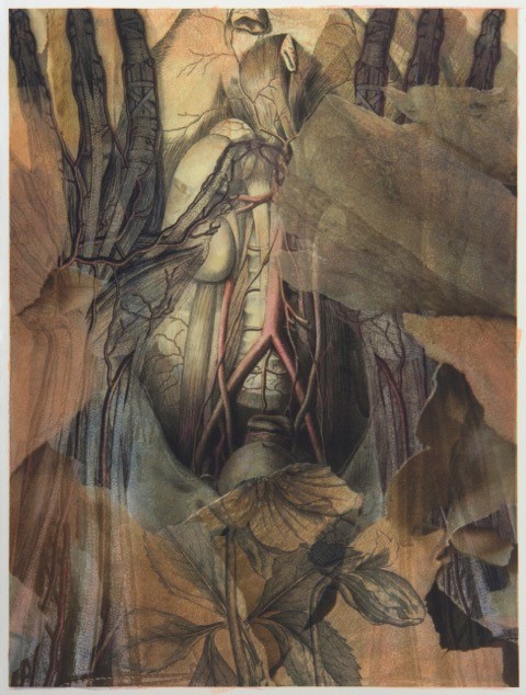
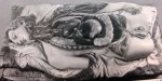
One work to come out of this residency is Holding On, which deals with the interwoven relationship of botany and medicine. I have incorporated empty Ginseng Royal jelly glass bottles as “fruit” on the vine, the red and blue wires refer to the arteries and the plastic tubing to intravenous drip tubing. The title refers to the notion that Ginseng root could be gnawed on in one’s last hours while waiting for the arrival of your children for the final goodbye.

3.Ting_Holding On_0170820_131928

Mary Ting, Holding On (detail), 2017, vine, wire, plastic tubing, glass, 80 x 24 x 10 inches.

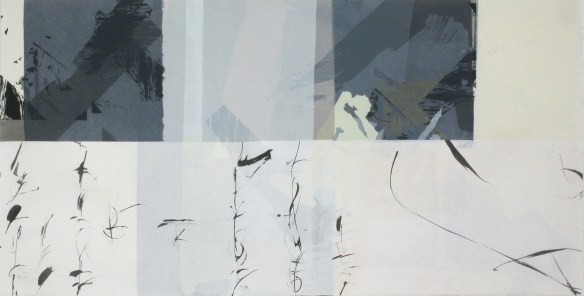
The library research also inspired The Gardener’s Medical Manual, a new rendition of an earlier series, The Other Garden.  Among outsized botanical specimens with eyes, one can find a woodblock image from the ancient Chinese classic, Mountains and Seas, 山海经, an early geography text that was meant to be neither factual nor allegorical. Centipedes also loom large, as my grandmother’s life was saved by the medical application of a poisonous centipede.

4.MaryTing04_Doctor'sGardenManual_300det2

Mary Ting, The Gardeners Medicinal Manual (detail), 2017, cut tyvek, silkscreen, rubberstamps, ink, 20 x 30 inches.

I am continually struck by how so much has changed outwardly, given technological developments, but our medieval notions of man’s dominion over nature and its ravaging remains unchanged. The lure of wild ginseng continues with its illegal harvesting and unsustainable consumption. Though I suspect that it functions primarily as a status gift and that many, like my family members, never utilize the roots and the children arrive too late for the final goodbye.

Artist Inspiration: Plant Cure (Part 2)

Todays’ guest post is introduced by Maddy Rosenberg, curator and founder of CENTRAL BOOKING. The New York Academy of Medicine Library and CENTRAL BOOKING collaborated on the exhibition Plant Cure.  For this exhibition, five artists were selected to do research at the Academy Library over six months to produce work with their own unique take on medicinal plants. The project will culminate with an exhibition at CENTRAL BOOKING on the Lower East Side from September 6-October 29, 2017. Part 1 can be read here.

The next two artists featured in the Plant Cure collaboration between CENTRAL BOOKING and the New York Academy of Medicine Library are Susan Rostow and C Bangs. Susan’s sculptural work is extremely textural and beckons to be touched, while with C it’s our eye that takes the journey over the surfaces. Both artists’ works engage us and demand closer scrutiny.

Susan Rostow

I spent many wonderful hours of my childhood reading the encyclopedia. A set of books from A to Z neatly organized on a shelf with the entire world’s information gave me great joy. I may be a romantic, waxing poetic and nostalgic about the past, but that has not stopped me from enjoying the present times of clicking and swiping through Google images and other websites. My ongoing fascination with information, books and images continued to grow through decades and is presently expressed in my sculptural books.

The first time I entered the New York Academy of Medicine Library and was surrounded by rare books dating from the 15th through the 18th centuries, I felt as though I traveled back in time and entered the Middle Ages. I was taken with the smell of the leather covers, amazed by the weight and size of some of the books, marveled at the odd titles on the bindings, and was captured by highly detailed and precise illustrations. Prodigiorum Ostentorum Chronicon (1557) by Konrad Lykosthenes and Osteographia, or, The Anatomy of the Bones (1733) by William Cheselden are a couple of my favorites.

3_Rostow_

Susan Rostow working in studio.

Feeling incredibly inspired, I took my excitement to the studio along with photos of the pictures from the various books I had observed. Armed with a plethora of images and plenty of ideas, I began to work on my vision. Images of medicinal mushrooms and text pertaining to plant cures were put to use by first making carborundum printmaking plates. This is a low tech method used for making plates by hand. This simple, but elegant technique allowed me to connect with some of the similar hand techniques used by the original artists. I printed them with an etching press, a simple press whose basic principle has not changed for centuries. Choosing to use this technique with an old style press made me feel connected to some of the reproductions from the New York Academy of Medicine Library’s rare book collection.

1_Rostow

Susan Rostow, Bone Fungus. 2017, mixed media sculptural book with carborundum prints on paper, dried mushroom, wood, parabolic mirrors, real and plastic bones, sand, glass beads and pigments, 25 x 26 x 26 inches.

After printing hundreds of images of mushrooms and text on paper, the prints were bound together with dried mushrooms, mud, natural glues, and pigments. Paper, tree fungus, roots, soil, and casts from bones merged together creating sculptural books that look, smell and feel like unearthed relics secreted beneath the earth. Hopefully this synthesis captured some of the magic that I felt when I first viewed these incredibly illustrated books.

04_Rostow

Susan Rostow’s sculptural book Bone Fungus (left and center), and detail of Cheselden’s anatomical illustration (1733) (right).

2_Rostow

Prodigioky Ostentory Chronicon (left) William Cheselden’s anatomical illustration (1733) (center), and detail from Susan Rostow’s sculptural book Bone Fungus (right).

C Bangs

My art investigates frontier science combined with symbolist figuration from an ecological feminist point of view. A decade long collaboration with quantum consciousness physicist Dr. Evan Harris Walker has lead me to incorporate his equations in my paintings in a manner mutually agreed upon, designed to posit questions related to his theories. Functioning as design elements that often speak to the interconnectivity of everything in the cosmos, the equations parallel the sacred writings found in illuminated manuscripts. In recent collaboration with my partner, Dr. Greg Matloff, we investigate consciousness from the point of view of panpsychism philosophically, historically and scientifically.

The books I researched at the New York Academy of Medicine Library included Robert Fludd and Konrad Lykosthenes. What does humankind preserve and what do we eliminate? Fludd had a theory of cosmic harmony and Kepler correctly accused Fludd of being a theosophist. Additionally Fludd is remembered as an astrologer, mathematician, cosmologist, Quabalist and Rosicrucian. His writing centered around sympathies found in nature between man, the earth and the divine.

fpseedsfetus copy

Flowering Pavonis seeds used as an abortifacient with fetus studies. C Bangs (2017).

Maria Sibylla Merian’s Metamorphosis insectorum Surinamensium (1705) at the Brooklyn Botanic Garden ultimately lead me to contact the New York Botanical Garden. Merian wrote that slave women’s use of the peacock flower was deeply political, using it to abort pregnancies forced upon them by their slave owners. The history of abortifacients is nearly as old as the written word and the determination of pregnancy was left to the woman, who was not considered pregnant until she declared herself to be so. When the Catholic Church realized that they could not regulate abortifacients or convict the women who used them, they began persecuting midwives, declaring them witches.[1] The enforcement of religious law and witch burning was an effective tool for breaking a chain of knowledge about abortifacients that had been in circulation for over a thousand years. Despite Merian’s revelation about the peacock flower in her book, widely used by botanists and men of medicine, this knowledge was ignored. Merchants valued the plant’s looks and shipped large amounts of its seeds to their home countries, where the flower decorated many royal gardens.

Cosmic Pavonis Fludd mix copy

Flowering Pavonis and diagrams from Robert Fludd’s Utriusque cosmi majoris scilicet et minoris metaphysica (1617-1621). C Bangs (2017).

Ironically, when I wished to photograph the peacock flower at the Brooklyn Botanic Garden or the New York Botanical Garden, I found that it had been deaccessioned by Brooklyn and is kept in a section not available to the public at the New York Botanical Garden.

Pavoniscosmos1 copy

Flowering Pavonis and images from Konrad Lykosthenes’ Prodigiorum ac ostentorum chronicon (1557). C Bangs (2017).

Reference:
[1] Edwards, Stassa. The History of Abortifacients. Jezebel: 2014, November 18.

Artist Inspiration: Plant Cure (Part 1)

Todays’ guest post is introduced by Maddy Rosenberg, curator and founder of CENTRAL BOOKING. The New York Academy of Medicine Library and CENTRAL BOOKING collaborated on the exhibition Plant Cure.  For this exhibition, five artists were selected to do research at the Academy Library over six months to produce work with their own unique take on medicinal plants. The project will culminate with an exhibition at CENTRAL BOOKING on the Lower East Side from September 6-October 29, 2017.

I approached Lisa O’Sullivan, the Director of the New York Academy of Medicine Library, who I had first met when she participated in one of our panels at the gallery, with an idea for a collaborative project. An important component of CENTRAL BOOKING’s programming has always revolved around art and science as well as artist’s books, therefore a collaboration with the New York Academy of Medicine seemed only natural.

For the project, ultimately named Plant Cure, five artists were selected to do research at the Academy Library over six months to produce work with their own unique take on medicinal plants. The project will culminate with an exhibition at CENTRAL BOOKING on the Lower East Side from September 6-October 29, 2017, featuring the work of the artists in dialog with other artists who have also been intrigued by the theme in their own work. At the Academy, display cases document the research, source material, and working methods employed by each of the five artists in the process of creating their work for Plant Cure.

Over the next few weeks, I am pleased to be able to present here those five artists as they discuss their work and time at the Academy Library. This week we begin with James Martin and Nancy Campbell, both whose final project work is in printmaking, but through very different approaches and results.

James Martin

My questions: how have artists and anatomists from the past chosen to depict what lies beneath the surface of the body? How have botanists and artists portrayed the plants thought to have curative properties? What are the common design elements of these life forms? Have the different printing processes changed the nature of this visual information? And my creative query—how can I re-purpose these incredible pictures from the Academy Library and create something completely new?

I narrowed my focus to anatomical texts that explored arterial and venous networks, attracted to the obvious analogies to plant forms. Historical Collections Librarian Arlene Shaner was able to suggest many fascinating volumes, such as:

The crisp and stylized engravings of John Lizars (1825) use red and blue colors to graphically present the networks of veins and arteries. Antonio Scarpa’s large engravings on the subject of aneurysms are arranged with clarity and artfulness. Closeups of these lethal defects are beautifully abstract. Lithographs of arteries by Richard Quain and Joseph Maclise (1844) have a more poignant quality. The cadavers are not generic bodies, but individuals, often young. Instruments of dissection are part of the still life. Another completely different, but fascinating approach, is Wilhelm Braune’s Topographical Atlas (1888). The color lithographs are accurate renderings from frozen slices of cadavers. Our modern MRI imaging is the closest analogy. Some of these butcher shop portions produce a shiver of revulsion. But, the images are flat and the resulting shapes allow for alternate design opportunities.

Tabulae Arteriarum_watermark

Torso from Frederich Tiedemann’s Explicationes tabularum arteriarum corporis humani (1822).

For my exploration of medical botanicals, I began with the line woodcuts of Fuchs (1542). It could be used as a field guide today such is the clarity and accuracy of its observations. The engravings in William Woodville’s Medical Botany (1793) are even more detailed and nuanced. Structures are clear and complete from root to flower. The addition of color in the Henry Trimen and Robert Bentley’s Medicinal Plants (1880) imparts an even more lifelike quality to the illustrations.

Medical Botany_watermark

Hellebore from William Woodville’s Medical Botany (1793).

As part of my creative process, I took digital photographs of plates contained in the above described books. Back in my studio, I work with these photos with editing software. Beginning with anatomical images, I establish the “bones” of the composition.  These are layered with my photographs of tree bark to provide textures, shapes, and a non-specific context, with the relevant botanicals added to the mix. The finished piece was then printed via an inkjet printer on printmaking paper. I added another element with the application of monotype inks printed from mylar over the digital prints for a slight softening of the sharpness and more richness to the color.

Tree bark photograph used in Torso with Hellebore (Left). Monotype plate for Torso with Hellebore (Right).

My creative mash-ups of these historic images have been inspiring and fun. Thanks to all at the Academy for hosting this project and to Maddy Rosenberg of CENTRAL BOOKING for organizing this residency and the upcoming exhibition Plant Cure.

Torso with Hellebore

Torso with Hellebore by James Martin archival digital print with monotype.

Nancy Campbell

I absolutely adored my time spent in the Drs. Barri and Bobbi Coller Rare Book Reading Room at the New York Academy of Medicine. Handling objects so old, delicate, and precious was a rare treat, indeed.

While I enjoyed studying an array of different volumes in the Academy Library, Okamoto Ippo’s Jūshi kei ryaki wago (1693; 3 vol. book of Moxa-cautery) was a perfect match for me. Medieval Japanese picture scrolls have been a long fascination, and I have studied them in museum exhibitions in Japan and the USA. Of course, I have never held an actual medieval scroll and experienced the sequential unfolding of its story (scrolls being so incredibly fragile). Therefore, handling a 17th century Japanese book during my residence, with its ultra-thin, semi-transparent printed paper, was an amazing first-time experience for me and one that will surely affect my work for years to come.

01.Nancy Campbell.Artemisia

Artemisia by Nancy Campbell.

In my artwork I strive to evoke an Eastern sense of balance between fragility and strength while using a system of highly structured, intricate abstraction. My methods are slow and measured, but I work for a spontaneous result that inhabits an ambiguous realm between the visible and invisible, the logical and the intuitive, the representational and the abstract. Echoed in all of my work is a continuous play of opposites – often found at the heart of Japanese aesthetics.

02.Nancy Campbell, Meridian

Meridian by Nancy Campbell.

My work for the Plant Cure exhibition references text and diagrams that appear to be layered on top of one another. Each page in the Japanese books I viewed has hints of the previous page showing through the thin Japanese paper. I printed and painted on both sides of Japanese papers and used the method of collage (with Japanese glue) to layer multiple sheets together. A large screenprint based on a collage is still in process.

Who Practices “Visualizing Anatomy”?

Today’s guest post is written by Kriota Willberg, New York Academy of Medicine’s Artist-in-Residence.  Through graphic narratives, teaching, and needlework, Kriota explores the intersection between body sciences and creative practice. This May, Kriota taught a four-week workshop entitled “Visualizing and Drawing Anatomy,” which utilized live models as well as anatomical illustrations from the New York Academy of Medicine’s library. You can read more about Kriota’s work HERE.

Class

The class gets oriented before drawing practice.

The Visualizing and Drawing Anatomy workshop was held at the Academy Tuesday evenings in June.  Once again I was impressed by the participants willingness to practice looking underneath our models’ skin to draw the deep anatomical structures that give our bodies form.

iPad

Participants draw using their preferred medium, in this case, paper or an iPad.

Who benefits from this kind of drawing practice? Practically everyone. Trained artists sharpen their skills, and those new to art and drawing learn fundamental principles of anatomy that lay the foundation for drawing the human figure.

Debbie Rabina, who is new to art, took the workshop last year.  Since then she has kept a regular drawing practice and she occasionally incorporates anatomy into her work.

Rabina

Debbie Rabina’s drawing since taking “Visualizing Anatomy” in 2016.

Ellen Zaraoff is a photographer who has just started drawing. Until taking the classes this year she had been focusing on drawing portraits in charcoal.  She took the workshop to get an introduction to anatomy, structure, and proportion.

Sarah Wukoson has a BA in art, and works in medical research. She took the workshop this year because she’s interested in the intersection of art and medicine as well as “the interplay of different modes of understanding the body.”

Wukoson

Sarah Wukoson’s 2017 in-class sketches and exercises.

Jim Doolley is a “life-long art lover who decided a couple years ago to take a stab at producing, not just consuming.” His focus is drawing and painting. He took this class to improve his draftsmanship.

Dooley

Jim Dooley’s 2017 homework.

Susan Shaw is an artist.  She says, “I took the class (last year) because I found I was thinking 2 dimensionally when I was drawing and the figures seemed to have no life… I now think about how the body functions when I draw and it makes gesture and weighting much easier.”

Shaw

Susan Shaw’s figure drawing since taking “Visualizing Anatomy” in 2016.

The variety of participants: artists, illustrators, cartoonists and enthusiastic beginners – all interested in anatomy and the Library’s historical collection make this workshop one of my favorites to teach.

This September 14-October 5, Kriota is offering an “Embroidering Medicine Workshop,” which will take place at the Academy.  This four-week workshop explores The New York Academy of Medicine Library’s historical collections, examining relationships between medicine, needlework, and gender. Learn more and register HERE.


A Ceroplast at NYAM

In today’s guest post, the artist Sigrid Sarda tells us how historical collections inform her work. Visitors to our Festival of Medical History & the Arts may have seen her moulages in person, and be sure to visit her blog for information on exhibitions and more of her fabulous work.

Earlier this year, I began researching the collections at the Center for the History of Medicine and Public Health. I am an artist/ceroplast, which means wax modeler. The resources I discovered at the Center have been of great inspiration to my work.

Sigrid Sarda, "MRSA," 2013. Wax, Human Hair, Life-size.

Sigrid Sarda, “MRSA,” 2013. Wax, Human Hair, Life-size.

First, let me tell you about myself. For over 30 years I was a painter. Due to the death of my father and the psychological aftermath I experienced, I ceased painting. In its place, the obsession of the wax figure came into being. Since I was completely self-taught and only worked in this medium for a few years, it was necessary to learn more about its technique and history. Having always had a fascination with religious icons, the body (particularly skin), diseases, and later on death, as well as incorporating human remains such as teeth, bone, and hair in my work, I realized I needed a better understanding of the aesthetics and techniques of wax used in creating these life-size figures and medical moulages.

While exhibiting a waxwork in New York City, I met up with Lisa O’Sullivan, director of the Center for the History of Medicine and Public Health at NYAM, and Arlene Shaner, reference librarian and acting curator for historical collections. After discussing the exhibited piece and my ambitions working in wax Dr. O’Sullivan invited me to explore NYAM’s collections. This was an opportunity not to be missed!

Upon arriving at NYAM, I was directed to the 3rd floor of the massive Romanesque building where Arlene welcomed me. Delightful, funny, and knowledgeable, she made me immediately comfortable in the surroundings of the library and excited to view the books housed in the collection. She checked out my waxwork, we geeked out on ceroplasts, and spoke about other artists whose work dealt with death such as Joyce Cutler-Shaw.

On my second visit, filled with anticipation, I found myself greeted by marvelous books and an actual anatomical wax moulage of a diseased infant. After the initial perusal of my work, Arlene knew what was needed for my research: anatomical images and techniques, and had the books waiting for me in the formidable reading room. As a bonus she brought out the works of M. Gautier D’Agoty, the 18th-century French artist and anatomical illustrator. I pored over both heavily illustrated and non-illustrated books for hours, amassing information for future waxworks. There is truly nothing like the feel of a beautiful book in your hands. The library has become quite the addiction, what with the wonderful staff and superb collection!

Below are images from D’Agoty and various books consulted at NYAM, and above is one of my wax moulages.