
By Rebecca Pou, Archivist

The title page of Hooke’s Micrographia.
Robert Hooke was born on July 28 (O.S. July 18), 1635. To commemorate his birthday, we are featuring his book Micrographia as July’s item of the month.
Hooke published Micrographia in 1665 when he was 30 years old. At the time, Hooke was the curator of experiments for the Royal Society of London, which involved conducting several experiments a week and presenting them to the society. Hooke made many of the observations found in Micrographia through his activities for the society, and the Royal Society commissioned and printed the book.1
An extraordinary work, Micrographia details Hooke’s observations on objects as varied as the point of a needle, a louse, and the moon (he also utilized telescopes). The book includes 38 copperplate engravings of microscopic views based on Hooke’s drawings. Micrographia was not the first book of microscopic observations, but it was more successful and accessible than its predecessors. Who wouldn’t marvel at a close up shot of a flea?
Here is a selection of Micrographia’s plates (click to enlarge):
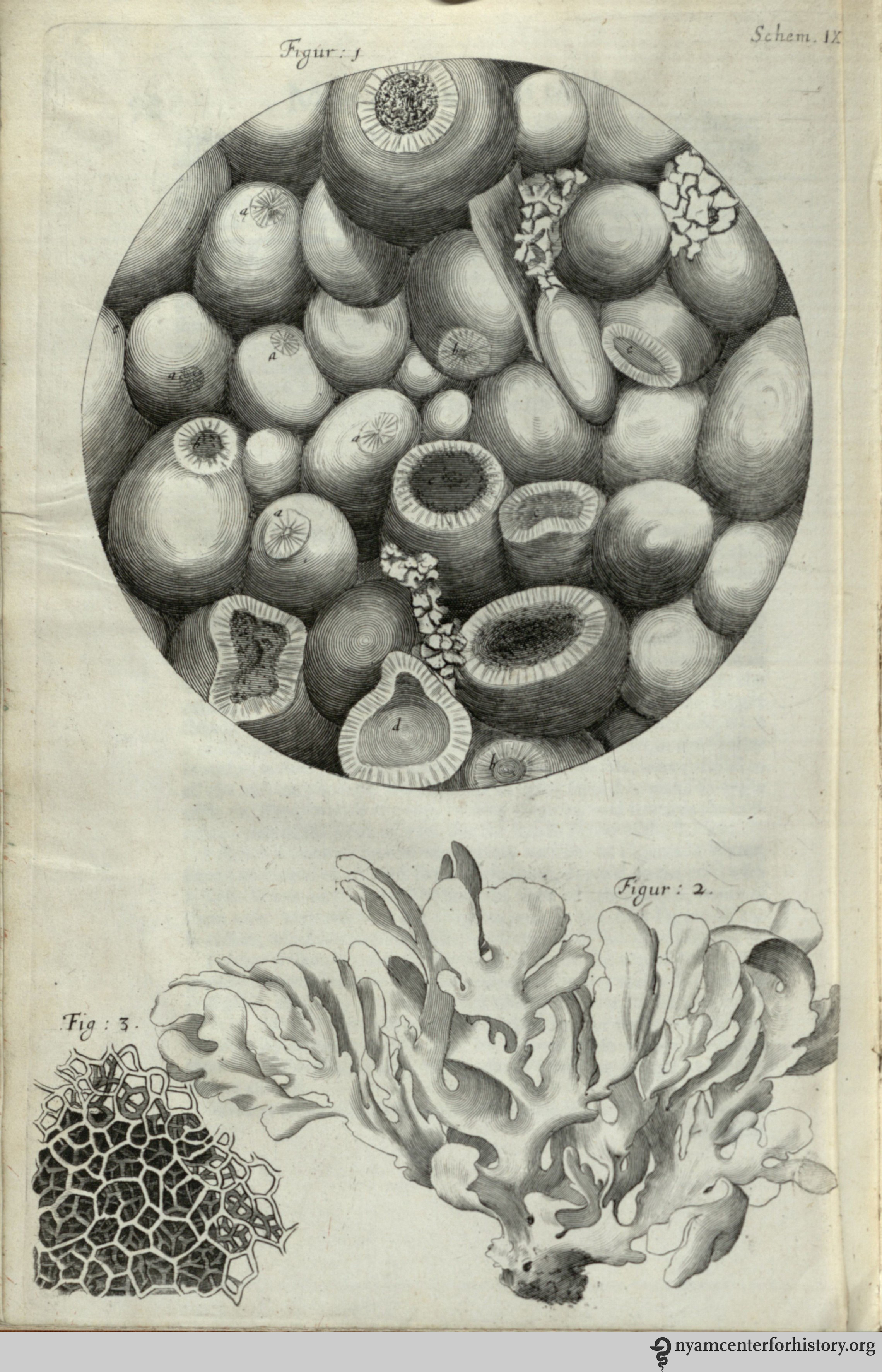
Fig. 1 shows a microscopic view of kettering-stone. In observation XV, Hooke notes, “We may here find a Stone by the help of a Microscope, to be made up of abundance of small Balls…and yet there being so many contacts, they make a firm hard mass…”

In his observation on cork, Hooke compared its structure to that of honeycomb. He discovered plant cells, “which were indeed the first microscopical pores I ever saw, and perhaps that were ever seen…,” and coined the term “cell.”
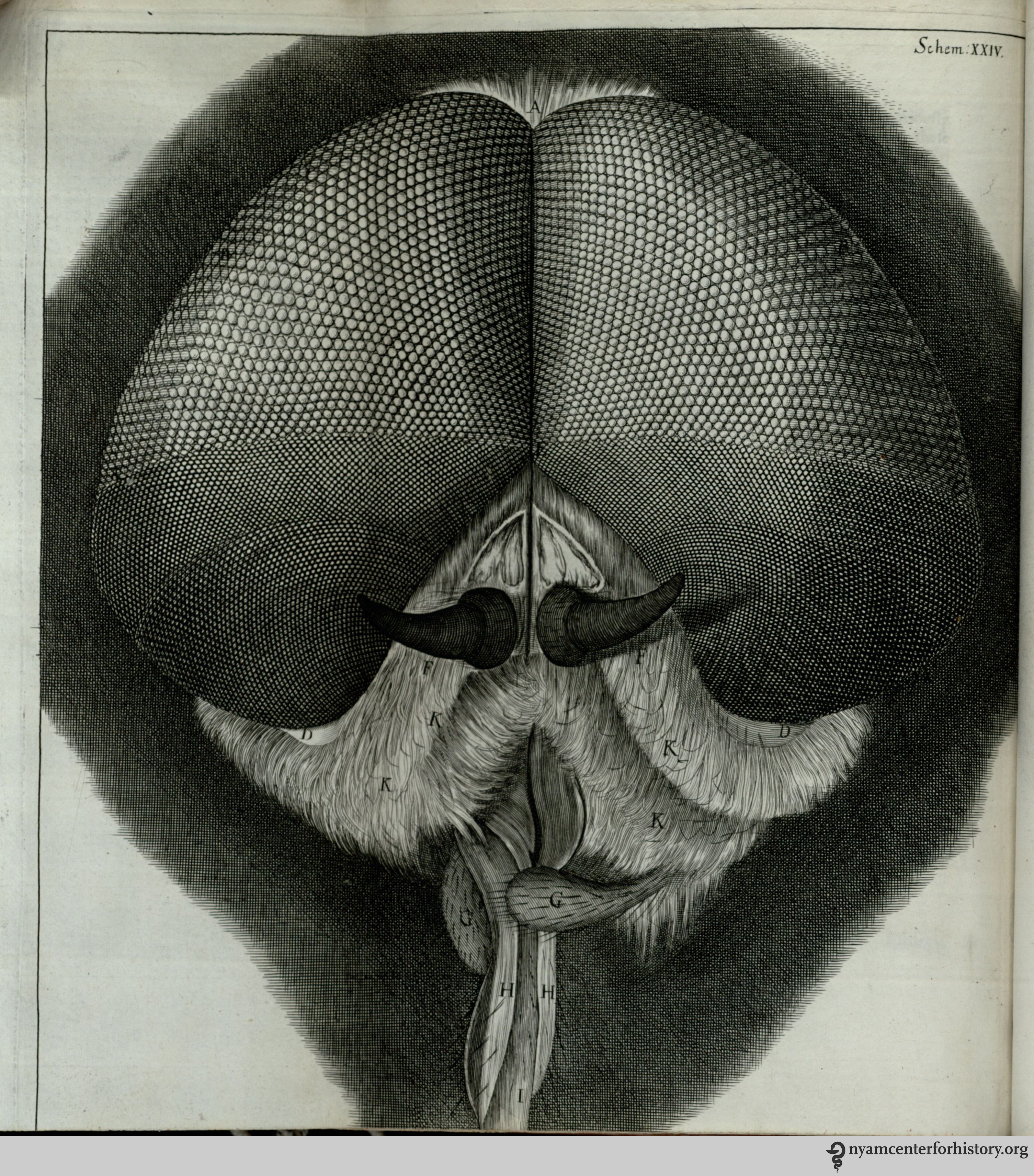
For observation XXXIV, Hooke examined the eyes and head of grey drone-fly.

Hooke seemed enamored with the white feather-winged moth, calling it a “pretty insect” and “a lovely object both to the naked Eye, and through a Microscope.”

The flea is one of several fold-out plates in the book. Again, Hooke has a scientist’s appreciation for the insect, commenting equally on its strength and beauty. He is particularly fascinated with the anatomy of its legs and joints, which “are so adapted, that he can…fold them short within another, and suddenly stretch, or spring them out to their whole length.”

In the last observations, Hooke turned his attention to celestial bodies. His study of the moon led him to surmise that the hills seen in Fig. 2 “may be covered with so thin a vegetable Coat, as we may observe the Hills with us to be, such as the short Sheep pasture which covers the Hills of Salisbury Plains.”
Reference
1. Espinasee, Margaret. Robert Hooke. London: Heinemann, [1956].

Hooke is always touted in biology classes as the one who used the word cell for a small, enclosed microscopic space, which eventually became the word for the living entity inhabiting those boundaries. But I had never seen any of his drawings except the one of cork.
When I took comparative anatomy in college, we had to do many dissections and draw what we saw. We had a cat dissection, a very detailed project that involved several layers of dissection. In the portfolio of my drawings, I included a microscopic view of one of the fleas from the cat fur (nowhere near as detailed as Hooke’s). The professor put several exclamation points on that drawing. Now I’m wondering if she was familiar with the drawings in Hooke’s Micrographia. (I did get an A on the project and for the course.)
Pingback: Whewell’s Gazette: Vol. 6 | Whewell's Ghost
Pingback: Whewell’s Gazette: Vol. 7 | Whewell's Ghost