Dr. William Edmund Aughinbaugh, Medical Adventurer (Item of the Month)

By Arlene Shaner, Historical Collections Reference Librarian

Dr. Aughinbaugh, circa 1915. In:

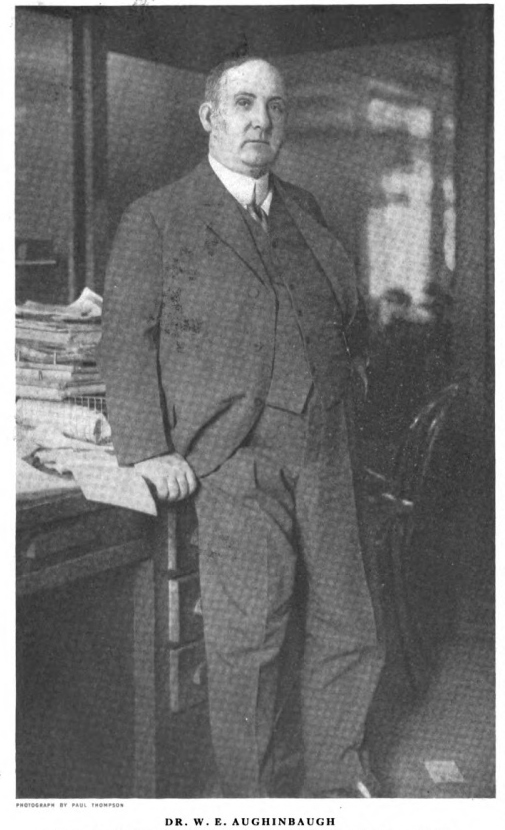
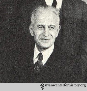
Dr. Aughinbaugh, circa 1915. In: “A Globe-Trotting Physician,” American Magazine, Nov. 1915, 34.

In the November 1915 issue of The American Magazine, the “Interesting People” section profiled an unusual physician. The article described Dr. William Edmund Aughinbaugh (18711940) as being “round like the earth; and he has rolled around it often. He has sawed bones and prescribed pills in every degree of latitude on both hemispheres.”1

As the article, and his autobiography, I Swear by Apollo, make clear, Aughinbaugh lived a life of adventure, traveling the globe for decades. Cuba, Venezuela, India, Peru, and Mexico were all early destinations where he treated lepers, studied the plague, and set up hospitals. He was a founder or early member of the Explorers, Adventurers, and Circumnavigators Clubs; taught courses about foreign trade at New York University and Columbia; and spent many years writing about and helping negotiate foreign trade agreements in Latin American countries and for South American natural resources.

The Academy’s manuscript collections contain a small album of photographs donated by the New York Public Library in 1952 (NYPL began sending items of medical interest that were given to them to the Academy in 1900). NYPL has a small collection of Aughinbaugh’s papers, mostly related to his work as the foreign editor of the New York Commercial. Aughinbaugh probably assembled the album between 1897 and 1906. Most of the photographs are unlabeled and trying to contextualize them has presented interesting challenges, demonstrating both the ways in which the written record helps us uncover more information and how much will probably remain forever unknowable.

It’s pretty clear that the first couple of photographs date to around 1895–97, when Aughinbaugh was a medical student at Columbian University (now George Washington) in Washington, D.C., and then an intern at Emergency Hospital there.

As both Aughinbaugh’s autobiography and his New York Times obituary attest, he helped finance his medical education by founding, with several other students, the Hippocratic Exhumation Corporation, essentially a grave-robbing operation. Aughinbaugh justified the less than savory labors of the corporation by assuring his readers that “care was always taken to undress the corpse and return the clothing to the grave…” as, according to court decree “a naked body belonged to no one—no crime would be committed by taking it.”2

Aughinbaugh insisted that he and his friends were not alone in this enterprise. Most medical students were desperate for bodies to dissect, and few legitimate ways to procure them existed. John Harley Warner and James Edmonson, in their recent book, Dissection: Photographs of a Rite of Passage in American Medicine: 1880-1930, corroborate this assertion, noting that in Washington, D.C., as well as in many states, there were no legal ways of obtaining bodies at that time, even though students were required to complete dissections to graduate. These two photographs, of Aughinbaugh (on the left) and two other students dissecting a body, and of Aughinbaugh and a fellow physician with a skeleton companion, fit right into the tradition of medical students posing with their cadavers in dissecting rooms.3

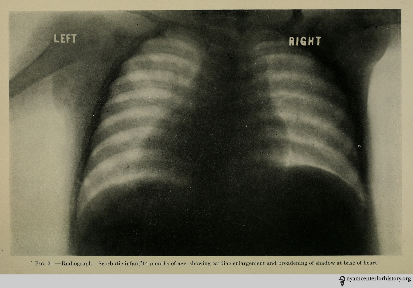
The album also contains posed portraits of patients suffering from diseases or showing the results of surgical operations. In some cases, Aughinbaugh pasted multiple photographs of the same patient into the scrapbook. The album dates from a time that witnessed the expanded use of photographs to document treatments and disease. While there is no way to be certain, these photographs may have been taken by Aughinbaugh himself.
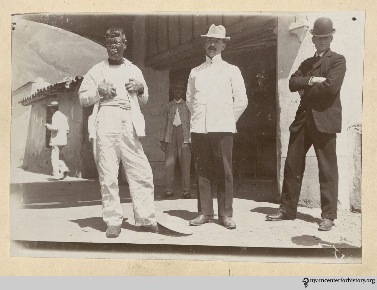
Another group of pictures shows groups of people that include Aughinbaugh himself (here in a white coat and hat). These images must date to Aughinbaugh’s years in Cuba. Having been denied the opportunity to enlist during the Spanish-American War in 1898 because of a heart condition, Aughinbaugh signed on as the ship’s surgeon for a vessel ferrying sick and wounded soldiers between Cuba and the United States. After the signing of the Treaty of Paris, he jumped ship while the boat was docked in Havana and stayed on as a civilian surgeon, working at the largest hospital in Cuba devoted to the care of leprosy patients, which, although he does not name it, must have been the hospital at San Lazaro, on the outskirts of Havana.4

Aughinbaugh’s autobiography provides real documentation for only a single photograph in his album. Aughinbaugh spent about four years (ca. 1902–1906) in India during a bubonic plague epidemic, working for the Indian Plague Commission. The picture shows an Indian ascetic suspended upside down over a fire. “I photographed one man who hung suspended by his feet from a banyan tree, while his youthful assistant built a fire of dried cow dung within a foot of his head,” Aughinbaugh writes, “When he was lowered, I… could not detect one sign of a burn”.5 He later submitted the photograph to a contest run by the New York Herald, won a prize, and added the clipping to the album.
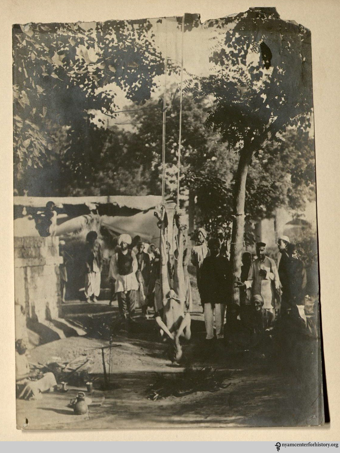
This album raises many questions, both about the use of photography by physicians to record information about medical practice and about the ways in which individuals choose to save images that document their own life experiences. Aughinbaugh’s choice to conflate the personal with the professional is part of what continues to make the album an intriguing part of our collections.

References

1. Barton, Bruce, “Globe Trotting Physician,” The American Magazine v.80 (Nov 1915), p. 34. Accessed online on July 29, 2015: http://hdl.handle.net/2027/coo.31924065598967?urlappend=%3Bseq=446

2. Aughinbaugh, W.E., I Swear by Apollo (New York: Farrar & Rineharrt, 1938), pp. 44-49. NYTimes obituary: http://query.nytimes.com/mem/archive/pdf?res=9C06E5D91F3CE73ABC4152DFB467838B659EDE Accessed online on July 29, 2015.

3. Warner, John Harley and James Edmonson, Dissection: photographs of a right of passage in American medicine, 1880-1930 (New York: Blast Books, 2009), pp. 17-19.

4. Aughinbaugh, pp. 103-113.

5. Aughinbaugh, p. 165.

How Air Conditioning Changed the NICU

By Johanna Goldberg, Information Services Librarian

We’ve entered the season of hot, humid, frizzy-headed misery outside and freezing temperatures from blasting office air conditioners inside. Which got me to thinking: What impact did air conditioning have on medicine?

Constantin P. Yaglou. From the Harvard School of Public Health 1955 yearbook.

Constantin P. Yaglou. From the Harvard School of Public Health 1955 yearbook.

One man did impressive work on this front. Constantin P. Yaglou (1897–1960) was not a physician, but a professor of industrial hygiene at Harvard’s School of Public Health. Born in Constantinople, he came to the United States in 1920 and earned a master’s degree from Cornell. He joined the Research Laboratory of the American Society of Heating and Ventilating Engineers in 1921, where he spent five years studying the influence of humidity, temperature, and air circulation on working and resting adults. In 1925, he joined the department of industrial hygiene at Harvard.1

His cross-disciplinary collaboration with Harvard Medical School’s pediatrics department, notably Dr. Kenneth Blackfan, proved innovative. Assisted by nurse Katherine MacKenzie Wyman, they published “The premature infant: A study of the effects of atmospheric conditions on growth and on development” in the American Journal of Diseases of Children in 1933.

The air conditioning unit in a nursery for premature infants. In “The premature infant: A study of the effects of atmospheric conditions on growth and on development,” American Journal of Diseases of Children, 1933, 46(5).

The air conditioning unit in a nursery for premature infants. In “The premature infant: A study of the effects of atmospheric conditions on growth and on development,” American Journal of Diseases of Children, 1933, 46(5).

They studied the effects of Harvard’s newly air conditioned nursery from 1926–1929, and compared their measurements to those from pre-air conditioned 1923–1925. (From 1926–1929, they controlled for variables like diet and dress.) They found that premature infants were less able to stabilize their body temperatures than infants born at term. Even among premature infants, ability to regulate temperature changed depending on birth weight. They determined the ideal temperature for premature newborns to be 75-100 degrees Fahrenheit with 65% humidity.2 These influential findings lay a foundation for the development and use of temperature-controlled incubators.3

Yaglou published a figure neatly summarizing the study’s major results in JAMA in 1938:

A summary of the results of the premature infant study. In "Hospital air conditioning," JAMA 1938, 110(24).

A summary of the results of the premature infant study. In “Hospital air conditioning,” JAMA, 1938, 110(24).

This figure comes from Yaglou’s broad-reaching “Hospital Air Conditioning,” which brought together studies on air conditioning’s effects in the operating room, recovery wards, premature nurseries (summarizing his prior work, as in the figure above), fever cabinets, allergen-free rooms, and oxygen chambers.4

According to the article, not only did air-conditioned operating rooms help those involved in surgery feel more comfortable, it also reduced “the risk of explosion of certain anesthetic gases.”4 In the post-operative recovery rooms, air conditioning reduced the risk of heat stroke and improved the body’s ability to recuperate, though Yaglou did not recommend a particularly cool temperature. “With a relative humidity of about 55 per cent,” he wrote, “a temperature of about 80 will probably prove acceptable.”

But even with the benefits of air conditioning discussed in the article, it was difficult to employ at a large scale in the late 1930s. Yaglou concluded, “High cost precludes cooling the entire hospital, but the needs of the average hospital may be satisfactorily fulfilled by the use of built-in room coolers in certain sections of the hospital and a few portable units which can be wheeled from ward to ward when needed.”4

In addition to his work in medical settings, Yaglou also performed military research on extreme climates like the Yukon, the tropics, and the Arizona desert, “working with volunteers to determine the limits of human endurance under severe heat, cold and humidity.”5 Perhaps it should come as no surprise that the UK Antarctic Place-names Committee christened Yaglou Point in his honor in 1965.6

References

1. Whittenberger JL, Fair GM. Constantin Prodromos Yaglou. Arch Environ Heal An Int J. 1961;2(2):93–94. doi:10.1080/00039896.1961.10662820.

2. Blackfan KD, Yaglou CP, Wyman KM. The premature infant: A study of the effects of atmospheric conditions on growth and on development. Am J Dis Child. 1933;46(5):1175–1236. doi:10.1001/archpedi.1933.01960060001001.

3. Rutter TL. Comfort zone. Harvard Public Heal Rev. 1997:29.

4. Yaglou CP. Hospital air conditioning. J Am Med Assoc. 1938;110(24):2003–2009. doi:10.1001/jama.1938.62790240003010.

5. Constantin Yaglou, Harvard Professor. New York Times. http://query.nytimes.com/gst/abstract.html?res=9A06E6DF123DE333A25757C0A9609C946191D6CF. Published June 4, 1960. Accessed July 21, 2015.

6. Yaglou Point, Antarctica – Geographical Names, map, geographic coordinates. Available at: http://www.geographic.org/geographic_names/antname.php?uni=16847&fid=antgeo_126. Accessed July 21, 2015.

X-raying Orphans: Fictionalizing Medical History in Orphan #8

Guest author Kim van Alkemade has a doctorate in English from the University of Wisconsin-Milwaukee and is a professor at Shippensburg University in Pennsylvania. Orphan #8 is her first novel.

“They weren’t treatments,” I interrupted, surprising both of us with my vehemence. “It was an experiment. I was experimented on, not treated.”1

The premise of my historical novel Orphan #8 is this: in 1919, four-year-old Rachel Rabinowitz is placed in a Jewish orphanage in New York where the fictional Dr. Mildred Solomon is conducting X-ray research using the children as her subjects. Years later, Rachel, who has become a nurse, is given the opportunity for a reckoning with her past when old Dr. Solomon becomes her patient. While the novel is fiction, medical research on children in orphanages was a common practice, and a child like Rachel Rabinowitz would not have been unique at the time. Not only were children “used as subjects in a number of experiments involving X-rays”2 but a “preponderance of the children subjects were poor, institutionalized, mentally ill, physically disabled, or chronically ill.”3

A dormitory in the Hebrew Infant Asylum. From Annual Report 1914 Hebrew Infant Asylum of New York.

A dormitory in the Hebrew Infant Asylum. From Annual Report 1914 Hebrew Infant Asylum of New York.

The inspiration for the novel arose from research I was doing about Jewish orphanages for a family history project. In the archives of the American Jewish Historical Society, I read that Dr. Elsie Fox, a graduate of Cornell Medical School, X-rayed a group of eight children at the Home for Hebrew Infants in New York City, resulting in persistent alopecia. Upon the transfer of these children to the Hebrew Orphan Asylum in October 1919, the Board of Trustees discussed what to do in the “matter of the children received with bald heads.” On November 9, 1919, they entered into their meeting minutes a letter from the Home for Hebrew Infants “assuming responsibility… for the condition of these children.” The letter refers to an enclosure of data about the eight children, as well as a letter from Dr. Fox detailing her X-ray treatments. Unfortunately, the enclosures were not entered into the minutes. On May 16, 1920, the matter was put to rest when the Trustees “ordered that children afflicted with alopecia should have wigs made, and be boarded out, if possible.”4

Detail of the Meeting Minutes of the Board of Trustees of the Hebrew Orphan Asylum. Courtesy of the American Jewish Historical Society.

Detail of the Meeting Minutes of the Board of Trustees of the Hebrew Orphan Asylum. Courtesy of the American Jewish Historical Society.

Dr. Solomon blinked, confused. She stared at me as if trying to focus on print too small to read. “You were one of my subjects?”

I nodded, imagining for a moment that she recognized me: her brave, good girl. She lifted her hand to my face, bent my head back to expose the underside of my chin. Her thumbnail circled the scars there, tracing the dimes of shiny skin. Then she placed her fingers against my drawn eyebrows and wiped away the pencil. Finally, she reached up to my hairline and pushed along the brow. My wig shifted. She pulled her hand back in surprise. It wasn’t tenderness I saw in her face, not even regret. Fear, maybe? No, not even that.

“So the alopecia was never resolved? I was curious about that, always meant to follow up. What number were you?”

I adjusted my wig. “Number eight.”5

Though I invented the character of Dr. Mildred Solomon before I discovered more about Dr. Elsie Fox, it turned out the real person was similar to my fictional character. Elsie Fox was born in Vienna, Austria, in 1885. When she graduated from Cornell with her medical degree in 1911, she was one of 8 women in a class of 53 graduates. She became a fellow of the New York Academy of Medicine in 1916, and was a member of the Bronx Roentgen Ray Society.6 A published medical researcher, she went on to become the Director of the Harvey School for the Training of Analytical and X-ray Technicians in Manhattan and was a Roentgenologist at City Hospital. She was 58 when she died in June 1943.

From Hess, Alfred F., M. D. Scurvy, past and present. Philadelphia, J.B. Lippincott Company, 1920.

From Hess, Alfred F., M. D. Scurvy, past and present. Philadelphia, J.B. Lippincott Company, 1920.

In my novel, I paired the fictional Dr. Solomon with a character closely based on a real orphanage pediatrician of the time. Dr. Alfred F. Hess was attending physician to the Hebrew Infant Asylum and a renowned researcher into childhood nutritional diseases. He was the innovator of an infant isolation ward at the orphanage in which babies were kept in separate glassed-in rooms to avoid the spread of disease. Hess is well-known for a quote in which he extolled the advantages of conducting research on “institutional children” who provided the advantage of belonging to “the same stratum of society,” being “reared within the same walls,” and having the “same daily routine, including similar food and an equal amount of outdoor life.” He concluded: “These are some of the conditions which are insisted on in considering the course of experimental infection among laboratory animals, but which can rarely be controlled in a study in man.”7

Glassed-in babies, from Annual Report 1914 Hebrew Infant Asylum of New York.

Glassed-in babies, from Annual Report 1914 Hebrew Infant Asylum of New York.

Dr. Hess’s approach to the study of scurvy, which involved inducing the condition in children and then experimenting with various cures, was controversial even in his lifetime. In 1921, Hess was criticized “for using ‘orphans as guinea pigs’ in studies of the dietary factors in rickets and scurvy” by “withholding orange juice from institutionalized infants until they developed the characteristic small hemorrhages associated with the disease.”8

From Hess, Alfred F., M. D. Scurvy, past and present. Philadelphia, J.B. Lippincott Company, 1920.

From Hess, Alfred F., M. D. Scurvy, past and present. Philadelphia, J.B. Lippincott Company, 1920.

“My name is Rachel, I’ve told you that. But you don’t care, do you? Even now, I’m just a number to you. All the children at the Infant Home were nothing more than numbers to you.” I thought of the tattoo on Mr. Mendelsohn’s frail arm. “Just numbers, like in the concentration camps.”

She gripped the sheets. “How can you say such a thing? You were in an orphanage, not some concentration camp. They took care of you, fed you, clothed you. Jewish charities support the best orphanages, the best hospitals. Even this Home is as good as it gets for old people like me. You have no right to even mention the camps.”

Of course the orphanage wasn’t a death camp, I knew that, but I wasn’t backing down. “You came into a place where we were powerless, you gave us numbers, subjected us to experiments in the name of science. How is that different?”9

When I would tell people about the medical experimentation on children depicted in my novel, they would often say it sounded like something the Nazis would do. As first I was impatient with the comparison: these experiments were conducted well before the rise Hitler in Germany, and the doctors conducting the research, many of them Jewish themselves, intended to advance medicine for the benefit of all children. Yet, as I thought about it from the point of view of one of the child subjects, I wondered if that distinction would matter.

It is easy for contemporary readers to conflate all medical experimentation on children with the atrocities of the Holocaust, but even after “the world was outraged at the murders carried out in the name of science by Nazi physicians during World War II,”10 some American doctors continued to use orphans, prisoners, and other disenfranchised populations in medical research without their consent. In my novel Orphan #8, I bring this aspect of medical history to general readers through the use of narrative and story. Medical students and physicians may also find that fiction provides an opportunity to explore these complex issues with empathy and imagination and to engage a wider community in the discussion of medical ethics.

References

1. van Alkemade, Kim. Orphan #8 (New York: William Morrow, 2015), 232.

2. Lederer, Susan E. and Michael A. Grodin. “Historical Overview: Pediatric Experimentation.” In Grodin, Michael A. and Leonard H. Glantz. Children as Research Subjects: Science, Ethics, and Law (New York: Oxford University Press, 1994), 10.

3. Lederer and Grodin, 19-20.

4. Executive Committee Minutes 1909-1930. Hebrew Orphan Asylum Collection, Archives of the American Jewish Historical Society, Center for Jewish History, 15 West 16th Street, New York, NY.

5. van Alkemade, 173.

6. The Bulletin of the New York Academy of Medicine. September 19 (1943): 676.

7. Lederer, Susan E. “Orphans as Guinea Pigs: American Children and Medical Experimenters, 1890-1930.” In Roger Cooter, ed. In The Name of the Child: Health and Welfare, 1880-1940 (New York: Routledge, 1992), 115.

8. Lederer and Grodin, 13.

9. van Alkemade, 282.

10. Lederer and Grodin, 16.

Adventures in Rare Book Cataloging

By Tatyana Pakhladzhyan, Rare Book Cataloguer

At the October festival celebrating the 500th birthday of anatomist Andreas Vesalius, The Drs. Barry and Bobbi Coller Rare Book Reading Room exhibited seven anatomical works drawn from the library’s extensive rare book holdings. Anatomy is one of the library’s major collecting strengths, including works by and related to Andreas Vesalius.

Visitors looking at books on display at 2014's Vesalius 500 festival.

Visitors looking at books on display at 2014’s Vesalius 500 festival. Photograph by Charles Manley.

Since the exhibited materials have been in the library’s collection for decades, I was curious to see how their online bibliographic records looked. As card catalogs turned into online catalogs at the end of last century, collection holdings became increasingly findable from far away. But in the process of converting card catalog records into online records, some items ended up with incomplete or incorrect information reflected in the online catalog. I found that the records of the seven anatomical holdings required some attention.

The purpose of rare book cataloging is to create elaborate catalog records for books printed during the hand-press period (c.1455c.1830) and to describe and record copy-specific information that would uniquely identify the library’s holding from other copies of the same title. Descriptive cataloging should be sufficiently detailed to represent the work.

Female flap anatomy from The Academy's copy of the 1559 English edition of Geminus’ Compendiosa.

Female flap anatomy from The Academy’s copy of the 1559 English edition of Geminus’ Compendiosa.

Rare book cataloging requires complete and faithful transcription of the title page in its original language, greater detail in the physical description area, and careful and thorough recording of various distinguishing points in the note area, including signature statements, identification of bibliographic format, annotations, pagination errors, illustration techniques and creators, printing method, binding style, and provenance. Full and accurate descriptions allow researchers to find materials in online catalogs. Adding images or links to digital copies is another catalog feature that allows for more sophisticated experience for rare material users.

I was particularly delighted to update the catalog record for the 1559 edition of Geminus’ Compendiosa totius anatomiae delineatio, aere exarata (A complete delineation of the entire anatomy engraved on copper). This beautiful folio is simply a work of art! Read more about the work in a recent blog post.

Male flap anatomy from The Academy's copy of the 1559 English edition of Geminus’ Compendiosa.

Male flap anatomy from The Academy’s copy of the 1559 English edition of Geminus’ Compendiosa.

The title page is an engraved plate, with a hand-colored portrait of Queen Elizabeth at center and the royal motto “Dieu et mon droit” under the portrait. Facing the title is the leaf with arms of the Order of the Garter “Honi soit qui mal y pense,” decorated with jewels. (Thanks to my library colleagues for helping me prove that “Honi soit qui mal y pense” motto is, in fact, the motto of the Order of the Garter.)

The coat of arms, left, and title page, right, of the Academy's copy of the 1559 English edition of Geminus’ Compendiosa.

The coat of arms, left, and title page, right, of the Academy’s copy of the 1559 English edition of Geminus’ Compendiosa.

Checking standard bibliographies for corresponding period and making identifying references is an essential step to rare book cataloging. While consulting A Bio-Bibliography of Andreas Vesalius by Harvey Cushing, (1943, no. VI.C-4, p. 128), I found his comment about known copies at that time, stating that the “leaf before title bearing royal arms and ‘Honi soit qui mal y pense,’ is missing in all copies but London (BM [British Museum]).” Our copy has this leaf, seen above left.

Rare book cataloging also requires pointing out differences between printings, or manifestations, of a particular work. While consulting the English Short Title Catalogue (ESTC) that lists more than 480,000 items published between 1473 and 1800, I found that the entry for this work has a note, “a variant state has B7 unsigned.” In the hand-press era, books were printed as sheets with varying numbers of pages per side, with signature marks as letters, numbers, or symbols at the bottom of each leaf to help binders assemble the sheets of a book into the right order. I was curious to find out if the NYAM copy was a variation with signature B7 unsigned, but it is signed, although not on the bottom of the page.

Note "B.vii" hiding at the bottom right of the page. The Academy's copy of the 1559 English edition of Geminus’ Compendiosa.

Note “B.vii” hiding under the text at the right of the page. The Academy’s copy of the 1559 English edition of Geminus’ Compendiosa.

The library’s 1559 edition, the English translation by Nicholas Udall, is a reissue of the 1553 edition, with a slightly different title page, a dedication, and a colophon leaf. Bookseller information from the colophon at foot of last leaf reads: “Imprinted at London within the blacke fryars: by Thomas Gemini. Anno Salutis. 1559. Mense Septemb.”

Final leaf with colophon. The Academy's copy of the 1559 English edition of Geminus’ Compendiosa.

Final leaf with colophon. The Academy’s copy of the 1559 English edition of Geminus’ Compendiosa.

Cataloging rare books is an exciting process and sometimes even an adventure, as older books are unique and carry impressions of their formal owners. Our copy’s provenance includes bookplate of bibliophile George Dunn, “From the Library of George Dunn of Woolley Hall near Maidenhead.” It was a generous gift to the Academy library from Mrs. George S. Huntington, the wife of a prominent anatomist.