Color Our Collections 2024


by Anthony Murisco, Public Engagement Librarian

On Monday, February 5th, we kicked off the eighth annual “Color Our Collections!” This initiative asks libraries, museums, and other cultural institutions to submit coloring books based on their collections. These coloring books don’t just live on our site for that one week; you can access them whenever you want! Over the years we have collected over 800 free coloring books for everyone to enjoy.

Maybe you need an excuse to color? Well, besides just being a fun activity, coloring has been shown to provide other benefits. Research has shown that it makes your brain feel a sense of relaxation. You don’t have to be an artist to break out crayons or colored pencils. The “low stakes” nature of the activity lets you enjoy it more. There may also be the subconscious thought of nostalgia lingering as you color; this used to be something you enjoyed! These factors combine to provide therapeutic effects to coloring (though it is important to keep in mind “therapeutic” is not the same as actual therapy!).

For the NYAM coloring book this year, we chose images of what it was like to be healthy in the 19th and 20th centuries. “Living Well in the 19th and 20th Century” shows activities meant to relax and help people taking care of one another, a woman ready for a bike ride, and acquaintances stopping to shoot the breeze on the grass. These are just some routes that you may take when practicing your own wellness routines.

You can use the hashtag #ColorOurCollections to show off your own work and see what others are doing.

A huge thank-you to our colleagues worldwide who have contributed coloring books to the collection. We couldn’t do it without you!

References:
Ali, Shainna. “Are Adult Coloring Books Actually Helpful?” Psychology Today, 27 Mar. 2018, http://www.psychologytoday.com/us/blog/modern-mentality/201803/are-adult-coloring-books-actually-helpful, accessed February 5th, 2024.

“3 Reasons Adult Coloring Can Actually Relax Your Brain.” Cleveland Clinic healthessentials, 26 May 2020, http://health.clevelandclinic.org/3-reasons-adult-coloring-can-actually-relax-brain, accessed February 5th, 2024.

Maker’s Mark: A Look at Early Modern Printers’ Devices

By Emily Miranker, Events & Project Manager

Did you know that required trademarks go back to 1266? In England, bakers were required by parliament to use a distinctive mark on the bread they sold.[i] Fun design/history/bibliographic fact, the Drs. Barry and Bobbi Coller Rare Book Reading Room here at the Library features trademarks in its décor. More specifically, the room’s chandelier have printers’ marks. As an homage to book history and the art of the book, the chandeliers of our reading room are decorated with printers’ marks.

RBR chandelier

I got to know these marks beyond “those pretty design bits on the lights” when we created special bookplates (another age old way to ‘mark’ your stuff) for Adopt-a-Book donors. The virtual bookplates that donors receive features four of these marks keeping them connected with the legacy and art of the book.

TIna's first demo of bookplate sketches

Our incredibly talented graphic designer sharing sample sketches for the adoption bookplates; artistic inspiration courtesy of early modern printers, the architecture of the rare book room and the Academy building.

As the name suggests, printers’ marks are a device or emblem, like a logo, that early printers used to make clear the source of the item. According to Printer’s Marks, the first of these is Johann Fust and Peter Schöffer’s Mainz Psalter of 1457. Among the best well-known of these old printers’ marks is one that you will find on our library’s custom designed chandeliers and on our adoption bookplates (upper righthand corner) the device of Aldus Manutius: the dolphin and anchor.


Hippocrates_Omnia Opera_1526-printers mark_watermark

The dolphin twined around an anchor predates Manutius. Going back to Roman times, this pair symbolizes the adage, “Make haste slowly.” (The dolphin is haste, and the anchor is slow.)

Next to Aldus in the upper left corner of the bookplate, the ethereal hand manipulating the compass with the Latin motto Labore et Constantia (Work and Constancy) belongs to Dutch publisher Christophe Plantin (1520-1589). During his life, he used a large number of devices and they could vary in appearance. There are three primary types; the first features a tree and the second a scroll with a Latin motto twined around a grape vine; the third is the hand and compass and first appeared in 1557.[ii] The compass is symbolic of the motto: the leg of the compass turning around is work while the stationary point is constancy.

Below Plantin’s mark on the lower left, is the printer’s mark of Paris printer and bookseller Poncet Le Preux (1508 – 1551). His initials P L P are ‘tethered’ together by a tasseled cord.

Lastly, the monogram in the lower right corner of the bookplate that also adorns our chandeliers belongs to Badius Ascensius or Jodocus Badius (1462 – 1535). Originally from Flemish town of Asche, he set up a print shop in Paris, Prelum Ascensianum, in 1503. The initials in the monogram are I V A B, the A and V intersecting to form the diamond shape at the center, which stand for his Latinate name Iodocus Van Asche Badius.

Your Name Here bookplateWe invite you to come look at these gorgeous marks on the chandeliers and in the books themselves at our First Monday tours. The first Monday of every month at 12 pm we do a free tour of the Rare Book Room. Or adopt a book in our collection and receive a copy of these marks in the custom designed donor bookplate.

Bonus mark! This is the mark used by Badius’ printing house, Prelum Ascensianum (his monogram featuring at the bottom center, the shop’s name visible on the center crossbeam of the press itself) and my personal favorite because it is a printer in action.Beroaldi_Opvscvlvm _1511-tp-ornament_watermark

References:
[i] https://en.wikipedia.org/wiki/Trademark#History Accessed 4/18/18
[ii] Roberts, William. Printer’s Marks: A Chapter in the History of Typography (New York: George Bell & Sons, 1893).
printermark shop ad

Embroidering Medicine, Re-imagining Embroidery

Today’s guest post is written by Kriota Willberg, New York Academy of Medicine’s Artist-in-Residence researching the history of sutures and ligatures.  Through graphic narratives, teaching, and needlework, Kriota explores the intersection between body sciences and creative practice. This past September, Kriota taught a four-week workshop entitled “Embroidering Medicine,” which explored the relationships between medicine, needlework, and gender.

Historically, needlework has been used to enforce stereotypes of women as docile, obedient, and incapable of creative or original thought.  Embroidery as a skill and a decorative medium was a required part of a woman’s education for centuries.  Across different periods of time, embroidery techniques (stump work, black work, etc.) and themes (religious, botany, etc.) in vogue provided only very narrow focus for creative outlets for women.  From the 16th century onwards, embroidery publications encouraged the use of pre-drawn patterns.  Images generally included geometric stitch motifs, flowers, plants, and animals. Many women, including Mary Queen of Scots translated images from books such as the Icones Animalium (1560) and La Nature et Diversité des Poissoins (1555).[1]

Image1

Workshop participants discuss their work.

The cultural pressure to recreate existing patterns and images has been the basis of the argument that a craft such as embroidery is inherently unoriginal. In the 16th century and later, many people considered women incapable of original creative thought, proved by the adherence of women to unimaginative media such as embroidery.

As the Academy’s Artist-in-Residence, I wanted to facilitate an embroidery workshop that connected the histories of women, medicine, and embroidery. In my own work I have done this by looking at the broad scope of health and medical literature available at the Academy Library, exploring feminist histories of medicine and of needlework, and using drawing and needlework to identify and describe intersections of medical and textile arts. In the workshop I encouraged participants to re-imagine the tradition of embroidery pattern translation by using historical medical imagery mostly created by male artists.

Image2

Dental anatomy after an image by John Hunter by Stephanie Russell.

The workshop was a great way to explore history on one’s own terms. Cultural forces limiting women’s economic and artistic independence have been at work for centuries in Europe and the United States. The establishment and regulation of craft and medical guilds in Medieval Europe began the limitation of women from the professional practices of medicine and needlework.[2] While working with materials from the historical collection, the status of women as medical professionals during different periods was a topic of conversation as we stitched our embroidery.

Image3

An embroidery in progress by Abby Tannenbaum, stitched from an image in the Hortus Sanitatis.

Our group (coincidentally all women) explored the library images, learned about the history of medicine, examined books about home economics and sciences, found images to work with that were interesting or personally relevant, explored historical descriptions of medical needlework (i.e. suturing the body), and practiced embroidery stitches. The products of this workshop embody the stitcher’s process and creative experimentation combining the history of medicine with the histories of feminism and the decorative art of embroidery.

Image4

Kriota Willberg (third from left) and embroidery participants in the Drs. Barry and Bobbi Coller Rare Book Reading Room.

There is a lot of creativity that can go in to re-imagining a pre-existing image. In addition to working with line, filling, and color, embroidery can add texture and depth through its use of various types of threads and yarns. The embroiderer studies the image she is recreating. Is it educational? Entertaining? Are certain anatomical structures or pathological states emphasized? The embroiderer interprets the original artist’s intentions, determines her own interests in the image, and re-works the image using color, texture, stitch variety, and fabric, to create a new work. To practice basic stitches, an embroiderer might choose to follow an original pattern closely or experiment and make “mistakes,” as one might when drawing in a sketchbook.

Image5

Anatomized genitalia from an anatomy book by Adriaan van de Spiegel (aka Spiegalius) and Giulio Cesare Casseri (aka Casserius) published in the 17th century. Stitched by Susan Shaw.

Embroidery is becoming a popular medium again and still provokes associations with hominess and winsome imagery. I was delighted to see workshop embroiderers re-imaging pictures of dental anatomy and dissected genitalia into images that are simultaneously comforting and disturbing. The embroiderers in this workshop are taking needlework in new and exciting directions!

Endnotes:
[1] Swain, Margaret. The Needlework or Mary Queen of Scots. New York, London: Man Nostrand Reinhold Company, Inc., 1973.
[2] I recommend reading The Subversive Stitch by Rozsika Parker and Women and the Practice of Medical Care in Early Modern Europe, 1400-1800 by Leigh Ann Whaley. Reading these books simultaneously has been a revelatory experience!

Who Practices “Visualizing Anatomy”?

Today’s guest post is written by Kriota Willberg, New York Academy of Medicine’s Artist-in-Residence.  Through graphic narratives, teaching, and needlework, Kriota explores the intersection between body sciences and creative practice. This May, Kriota taught a four-week workshop entitled “Visualizing and Drawing Anatomy,” which utilized live models as well as anatomical illustrations from the New York Academy of Medicine’s library. You can read more about Kriota’s work HERE.

Class

The class gets oriented before drawing practice.

The Visualizing and Drawing Anatomy workshop was held at the Academy Tuesday evenings in June.  Once again I was impressed by the participants willingness to practice looking underneath our models’ skin to draw the deep anatomical structures that give our bodies form.

iPad

Participants draw using their preferred medium, in this case, paper or an iPad.

Who benefits from this kind of drawing practice? Practically everyone. Trained artists sharpen their skills, and those new to art and drawing learn fundamental principles of anatomy that lay the foundation for drawing the human figure.

Debbie Rabina, who is new to art, took the workshop last year.  Since then she has kept a regular drawing practice and she occasionally incorporates anatomy into her work.

Rabina

Debbie Rabina’s drawing since taking “Visualizing Anatomy” in 2016.

Ellen Zaraoff is a photographer who has just started drawing. Until taking the classes this year she had been focusing on drawing portraits in charcoal.  She took the workshop to get an introduction to anatomy, structure, and proportion.

Sarah Wukoson has a BA in art, and works in medical research. She took the workshop this year because she’s interested in the intersection of art and medicine as well as “the interplay of different modes of understanding the body.”

Wukoson

Sarah Wukoson’s 2017 in-class sketches and exercises.

Jim Doolley is a “life-long art lover who decided a couple years ago to take a stab at producing, not just consuming.” His focus is drawing and painting. He took this class to improve his draftsmanship.

Dooley

Jim Dooley’s 2017 homework.

Susan Shaw is an artist.  She says, “I took the class (last year) because I found I was thinking 2 dimensionally when I was drawing and the figures seemed to have no life… I now think about how the body functions when I draw and it makes gesture and weighting much easier.”

Shaw

Susan Shaw’s figure drawing since taking “Visualizing Anatomy” in 2016.

The variety of participants: artists, illustrators, cartoonists and enthusiastic beginners – all interested in anatomy and the Library’s historical collection make this workshop one of my favorites to teach.

This September 14-October 5, Kriota is offering an “Embroidering Medicine Workshop,” which will take place at the Academy.  This four-week workshop explores The New York Academy of Medicine Library’s historical collections, examining relationships between medicine, needlework, and gender. Learn more and register HERE.


Robert L. Dickinson: Doctor and Artist

Today’s guest post is written by Rose Holz, Ph.D., historian of medicine and sexuality at the University of Nebraska – Lincoln where she serves as the Associate Director of the Women’s & Gender Studies Program and Director of Humanities in Medicine.  She is the author of The Birth Control Clinic in a Marketplace World (Rochester, 2012). Her current project investigates the intersection of medicine and art by way Dr. Robert L. Dickinson (1861-1950) — gynecologist, sexologist, and artist extraordinaire — and his prolific ten-year collaboration with fellow artist Abram Belskie (1907-1988). Not only did it yield in 1939 the hugely influential Birth Series sculptures but also hundreds of medical teaching models about women’s and men’s sexual anatomies. On Thursday, April 13, Rose will give her talk, “Art in the Service of Medical Education: The Robert L. Dickinson-Belskie Birth Series and the Use of Sculpture to Teach the Process of Human Development from Fertilization Through Delivery.” To read more about this lecture and to register, go HERE.

My interest in Dr. Robert L. Dickinson began many years ago when I was in graduate school, working on my Ph.D. in history and writing my dissertation on the history of birth control clinics in America. And, as has been the case with so many other scholars who have written about matters related to women, medicine, and sexuality in the twentieth century U.S., Dickinson made his brief cameo entrance into my story, though not without leaving behind a lasting impression.

For me it was the images — because, like me, Dickinson was compelled to color and draw. Early on, while pouring over Planned Parenthood records, I remember chuckling over a letter he had written to a contraceptive manufacturer complaining about the poor quality of one of their products, to which he then attached a drawing to illustrate his case.

Then there were the birth control manuals Dickinson wrote in the 1930s. Not only did he illustrate all the contraceptive methods then available, but he also offered birds-eye-view, architectural-style drawings to visualize how best to lay out gynecological clinics. More intriguingly still was what he included at the center of this architectural drawing, a tiny woman lying on the gynecological table with her legs spread wide open as the doctor conducted the physical exam.

IMG_4813

Pages from “Control of Contraception (2nd edition)” by Robert L. Dickinson.

As somebody who also loves small things—especially miniature worlds populated by miniature people—I could not help but find myself be smitten by this unusual man. However, at the time I had a different story to tell, a Ph.D. to defend, and a new job as a professor to pursue. And as the years passed, Dickinson slowly receded into the background.

IMG_1603

Drawings of the location of Embryo and size of Fetus. Source.

But Dickinson is not one to be denied, and that he has remained in obscurity for so long somehow explains to me why he has resurfaced—with a glorious vengeance—into my imagination. Indeed, he has made it clear to me that his story will be told; his skills as a doctor and artist properly recognized. And he has made it further clear that this story will begin with what he created in the twilight of his life: The 1939 Birth Series sculptures.

IMG_2449

Dickinson and Belskie’s “Sculptured Teaching Models Collection.” From the unprocessed Abram Belskie Papers, Belskie Museum, Closter, NJ.

Join us on Thursday, April 13 to learn more about Dr. Robert L. Dickinson and his Birth Series sculptures. To RSVP to this free lecture, click HERE.

Charles Bell: Artistry and Anatomy

By Anne Garner, Curator, Rare Books and Manuscripts

1bell_engravingsofthearteries_1801-plate1-crop_watermark

Close-up of plate 1 showing the heart, from Charles Bell’s Engravings of the Arteries, 1801.

Imagine you’re a medical student, and inside your go-to study text is this astonishing, hand-colored image of the heart (above). Twenty-first century medical students should be so lucky!

Engravings of the Arteries by the Scottish anatomist, Charles Bell (1774-1842), was first published in 1801, some 57 years before the first edition of Henry Gray’s Anatomy. Devised for medical students, it aimed to offer accurate and simply-rendered illustrations of the arteries to “present to the student at one glance the general distribution of the vessels and to fix them in his memory.”  The book was used by students as a preparatory text for surgical study and practice.

The ten beautifully-rendered engravings in this volume were delicately colored by hand, and labelled with letters corresponding to explanatory descriptions of the arteries on the opposite page.  In the preface to this work, Bell is explicit in his instructions on how the book was to be used:

In studying the arteries, or any part of anatomy, we should, in the first place, run the eye over the corresponding plate, then read the general description in the text; and lastly, proceed to study more closely, step by step.

For Bell, true anatomical understanding was aided in pairing accurate drawing with thorough description.  Bell also believed that a variety of bodies should be used as subjects, and that the artist must choose the most typical anatomical examples to copy accurately. Any deviation from usual forms would be preserved in the illustration, but noted and explained in the description.

3bell_engravingsofthearteries_1801_3_watermark

Plate 3, showing the carotid artery, the lower thyroid artery and the upper thyroid artery in Charles Bell’s Engravings of the Arteries, 1801.

Engravings of the Arteries was the second volume of two; Charles’ brother, John Bell, an eminent surgeon, had written the first volume of this companion set, Engravings, Explaining the Anatomy of the Bones, Muscles, and Joints, in 1794. The elder brother mentored the younger in medicine, though Charles’s formal training occurred at Edinburgh University (he earned his medical degree there in 1798.)  Both Charles and John taught anatomy at the Royal College of Surgeons, until 1804, when the brothers were banned from practicing medicine in Edinburgh by the faculty there, jealous of the success of their anatomy classes.  They moved to London that year and established a new anatomy school, as well as a thriving surgical practice.

On the left: Plate 6, showing the arteries of the arm, and on the right, plate 9, showing the arteries of the lower extremity, both from Charles Bell’s Engravings of the Arteries, 1801.

Trained as an artist, Charles Bell’s skill in this respect is a landmark of his considerable body of anatomical work.  In 1806, Bell wrote an important book, Essays on the Anatomy of Expression in Painting, combining his interest in art and medicine in a book rich in information.  This new book, for an audience of visual artists, advanced ideas he’d first voiced in Engravings, arguing for great attention to anatomy to render the human body accurately (this time, for art’s sake.)

6bell_anatomyofexpression_1806_36_watermark

Bell argued in Essays on the Anatomy of Expression in Painting (1806) that the ancient models often imitated by painters did not accurately reflect anatomical realities.

During his career, Bell also made important inroads in determining the sensory functions of the nervous system.  He was an early advocate of the idea that different parts of the brain controlled different functions; his pioneering work on the brain and cranial nerves influenced the work of other important brain researchers for decades[1].  Bell’s palsy, or facial paralysis caused by nerve dysfunction, is named after him.

For art lovers–and heart lovers too, on our minds this National Heart Month of February–Bell’s gossamer drawings have a special place in the history of anatomical illustration.  Bell understood that image and text could work in concert to save laborious use of words to convey anatomy.  But he also understood the added value of drawing the body with attention, skill, and reverence.  Bell knew that accurate and clear drawings could dramatize with nuance the systems of the body, in all their astonishing perfection. That idea is very much on display in this beautiful book.

Reference:

[1] Bell C. On the nerves; giving an account of some experiments on their structure and functions, which lead to a new arrangement of the system. Philosophical Transactions of the Royal Society of London. 1821 Jan 1;111:398-424.

ad_bell_heartcompact

Many Anatomy Lessons at the New York Academy of Medicine

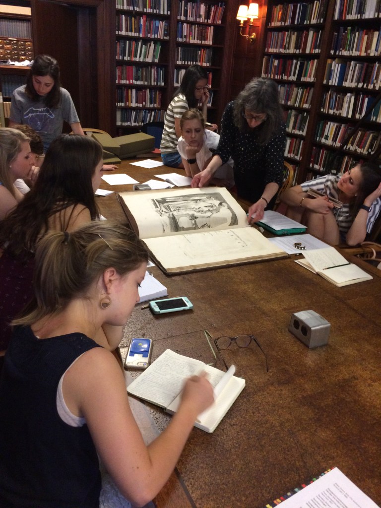
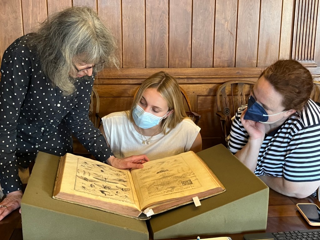
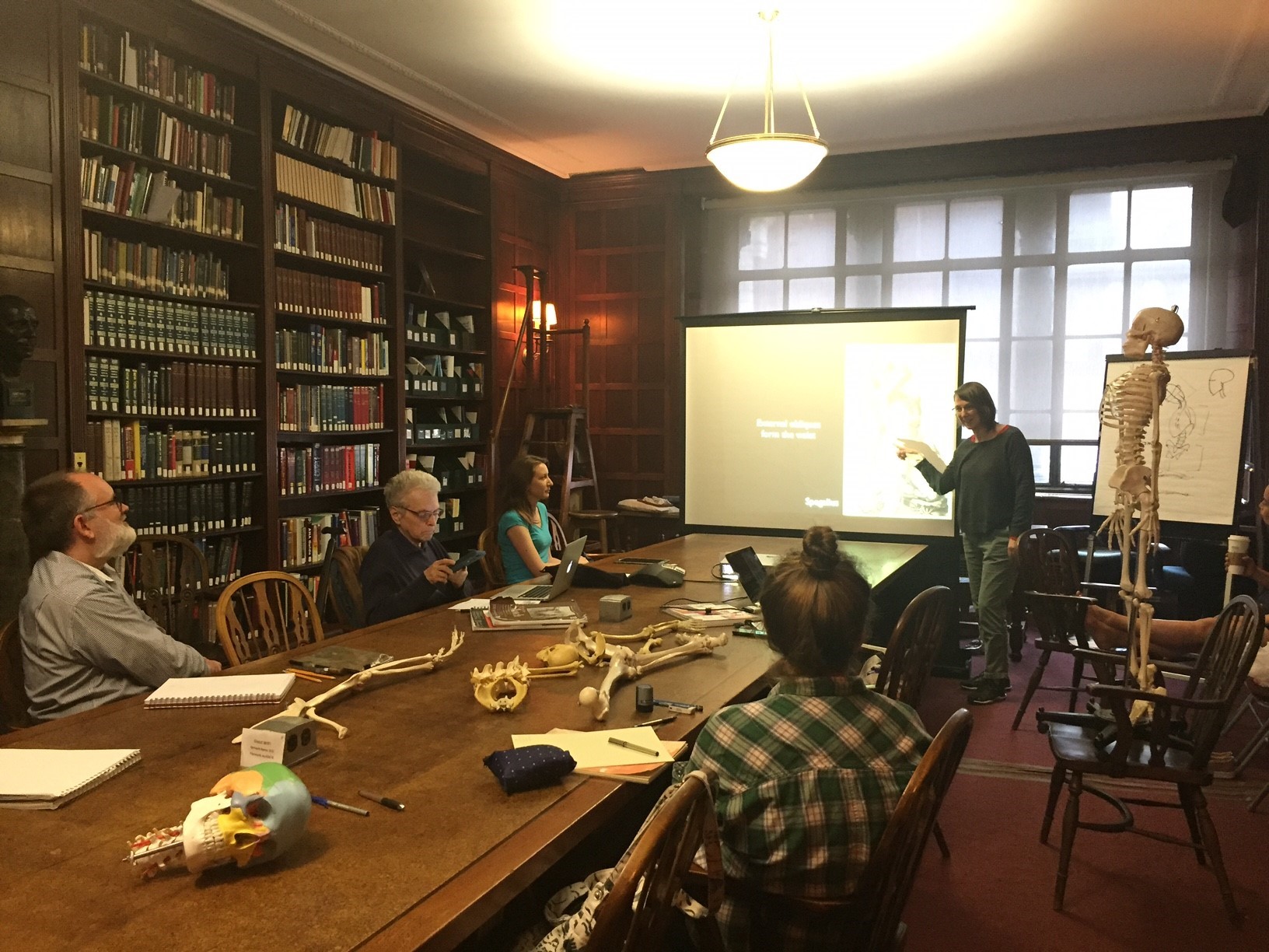
Kriota Willberg, the author of today’s guest post, explores the intersection of body sciences with creative practice through drawing, writing, performance, and needlework. She is offering the workshop “Visualizing and Drawing Anatomy” beginning June 6 at the Academy. Register online.

Cheselden's Osteographia, 1733, opened to the title page and frontispiece.

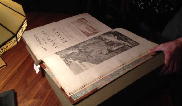
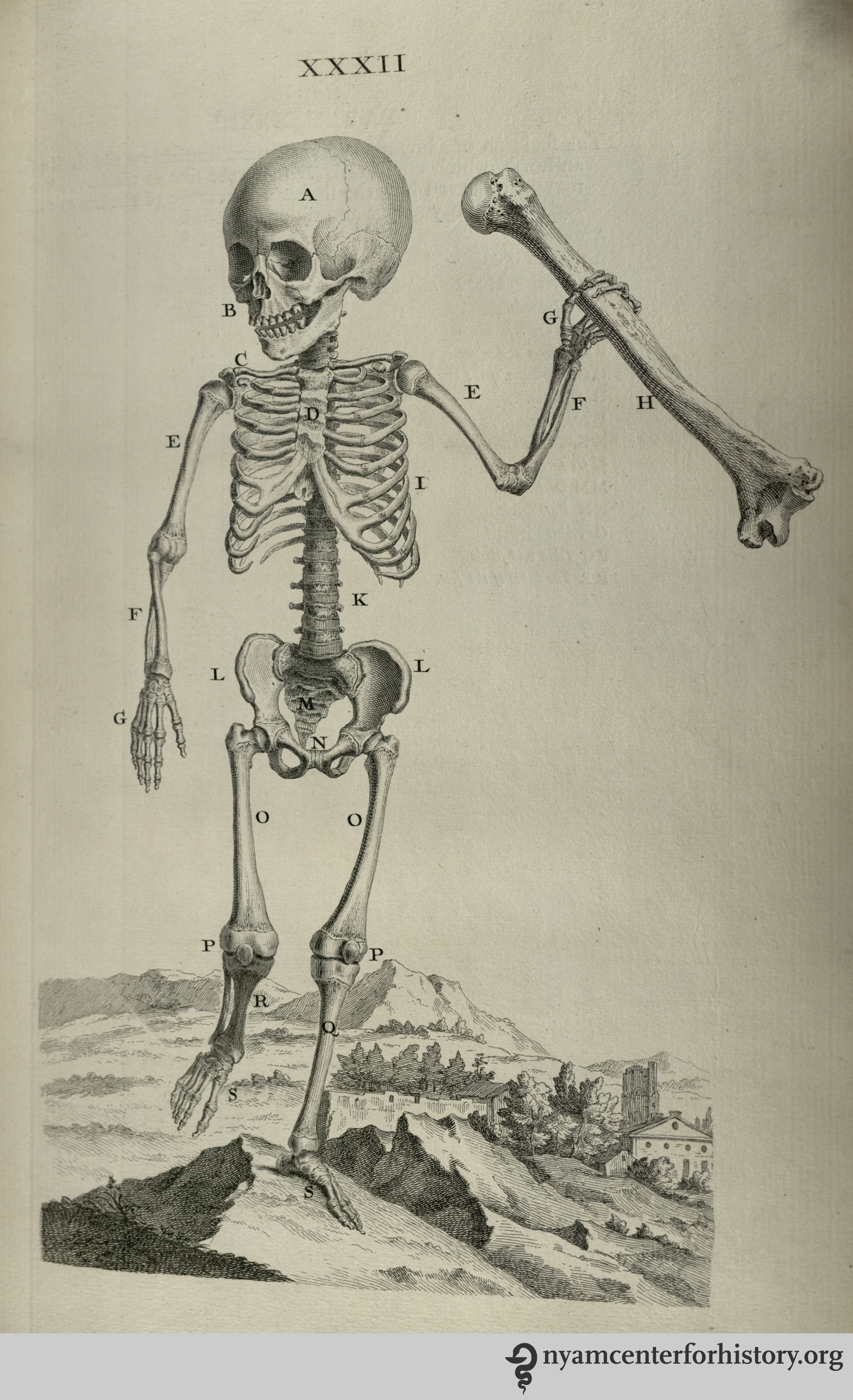
Cheselden’s Osteographia, 1733, opened to the title page and frontispiece.

Different Disciplines, Same Body

I teach musculoskeletal anatomy to artists, dancers, and massage therapists. In my classes the students study the same raw material, and the set of skills each group acquires can be roughly organized around three distinct areas: representation of the body, kinesiology (the study of movement), and palpation (feeling the body).

As an anatomy teacher I am constantly on the prowl for images of the body that visually reinforce the information my students are learning. The Internet has become my most utilized source for visual teaching tools. It is full of anatomy virtual galleries, e-books, and apps. 3D media make it ever easier to understand muscle layering, attachment sites, fiber direction, and more.

In spite of the overwhelming volume of quality online cutting-edge anatomical imagery, I find myself drawn to historical 2D printed representations of the body and its components, once the cutting-edge educational technology of their respective centuries. Their precision, character, size, and even smell enhance my engagement with anatomical study. Many of these images emphasize the same principles as the apps replacing them centuries later.

The Essential Structure Of The Body

Different artists prefer different methods of rendering bodies in sketches. One method is to organize the body by its masses, outlining its surface to depict its bulk. Another method is to draw a stick figure, organizing body volume around inner scaffolding.

Plate XXXIII in Cheselden, Osteographia, 1733.

Plate XXXIII in Cheselden, Osteographia, 1733.

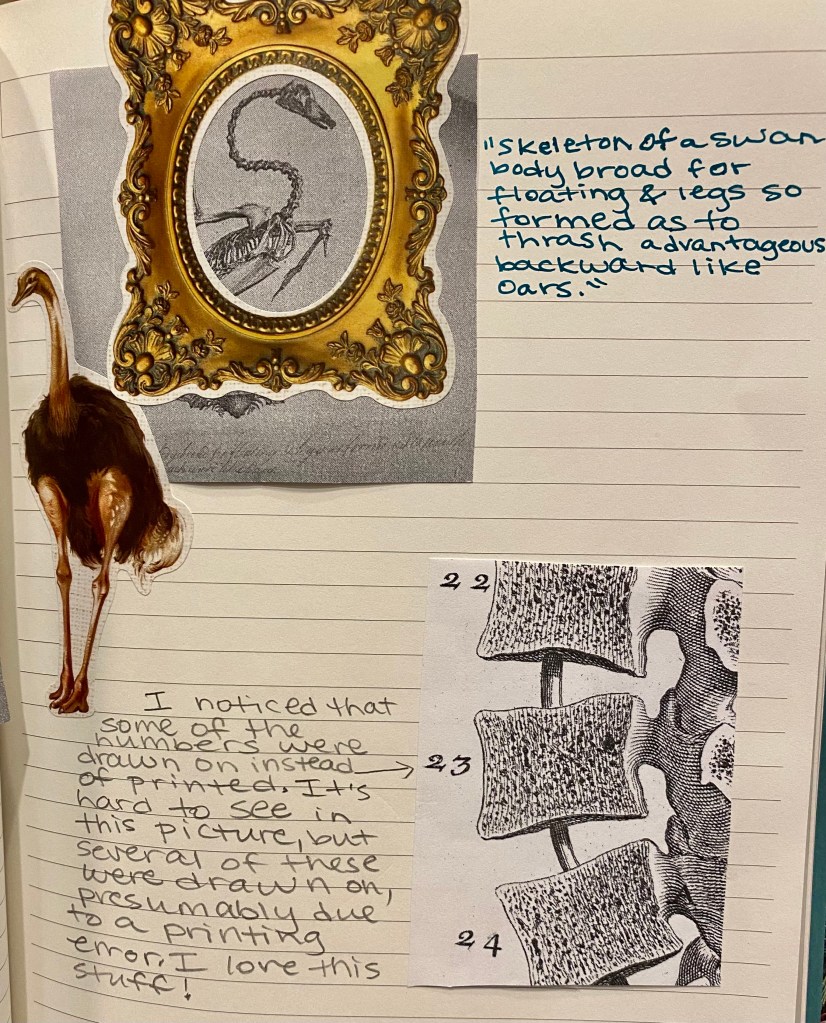
And what is a skeleton but an elaborate stick figure? William Cheselden’s Osteographia (1733) presents elegant representations of human and animal skeletons in action. These images remind us that bones are rigid and their joints are shaped to perform very specific actions. The cumulative position of the bones and joints gives the figure motion. In Cheselden’s world of skeletons, dogs and cats fight, a bird eats a fish, a man kneels in prayer, and a child holds up an adult’s humerus (upper arm bone) to give us a sense of scale while creating a rather creepy theatrical moment.

Muscle Layering

3D apps and other imaging programs facilitate the exploration of the body’s depth. One of the challenges of artists and massage therapists studying anatomy is transitioning information from the 2D image of the page into the 3D body of a sculpture or patient.

Planche 11 in Salvage, Anatomie du gladiateur combattant, 1812.

Planche 11 in Salvage, Anatomie du gladiateur combattant, 1812.

Salvage’s Anatomie du gladiateur combattant: applicable aux beaux artes… (1812) is a 2D examination of the 3D Borghese Gladiator. Salvage, an artist and military doctor, dissected cadavers and positioned them to mimic the action depicted in the statue. His highly detailed images depict muscle layering of a body in motion. The viewer can examine many layers of the anatomized body in action from multiple directions, rendered in exquisite detail. Salvage retains the outline of the body in its pose to keep the viewer oriented as he works from superficial to deeper structures.

Tab. VIII in Albinus, Tabulae sceleti et musculorum corporis humani, 1749.

Tabula VIII in Albinus, Tabulae sceleti et musculorum corporis humani, 1749 edition.

Bernhard Siegried Albinus worked with artist Jan Wandelaar to publish Tabulae sceleti et musculorum corporis humani (1749). Over their 20-year collaboration, they devised new methods for rendering the dissected body more accurately.  The finely detailed illustrations and large size of the book invite the reader to scrutinize the dissected layers of the body in all their detail. Although there is no superficial body outline, the cadaver’s consistent position helps to keep the reader oriented. On the other hand, cherubs and a rhinoceros in the backgrounds are incredibly distracting!

Fiber Direction

Familiarity with a muscle’s fiber direction can make it easier to palpate and can indicate the muscle’s line of pull (direction of action).

Figure in Berengario, Anatomia Carpi Isagoge breves, 1535.

Figure in Berengario, Anatomia Carpi Isagoge breves, 1535.

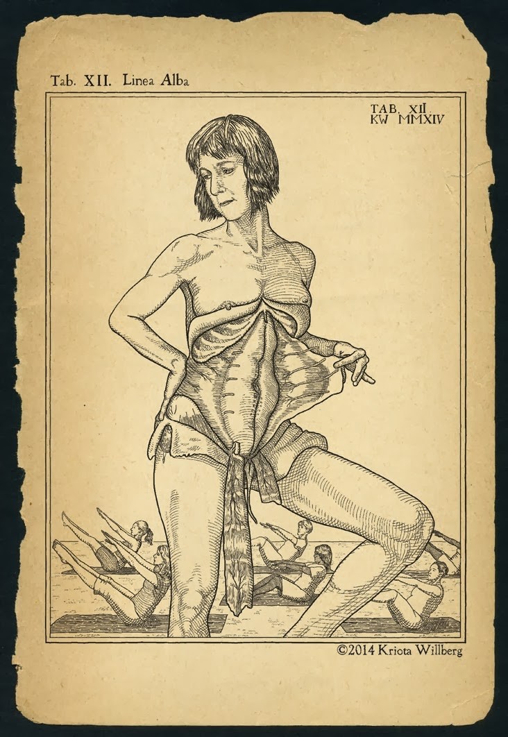
The images of Jacopo Berengario da Carpi’s Anatomia Carpi Isagoge breves, perlucide ac uberime, in anatomiam humani corporis… (1535) powerfully emphasize the fiber direction of the muscles of the waist. This picture in particular radiates the significance of our “core muscles.” Here, the external oblique muscles have been peeled away to show the lines of the internal obliques running from low lateral to high medial attachments. The continuance of this line is indicated in the central area of the abdomen. It perfectly illustrates the muscle’s direction of pull on its flattened tendon inserting at the midline of the trunk.

The Internal Body Interacting with the External World

One of the most important lessons of anatomy is that it is always with us. Gluteus maximus and quadriceps muscles climb the stairs when the elevator is broken. Trapezius burns with the effort of carrying a heavy shoulder bag. Heck, that drumstick you had for lunch was a chicken’s gastrocnemius (calf) muscle.

Tab. XII in Speigel, De humani corporis fabrica libri decem, 1627.

Tab. XII in Speigel, De humani corporis fabrica libri decem, 1627.

Anatomists from Albinus to Vesalius depict the anatomized body in a non-clinical environment. One of my favorites is Adriaan van de Spiegel and Giulio Casseri’s De humani corporis fabrica libri decem (1627). In this book, dissected cadavers are depicted out of doors and clearly having a good time. They demurely hold their skin or superficial musculature aside to reveal deeper structures. Some of them are downright flirtatious, reminding us that these anatomized bodies are and were people.

Kriota Willberg's self portrait. Courtesy of the artist.

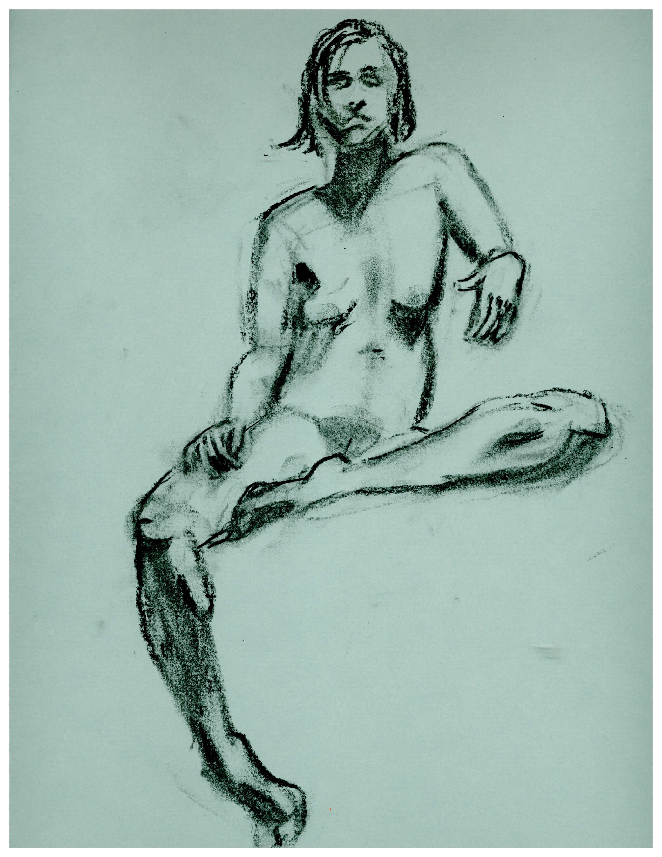
Kriota Willberg’s self portrait. Courtesy of the artist.

I am so enamored of van de Spiegel and Casseri that I recreated page 24 of their book as a self-portrait. After my abdominal surgery, the image of this cadaver revealing his trunk musculature resonated with me. In my portrait I assume the same pose, but if you look closely you will see stitch marks tracing up my midline. I situate myself in a “field” of women performing a Pilates exercise that challenges abdominal musculature. And of course, I drew it in Photoshop.

Revisiting the Fabrica Frontispiece

Jeffrey M. Levine, MD, AGSF, author of today’s guest post, will present “Revisiting the Frontispiece: Vesalius’s Jewish Friend and the Impact of the Inquisition” with Michael Nevins, MD, at our October 18th festival, Art, Anatomy, and the Body: Vesalius 500.

Between the first edition in 1543 and the second edition in 1555, the frontispiece of Andreas Vesalius’ classic masterpiece, De humani corporis fabrica, was recut with many subtle variations in both style and content. I am thrilled to be presenting “Revisiting the Frontispiece: Vesalius’s Jewish Friend and the Impact of the Inquisition” at Art, Anatomy, and the Body: Vesalius 500 with my colleague and mentor, Dr. Michael Nevins. Together we will examine and compare the frontispieces and offer theories as to why differences appear. We propose, for example, that some changes to the second edition were in reaction to the Inquisition, which was revived by Pope Paul III.1

Today’s guest post introduces selected features of the frontispiece of the Fabrica’s first edition. This intricate and multilayered composition features the master Vesalius dissecting a young female corpse, her abdomen flayed open. They are surrounded by a multitude of spectators crowded into a three-tiered wooden scaffold built into a semicircular amphitheater of Corinthian columns. At top-center is the decorative escutcheon that bears the name of the book and the author. Above is the family coat-of-arms of Andreas Vesalius flanked by two putti, the chubby male children who were often a feature of Renaissance art, and two gargoyles. Below is the face of Jupiter, the Roman king of gods.

The frontispiece to the 1543 Fabrica in our collection.

The frontispiece to the 1543 Fabrica in our collection. Click to enlarge.

The frieze sitting atop the columns contains symbols including a bucranium, or ox skull with garlands hanging from its horns, which was the symbol of the University at Padua,2 and a winged lion representing the evangelist St. Mark, the symbol of neighboring Venice.3 The columns are flanked by two men, one naked with tense muscles and a worried look, the other relaxed and smartly dressed.

To the right of the skeleton bearing a risus sardonicas, a man in a truncated conical hat recoils as if in terror, squinting and raising his left hand in a defensive gesture. In his 1964 biography of Vesalius, Charles O’Malley identifies this figure as Vesalius’s Jewish friend, Lazaro de Fregeis, who assisted with the Hebrew nomenclature in the Fabrica.4 The only woman other than the corpse appears as a mysterious figure peeking between the columns. There are two Franciscan Monks among the spectators, neither exhibiting much interest in the dissection. Below right is a pickpocket caught in the act. On the opposite side, a leashed monkey screams in protest, and under the table two men fight over the dissecting tools.

There is much more to learn about the frontispiece of the first edition of the Fabrica, and even more when compared to the second edition. To find out more about the changes to the second edition frontispiece, and how they may have contained coded messages reflecting tensions of 16th-century Italian society, particularly in the context of the situation of European Jewry, come to our presentation at the New York Academy of Medicine’s Vesalius 500 celebration on October 18.

References

1. Historical overview of the Inquisition. 2001. Available at: http://galileo.rice.edu/lib/student_work/trial96/loftis/overview.html. Accessed September 23, 2014.

2. Padova Terme Euganee. University of Padua – Palazzo Bo. Available at: http://www.turismopadova.it/en/university-padua-palazzo-bo. Accessed September 23, 2014.

3. Imboden D. Winged Lion of St. Mark. Durant Cheryl Imboden’s Venice Visit. Available at: http://europeforvisitors.com/venice/articles/winged_lion_of_st_mark.htm. Accessed September 23, 2014.

4. O’Malley C. Andreas Vesalius of Brussels, 1514-1564. Berkeley: Univ. of California Press; 1964.

Introducing Graphic Medicine

Ian Williams and MK Czerwiec, authors of today’s guest post, co-run the website GraphicMedicine.org. They will present “Graphic Medicine and the Multiplanar Body” at our October 18th festival, Art, Anatomy, and the Body: Vesalius 500.

The 2010 Comics & Medicine gathering before Senate House.

The 2010 Comics & Medicine gathering before Senate House.

In the summer of 2010 a group of scholars, health care professionals, and comics artists gathered in Senate House, London. This brutal-looking art deco building, said to have inspired George Orwell’s “Ministry of Truth,” represented Gotham City Courts in the films Batman Begins and The Dark Knight. Those gathered, however, were not particularly interested in superheroes. They focused on graphic memoirs of illness, a modern phenomenon born of the counterculture in the 60s and 70s that has gathered momentum over the last 20 years.

Among the 75 delegates from around the world were the authors of this blog entry. The lead organizer of the conference was Ian Williams, a doctor and comics artist, creator of The Bad Doctor (2014, Myriad Editions). MK Czerwiec (pronounced sir-wick), aka Comic Nurse, has been making comics about her work in HIV/AIDS and hospice care since the late 1990s as a way of processing these caregiving experiences. We have now worked together for four years, talking and writing about the interplay between the comics and health care. We make comics, collaboratively and separately, and will give a talk on October 18th at “Art, Anatomy, and the Body: Vesalius 500” about Graphic Medicine, the field we helped pioneer.

The Bad Doctor. Cover by Ian Williams.

The Bad Doctor. Cover by Ian Williams.

MK Czerwiec teaching at Northwestern Feinberg Medical School. Still from BBC story by Katie Watson.

MK Czerwiec teaching at Northwestern Feinberg Medical School. Still from BBC story by Katie Watson.


Often when we describe Graphic Medicine, people say that comics must make an excellent educational medium for patients, especially those with poor literary skills and marginalized groups such as drug addicts, teenage mothers, or the mentally ill. While comics have certainly been used to reach these audiences, the idea behind this response is freighted with assumptions about comics, their target demographics, and the literacy skills or aesthetic proclivities of the social groups so named.

Stack of medically-themed graphic novels. Photo by Ian Williams.

Stack of medically-themed graphic novels. Photo by Ian Williams.

We regard comics as a sophisticated, rich, and adaptable system through which to explore the complex issues of health care. Our primary interest has been the use of graphic illness narratives to provide new knowledge about the illness experience and commentary on the pervading cultural conceptions of disease and health care. We are also interested in the psychological process of making comics. We have also been teaching using comics—both making them and reading them—in medical schools in the US and UK.

Binky Brown Meets The Holy Virgin Mary cover by Justin Green

Binky Brown Meets The Holy Virgin Mary cover by Justin Green

In 1972 Justin Green became the first comics artist to unburden his psychological troubles onto the page, creating Binky Brown Meets the Holy Virgin Mary. This inspired subsequent generations of artists to articulate their corporeal experiences in words and pictures, a process that Elisabeth El Refaie refers to as “pictorial embodiment.”1 More than 40 years later, the myriad comics titles that appear each year include stories of disease or trauma, known as “graphic pathographies,”2 in which the authors give highly subjective accounts of their own illnesses or caregiving experiences. The production of these works involves the repeated drawing of the author’s or subject’s body over a prolonged period, which may have interesting effects on how the artist perceives the body. The relentless decision-making process forces the artist to examine fears, suffering, anger, disgust, disappointment, and grief and distill the whole into a succinct series of sequential panels through which to transfer the narrative to the reader.

2014 Comics & Medicine poster. Art by Lydia Gregg.

2014 Comics & Medicine poster. Art by Lydia Gregg.

Since the London gathering, we have held international conferences in Chicago, Toronto, Brighton, and Baltimore. The movement is growing and what was initially viewed by some as a novelty interest is gaining respect in academia. As the nature of literacy changes, moving from the textual towards the image, comics is once again in ascendance, gaining new readers who might have previously dismissed the medium.


References

1. El Refaie, E. (2012). Autobiographical comics: Life writing in pictures. Jackson: University Press of Mississippi.

2. Green, M. J., & Myers, K. R. (2010). Graphic medicine: Use of comics in medical education and patient care. BMJ, 340, c863.

Registration Open for Vesalius 500 Workshops

Registration is now open for our hands-on art and anatomy workshops, presented as part of our Vesalius 500 celebrations on October 18, 2014. Create your own articulated anatomical figure or “exquisite corpse” at the Gladys Brooks Book & Paper Conservation Laboratory; learn Renaissance drawing techniques with medical illustrator Marie Dauenheimer; or explore the anatomy and art of the hand with physical anthropologist Sam Dunlap.

Spaces are strictly limited so register soon. Registration at one of the workshops includes free entry to the Festival. You can register for the Festival (without workshop attendance) here.

From the Cradle to the Grave: Session One: The Cradle

Moveable baby and female pelvis from one of NYAM’s 19th century obstetrics texts, Dr. K. Shibata's Geburtschülfliche Taschen-Phantom, or the Obstetrical Pocket-Phantom.

Moveable baby and female pelvis from one of NYAM’s 19th century obstetrics texts, Dr. K. Shibata’s Geburtschülfliche Taschen-Phantom (Obstetrical Pocket-Phantom).

Working with NYAM’s conservation team, celebrate Vesalius’s life with a hands-on workshop producing your own articulated anatomical figures in the Gladys Brooks Book & Paper Conservation Laboratory.

Time: 11am-1pm
Cost: $55
Includes: All materials, and free entry to the Festival.
Maximum participants: 12
Register here

During the morning’s Cradle workshop, we will construct paper facsimiles of a moveable baby and female pelvis from one of NYAM’s 19th century obstetrics texts, Geburtschülfliche Taschen-Phantom (or the Obstetrical Pocket-Phantom). The book was written by Dr. K. Shibata, a Japanese author studying in Germany, and was published first in German before being translated into English and Japanese.

Participants will have time to make at least one paper baby and pelvis, which can be produced as paper dolls or magnets.

From the Cradle to the Grave: Session Two: The Grave

An exquisite corpse made by staff of the Gladys Brooks Book & Paper Conservation Laboratory.

An exquisite corpse made by staff of the Gladys Brooks Book & Paper Conservation Laboratory.

Working with our conservation team, celebrate Vesalius’s life with a hands-on workshop producing your own “exquisite corpse” in the Gladys Brooks Book & Paper Conservation Laboratory.

Time: 2:30pm-4:30pm
Cost: $55
Includes: All materials, and free entry to the Festival.
Maximum participants: 12
Register here

During the afternoon’s Grave workshop, we focus on producing a Vesalian-themed exquisite (or rotating) corpse. Loosely based on the surrealist parlor game in which a picture was collectively created by assembling unrelated images, this workshop will employ a special, rotating binding structure and mix-matched facsimile images from NYAM’s rare book collections to allow students to create their own unique, moveable pieces of art.

Renaissance Illustration Techniques Workshop with Marie Dauenheimer, Medical Illustrator

Students at medical illustrator Marie Dauenheimer's workshop at last fall's Festival.

Students at medical illustrator Marie Dauenheimer’s workshop at last fall’s Festival.

Time: 10am-1pm
Cost: $85
Includes: All materials, and free entry to the Festival.
Maximum participants: 15
Register here

Artists and anatomists passionate about unlocking the mysteries of the human body drove anatomical investigation during the Renaissance. Anatomical illustrations of startling power vividly described and represented the inner workings of the human form. Leonardo da Vinci’s notebooks were among the most magnificent, merging scientific investigation and beautifully observed drawing.

Students will have the opportunity to learn and apply the techniques used by Renaissance artists to illustrate anatomical specimens. Using dip and technical pens, various inks and prepared paper students will investigate, discover, and draw osteology, models, and dissected specimens from various views creating an anatomical plate.

Understanding the Hand, physical anthropology workshop with Sam Dunlap, Ph.D.

Dr Sam Dunlop leading a workshop at last year's Festival.

Dr Sam Dunlop leading a workshop at last year’s Festival.

Time: 2:30pm-5:30pm
Cost: $75
Includes: All materials, and free entry to the Festival.
Maximum participants: 15
Register here

The hand as an expression of the mind and personality is second only to the face in the Renaissance tradition of dissection and illustration that continues to inform both art and science. Basic anatomical dissection, illustration, and knowledge continue to be fundamental in many fields from evolutionary biology to surgery, medical training, and forensic science. This workshop will offer participants the opportunity to explore the human hand and its anatomy, which will be demonstrated with at least three dissections.  Opossum (Didelphis virginiana) and orangutan (Pongo pygmaeus) forelimbs will be available along with other comparative skeletal material. We will discuss hand evolution, embryology, and anatomy, and the artistic importance of the hand since its appearance in the upper palaeolithic cave art. We will also analyze the hand illustrations of da Vinci, Vesalius, Rembrandt, and artists up to and including the abstract expressionists.