The History of Garlic: From Medicine to Marinara

Today’s guest post is written by Sarah Lohman, author of Eight Flavors: The Untold Story of American Cuisine (Simon & Schuster, 2016). On Monday, June 5, Lohman will give her talk, “The History of Garlic: From Medicine to Marinara.” To read more about this lecture and to register, go HERE.

Ms. Amelia Simmons gave America its first cookbook in 1796; within her pamphlet filled with sweet and savory recipes, she makes this note about garlic: “Garlickes, tho’ used by the French, are better adapted to the uses of medicine than cookery.” In her curt dismissal, she reflected a belief that was thousands of years old: garlic was best for medicine, not for eating. To add it to your dinner was considered the equivalent of serving a cough syrup soup.

There are records of ancient Greek doctors who prescribed garlic as a strengthening food, and bulbs were recovered from Egyptian pyramids. Garlic was being cultivated in China at least 4,000 years ago, and upper class Romans would never serve garlic for dinner; to them, it tasted like medicine.

In medieval Europe, garlic was considered food only for the humble and low.  While those that could afford it imported spices like black pepper from the Far East, lower classes used herbs they could grow. Garlic’s intense flavor helped peasants jazz up otherwise bland diets. It was made into dishes like aioli, originally a mixture of chopped garlic, bread crumbs, nuts and sometimes stewed meat. It was intended to be sopped up with bread, although it was occasionally served as a sauce to accompany meats in wealthier households.

woodville_medicalbotany_garlic_1790_Watermark

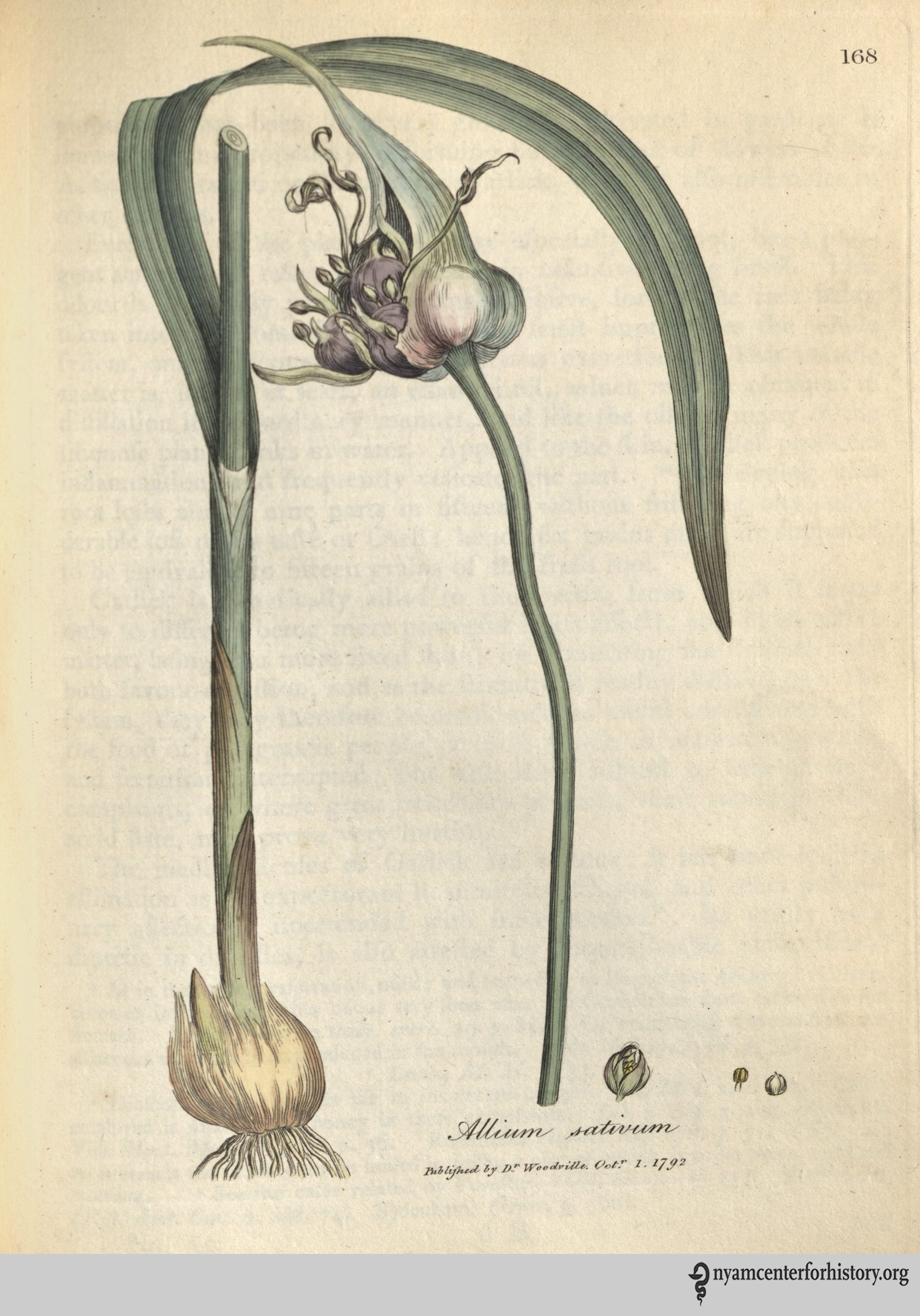
Garlic (Scientific name Allium Sativum) from Medical Botany (1790) by William Woodville.

The English, contrary to the stereotype about bland British cooking, seemed particularly enchanted by garlic. In the first known cooking document in English, a vellum scroll called The Form of Cury, a simple side dish is boiled bulbs of garlic. Food and medicine were closely intertwined in Medieval Europe, and garlic was served as a way to temper your humors. Humors were thought to be qualities of the body that affected on your health and personality. Garlic, which was thought be “hot and dry,” shouldn’t be consumed by someone who was quick to anger, but might succeed in pepping up a person who was too emotionally restrained. According to food historian Cathy Kaufman, a medieval feast might have a staggering amount of different dishes, all laid on the table at one time, so that different personality types could construct a meal that fit their humors.

Up through the 19th century, people also believed you got sick by inhaling bad air, called “miasmas.” Miasmas hang out by swamps, but also by sewage, or feet–I always imagined them as the puddles of mist that lie in the nooks between hills on dark country roads. Garlic can help you with miasmas, too. Ever see an image of plague doctors from Medieval Europe wearing masks with a long, bird-like beak? The beak was filled with odorous herbs, garlic likely among them, designed to combat miasmas.

In 18th-century France, a group of thieves may have been inspired by these plague masks. During an outbreak of the bubonic plague in Marseilles in 1726 (or 1655, stories deviate), a group of thieves were accused of robbing dead bodies and the houses of the deceased and ailing, without seeming to contract the disease themselves. Their lucky charms against the miasmas? They steeped garlic in vinegar, and soaked a cloth or a sponge in the liquid, then tied it like a surgical mask over their mouth and nose. In their minds, the strong smells would repel miasmas. This story is probably a legend, but I think there is some grain of truth to it: in modern studies, garlic has been shown to obfuscate some of the human smells that attract biting bugs. Since we now know bubonic plague was carried by fleas, it’s possible the thieves were repelling the insects. The plague is also a bacterial infection, and both vinegar and garlic are effective antimicrobials.

Garlic remained in the realm of medicine for most of the 19th century. Louis Pasteur first discovered that garlic was a powerful antimicrobial in 1858. In 1861, John Gunn assembled a medical book for use in the home, The New Domestic Physician, “with directions for using medicinal plants and the simplest and best new remedies.” Gunn recommends a poultice of roast garlic for ear infections:

“An excellent remedy for earache is as follows: Take three or four roasted garlics, and while hot mash, and add a tablespoonful of sweet oil and as much honey and laudanum; press out the juice, and drop of this into the ear, warm, occasionally.”

Salmon_EnglishHerbal_1710_408_garlic_watermark

Garlick from Botanologia: The English Herbal (1710) by William Salmon.

He also recommends garlic for clearing mucus from the lungs and reducing cough, given by the spoonful with honey and laudanum.  Gardening for the South: Or, How to Grow Vegetables and Fruits, an 1868 botanical guide, says the medicinal values of garlic include making you sweat, which,  like bloodletting, was believed to leach out disease; it will also make you urinate, and is an effective “worm destroyer,” for any intestinal hitchhikers you might have. By the late 19th century, scientists also used garlic to treat TB and injected it into the rectum to treat hemorrhoids.

Today, garlic is one of the most heavily used home remedies, and it is increasingly being studied in the medical field. Some of its historic uses have been proved as bunk–while others, like its efficacy as a topical antiseptic, hold up. But since the late 19th century, garlic has found an even more worthwhile home, thanks to French chefs and Italian immigrants, who spread their garlic heavy cuisine around the world, and made even garlic-reticent Americans a lover of this pungent plant.

Join us on Monday, June 5 to learn more about this topic.  Click HERE to register.

Let’s Digitize! Building the Library’s Digital Lab

By Robin Naughton, Head of Digital

If your materials cannot take a trip to an external digitization lab to be converted from analog to digital, then you do what all aspiring DIYers do: you bring the lab to your materials. The New York Academy of Medicine Library has an amazing and significant collection of rare and unique materials that will benefit both researchers and the general public once digitized.  Thus, our goal is to develop a robust digital infrastructure to support the creation and preservation of our digital assets internally, particularly rare, fragile and unique materials.

The Federal Agencies Digital Guidelines Initiative (FADGI), a collaborative started in 2007 to create sustainable guidelines for digitization, is the gold standard for cultural heritage digitization. In 2016, FADGI’s Still Image Working group released an updated “Technical Guidelines for Digitizing Cultural Heritage Materials,” which updates the specifications for being FADGI compliant.  Establishing a robust digital infrastructure means being FADGI compliant while integrating the needs of our users and the strategic goals of the Library.

Inventory & Solution

We began the process by conducting an inventory of our resources.  The Library has multiple scanner setups and each is good for its purpose.  However, no setup was best for digitizing rare materials.

  • Flatbed Scanner: Our Epson Perfection v700 Photo flatbed scanner, used in earlier digitization projects, was good for small flat materials. Prior to our digital setup, we used it to digitize additional items for our William H. Helfand Collection of Pharmaceutical Trade Cards.
  • Book Scanners: We have two Bookeye scanners: a Bookeye 3 and a Bookeye 4.  Our Bookeye scanners were used to create images for patrons, but posed complications when thinking about scanning rare books. Our Bookeye 3 scanner with a glass platen is best for large flat materials. Our Bookeye 4 scanner with v-cradle is a workhorse, but posed problems for items with tight bindings that were unable to open 90 degrees. Our Konica Minolta Scan Diva scanner stopped working due to a problem with the software and the company was unable to replace it.  As a result, the scanner was no longer usable.

The inventory revealed the need for a solution that followed FADGI guidelines for digitizing rare materials, considered the binding of the item, made sure digitization would not damage the item, and used equipment that could be easily maintained.

Library Digital Lab

Thanks to the generous support of the Gladys Brooks Foundation, we created a digital lab that combined the old and the new.

The digital lab is illuminated by two Profoto strobe lights that flank an old copy stand sitting on a production workbench. Attached to the copy stand is a refurbished Phase One 645DF camera with 80mm lens, and a Mamiya Leaf 50 megapixel digital back.  Just off to the right is the digital workstation, which includes a Mac Pro, Eizo monitor, and Capture One Cultural Heritage (CH) software used in the digitization process.

This slideshow requires JavaScript.

Photos courtesy of the Library’s Digital Team.


Discussion with our digitization working group and other cultural heritage organizations with digitization labs led to the digital camera setup that was modular and best for digitizing rare materials. Modularity offered the opportunity to grow and develop as the technology changed while making it easy to replace parts as necessary. Thus, if the lights needed to be replaced, we would not need to replace the entire setup, but rather just the lights.  The digital camera setup also offered flexibility because there was no set angle at which to open an item.  Using book cradles, we can adjust the angles based on the object for best shooting and handling.

A recent report from the Library of Congress, “Library of Congress Lab: Library of Congress Digital Scholars Lab Pilot Project” provides great recommendations for digital lab design.  The research conducted and the recommendations from the report are a model for a digital lab that can benefit us all in our own efforts to digitize and make rare materials accessible to a wider audience.

This blog post is the first of series of post from the digital lab.  We will share our process and activities, so stay tuned.

The Enduring Impact of the X-Ray

Today we have part two of a guest post written by Dr. Daniel S. Goldberg, 2016 recipient of the Audrey and William H. Helfand Fellowship in the History of Medicine and Public Health. Part one can be read here.

X-ray exhibitions were hugely popular all over the country, and the greater NY area was no exception.  At a February 1896 demonstration run by Professor Arthur Wright, director of the Sloan Laboratory at Yale University, a newspaper reported that despite the auditorium being literally jam-packed, students were still crawling through windows 30 minutes into the lecture — and all this despite the fact that none of the audience, save those in the first few rows, could even hear Wright’s discussion.  The deans of multiple Yale schools (Divinity, Law, and Science), the head of the Yale Corporation, and the chief medical examiner were all in attendance.

bostonmedsurgjournal_feb201896_rontgenprocess_watermark

Perhaps the first published X-ray in the United States of a clinical condition. In “Rare Anomalies of the Phalanges Shown by the Röntgen Process,” Boston Medical and Surgical Journal 134(8), February 20, 1896: 198–99.

The pressing question is “why”? Why did X-rays exert such tremendous power across a wide spectrum of social domains? (X-rays were a constant topic of conversation in sermons and religious journals, in women’s journals, in influential satirical periodicals like Punch, and were the subject of a seemingly endless number of political and non-political cartoons, to name but a few).  Although historians of the X-ray have offered a number of plausible answers, I believe there is a key element left unexplored in the historiography: the intellectual frameworks, or ideas, relating to changing ideas of truth, doubt, and objectivity in U.S. society at the time.

Two of these frameworks are most useful in unpacking the stunning impact of the X-ray: the rise of mechanical objectivity, and what can be called “somaticism” within medicine and science.  Historians of science Lorraine Daston and Peter Galison explain that a new model of ‘objectivity’ begins to take hold during the middle decades of the 19th century.  Under this new model, the truth-value of scientific knowledge is a function of the investigator’s ability to remove or eliminate human, subjective influence from the knowledge-making process.  The fact that this is more or less impossible, and that X-rays can be manipulated in all sorts of ways was well-known to contemporaries and remained a source of anxiety for some time.  The important point is the ultimate goal: to let the mechanical processes of nature speak for themselves and reveal their truths.  Ideas of objectivity, as Daston and Galison point out, have for over four hundred years been connected to scientific images, which makes media like photography and X-rays especially significant.

By the end of the 19th century, ideas of mechanical objectivity begin to fundamentally reshape ideas of what is known and what is certain.  This is especially crucial in a century that features so much intense change, including but not limited to governments, family and labor structures, migration patterns, and, of course, industrialization and urbanization.  Late Victorians were beset with anxieties connected to their changing world, and they were especially concerned with artifice and deception — that the world was not what it seemed.  As such, intellectual frameworks that shaped the criteria for truth were hugely influential, and traveled well beyond narrow networks of scientists and medical men.

Somaticism integrates in important ways with constructs of mechanical objectivity.  Historians of medicine have documented the influence of somaticism (or, “empiricism,” as it is also sometimes termed) within medicine over the long 19th century.  The core of the framework is that truths about disease and the body are to be found in pathological anatomical objects.  The existence of these objects can then be clinically correlated with the illness complaints the patient has, or more likely had given that pathological objects are most likely to be located precisely during a postmortem — until the X-ray.  The truths of the sick body are to be found in the natural objects of disease, which makes seeing those objects so essential.  Laennec himself explained that the point of the stethoscope was not to listen; listening was merely a means to an end.  The point, as Jacalyn Duffin explains, was “to see with a better eye.”

Collectively, these frameworks go a significant length in explaining the enormous and enduring social impact of the X-ray.

X Rays for Consideration_watermarked

Article from the New York Record. May 1896.

For example, Morton’s clippings contain a May 1896 article from the New York Record entitled “X Rays for a Consideration: Light in a Human Kidney.”  The article details what may be the first private X-ray laboratory opened in New York City, founded by Mrs. M.F. Martin, and located at 110 East 26th Street.  The lab was intended solely for the use of physicians and hospitals.  One of its first patients was a doctor named George McLane, who traveled from North Dakota to have his kidney X-rayed for evidence of a possible stone.  A surgeon removed McLane’s kidney, and Morton placed it on a plate and subsequently irradiated it with X-rays.  The procedure “revealed the absence of any stone in the organ, demonstrating the entire reliability of doctors to prove the absence of stone in the kidney.”

The X-ray shines its light into the hitherto dark spaces inside the human body, revealing the truth of a disputed question: whether McLane suffered from a kidney stone or not.  The truth resides in the natural object itself, and the mechanism of the X-ray supposedly insulates the production of medical knowledge from the whims and artifices of the investigator (as compared to illustrations and drawings, for example).

Or, as Dr. McLane himself stated at the Post Graduate Hospital (the primary hospital at which Morton cared for inpatients):

“Dr. McLane spoke modestly at the Post Graduate Hospital about the risk he had taken in the name of science . . . ‘Hitherto a great many mistakes have been made owing to the inability of doctors to prove the absence of stone in the kidney . . .’  Now, by a very simple process, the truth can easily be determined.”

It is difficult to imagine how powerful it must have been, in 1896, to witness an X-ray operator remotely anatomize the living body.  Seeing inside the body had been a dream of physicians for centuries prior, and there is every reason to believe that its achievement has not eroded much of its social power.  Americans still perform significantly more medical imaging procedures than virtually any of our comparator societies, and what is most interesting is the evidence that this utilization is driven both by supply and demand.  That is, it is not merely that we have expensive X-ray and medical imaging machines — so we use them.  Across a wide variety of illness paradigms, illness sufferers and patients request medical imaging; they want it to be performed on their bodies.  The history of the X-ray helps us understand the enduring power of these tools, of what it means to delve into the penetralium.

The Early Days of the X-Ray

Today we have part one of a guest post written by Dr. Daniel S. Goldberg, 2016 recipient of the Audrey and William H. Helfand Fellowship in the History of Medicine and Public Health. Dr. Goldberg is trained as an attorney, a historian, and a bioethicist.  He is currently on the faculty at the Center for Bioethics and Humanities at the University of Colorado Anschutz Medical Campus.

After news of Wilhelm Röntgen’s discovery of X-rays was cabled across the Atlantic late in 1895, evidence suggests X-ray experimentation was taken up eagerly all over the U.S. almost immediately.  While scientists and physicians scrambled to build their own X-ray machines, newspapers in major cities throughout the country eagerly reported on their progress, with stories small and large appearing in nearly every significant daily from New York and Philadelphia to Chicago and St. Louis to San Francisco and Los Angeles.  Historians of the X-ray estimate that within only a year of Röntgen’s discovery, literally thousands of articles had been published on the X-ray in both lay and expert periodicals.  Even in the fertile print culture of 1896, this is a significant accounting.

Therein lies the methodological difficulty for the historian of the X-ray.  So often, the craft of history is a tedious search for small scraps of information that may not even exist.  Yet, as to X-rays, the problem is one of feast, not famine.  With so much print material appearing in so many different sources in so many different places all at the same time, sifting through the morass to articulate coherent and important narratives is difficult.

What makes this task far easier is a remarkable collection held at the New York Academy of Medicine Library.  The William J. Morton Collection is a small holding, consisting of only two boxes.  The second box is the true treasure, containing a single folder, approximately six inches thick.  Inside is an unbound series of pages consisting solely of newspaper clippings related primarily to early X-ray use in the U.S.  These are Morton’s clippings, and as far as is known, the order and arrangement of the pages is original to Morton himself.  The collection is astounding, for it represents something of an index or a cipher for the ferment of X-ray use in NYC in the first half of 1896.

Clippings

Newspaper clippings from the William J. Morton Collection, New York Academy of Medicine Library.

There is no question that New York City played an important role in early X-ray use, if for no other reason than the enormous shadow cast by the inventor, Thomas Edison.  There were, however, many other important figures involved in early X-ray use in NYC, including Nikola Tesla[1], Michael Pupin[2], and Morton.  Morton, the son of William T.G. Morton of anesthesia fame, was a prominent physician, a fellow of the New York Academy of Medicine, and a respected neurologist and electro-therapeutic practitioner.

Telegrams_watermark

A telegram dated January 2, 1896 from Dyer & Driscoll, attorneys for none other than Thomas Edison, indicated that Morton visited Edison’s workshop for the purpose of conducting experiments (almost certainly with X-rays) several days earlier.

Because Morton was unquestionably at the forefront of early X-ray experimentation in NYC, his curation is a reasonable index as to important events and moments in the early use of X-rays in NYC.  There are limitations to this approach, of course.  Morton was obviously interested in his own role in early X-ray experimentation, so there is something of a selection bias at work (although it should be noted that there are no shortage of clippings pertaining to Pupin’s important work).

The collection is full of interesting and significant stories in the early history of X-ray use.  For example, in March 1896, strongman Eugene Sandow, considered the father of modern bodybuilding, turned to Morton in an effort to locate the source of a frustrating pain he was experiencing in his foot.  Apparently Sandow had stepped on some broken glass, but even his personal physician could not specify the location of the glass in his foot.  The potential for the X-ray must have seemed obvious, and Sandow reached out specifically to Morton to see if he could be of help.  Morton was eager to oblige.  He turned the X-rays on Sandow’s foot and located the shard of glass disturbing Sandow’s equanimity.  A surgeon subsequently operated and removed the glass, and the story made national news.

How the photograph was made_watermark

The X-Ray of Eugene Sandow’s foot in process.

Interestingly, Sandow was apparently impressed enough with the powerful rays to send an unsolicited telegram to Edison, offering his services as a human subject for any X-ray experiments Edison wished to undertake.

SandowLetter_watermark

Letter to Thomas Edison from Eugene Sandow.

It is difficult to imagine how powerful it must have been, in 1896, to witness an X-ray operator remotely anatomize the living body.  Seeing inside the body had been a dream of physicians for centuries prior, and there is every reason to believe that its achievement has not eroded much of its social power.  Americans still perform significantly more medical imaging procedures than virtually any of our comparator societies, and what is most interesting is the evidence that this utilization is driven both by supply and demand.  That is, it is not merely that we have expensive X-ray and medical imaging machines; so we use them.  Across a wide variety of illness paradigms, illness sufferers and patients request medical imaging; they want it to be performed on their bodies.  The history of the X-ray helps us understand the enduring power of these tools.

Footnotes:
[1] Tesla was heavily involved in early X-ray experiments in his laboratory at 46 East Houston Street; much to Edison’s likely chagrin, given the frostiness of their relationship by the time. The New York newspapers constantly asked Edison about Tesla’s progress.
[2] Pupin, a Columbia University physicist, would in short order — in 1896, in fact —  go on to discover a way of substantially reducing the exposure time needed to produce an X-ray image from hours to minutes.  The basics of Pupin’s method are still used today.

The Architecture of Health Care (Part 2)

Today’s guest post is written by Bert Hansen, Ph.D., professor emeritus of history at Baruch College of CUNY.  He is the author of Picturing Medical Progress from Pasteur to Polio: A History of Mass Media Images and Popular Attitudes in America (Rutgers, 2009), and other studies of medicine and science in the visual arts.  He is presenting an illustrated lecture about historic New York City buildings, followed by two walking tours-Uptown (May 13) and Downtown (May 20).  His 6 pm talk on Thursday, May 11, is entitled “Facades and Fashions in Medical Architecture and the Texture of the Urban Landscape.”  To read more about this lecture and to register, go HERE.

Part 1 introduced readers to the architectural firm of Sawyer and York and two of their medical buildings.  Part 2 now looks at Charles B. Meyers, who was responsible for dozens of major buildings in New York City and farther afield, including more than a dozen hospitals just in the city.  Still, he remains largely unknown outside of architectural history circles.

Readers of this blog are likely to know the red brick Psychiatric Hospital at Bellevue and Manhattan’s towering Criminal Court Building and House of Detention (New Deal WPA, 1938-41), sometimes called “The Tombs,” taking the name of an earlier building in neo-Egyptian style.[1]  Less familiar will be Morrisania Hospital in the Bronx and the Baruch College administration building (originally Family Court, 1939) on 22nd Street and Lexington Avenue.[2]  Some will have seen or visited the giant cube on Worth Street that housed the City’s Department of Health until 2011.  But it’s unlikely many could connect any of these with an architect’s name.  Even fifty years after his death, the imprint of Meyers on the look of New York is enormous while his name and career remain obscure.  Readily familiar buildings are seldom remembered as his elegant work.

Charles Bradford Meyers (ca.1875-1958) was an alumnus of City College and of Pratt Institute.  Early he worked in the office of Arthur Napier.  By the 1910s, he had began to specialize in schools, hospitals, and other public buildings.  Among about a dozen New York City hospitals he built, the Psychiatric building at Bellevue (1931) is one of the most familiar, in the red-brick and white-stone Beaux-Arts style that McKim Mead and White had established in their master plan for the Bellevue campus.

Fig1

The original Bellevue Psychiatric Hospital building (462 First Avenue). Source: Wikipedia.

His headquarters building for the New York City Department of Health (1935) at 125 Worth Street, right near two be-columned neo-classical courthouses, is a sleek, if monumental Art Deco cube with the names of famous healers inscribed on all four facades.  This building was one of many supported by federal infrastructure funding through the New Deal.  Nearby is another monumental work of his, the Manhattan Criminal Court Building of 1938-1941).  It, too, was a New Deal effort, one of thousands of such projects that are being documented in a crowd-sourced web-site, The Living New Deal.[3]

Fig2

New York City Department of Health (125 Worth Street). Source: Bert Hansen.

The former Morrisania Hospital (1929) in the Highbridge section of the Bronx is now an apartment cooperative, not generally accessible to architecture buffs or the public in general.  But I had an opportunity to visit last October during the weekend of Open House New York, when hundreds of generally private spaces are opened to the curious.

1-UH-Entrance_lr

The former Morrisania Hospital (East 168th Street between Gerard and Walton Avenues in the southern Bronx). Source: New York Housing Conference.

In the mid 1970s, Morrisania Hospital was closed at the time of the city’s fiscal crisis of the 1970s, and the building sat empty for about twenty-five years.  During the time when its future was in doubt and it might have been demolished and lost to posterity, Christopher Gray wrote about it in his “Streetscapes” column in the New York Times (15 July 1990) with his characteristic blend of reportage and criticism:

“The façades are generally straw-colored brick, although they range from a light beige to a deep orange.  They are ornamented with delicately molded Renaissance-style terra cotta in acanthus leaf, egg and dart, Greek key and similar patterns.  Red roof tiles provide a final accent.  Although the main elevation, facing 168th Street, is fussy and over-decorated, the bulk of the complex is an educated, tasteful design—above the norm for municipal architecture in this period.”[4]

Fig3

Façade of the former Morrisania Hospital building. Source: Bert Hansen.

Gray wrote this column weekly from 1987 to 2014, offering such stimulating insights over more than twenty-five years.  I was one of his readers and, in retrospect, I now realize how much he shaped my awareness of the visual pleasures of the New York City’s historic architecture.  After Gray’s death earlier this spring, another New York Times writer on architecture and urban life, David W. Dunlap, called to mind Gray’s distinctive approach:  “Gray did not serve up conventional architectural assessments. . . .  His columns were narratives of creation, abandonment, and restoration that lovingly highlighted quirky design and backstairs gossip from decades past.”  And Gray himself, perhaps thinking of overlooked treasures like Morrisania Hospital, had once remarked, “I am much more interested in minor-league, oddball structures than in tour-bus monuments like the Woolworth Building.”[5]

Meyers was a prolific architect with a career of nearly sixty years.  His buildings exhibited a remarkable range of uses and aesthetic styles.  Because they are scattered around the city (and beyond), one can’t do a Charles B. Meyers walking tour.  But the historically curious can still visit former hospital buildings like Morrisania and Bellevue Psychiatry as well as the elegant downtown Art Deco cube that he built for the Health Department (since relocated to Queens) and that is now called the Health, Hospitals, and Sanitation Departments Building.

References:
[1] Norval White, Elliot Willensky, and Fran Leadon, AIA Guide to New York City, fifth ed. (Oxford University Press, 2010), p. 80.
[2]Alex Gelfand, “The Development and Evolution of the Baruch Campus,” (including photographs of architectural decoration on the Meyers building).
[3] The Living New Deal. “Manhattan Criminal Court Building-New York NY.”
[4] Christopher Gray, “Streetscapes: Morrisania Hospital; A Tidy Relic of the 1920’s Looking for a New Use,” New York Times, July 15, 1990, p. R8.
[5] David W. Dunlap, “Christopher Gray, Who Chronicled New York Architecture, Is Dead at 66,” New York Times, March 14, 2017, p. B15.

The Architecture of Health Care (Part 1)

Today’s guest post is written by Bert Hansen, Ph.D., professor emeritus of history at Baruch College of CUNY.  He is the author of Picturing Medical Progress from Pasteur to Polio: A History of Mass Media Images and Popular Attitudes in America (Rutgers, 2009), and other studies of medicine and science in the visual arts.  He is presenting an illustrated lecture about historic New York City buildings, followed by two walking tours-Uptown (May 13) and Downtown (May 20).  His 6 pm talk on Thursday, May 11, is entitled “Facades and Fashions in Medical Architecture and the Texture of the Urban Landscape.”  To read more about this lecture and to register, go HERE.

Even people who are not architecture buffs usually recognize big contemporary names in architecture like I. M. Pei (the Louvre pyramid) of Pei Cobb Fried and Partners (Bellevue’s new Atrium Pavilion, 2005) or Skidmore Owings and Merrill (New York University Medical School buildings in the 1950s and Mt. Sinai’s Annenberg Pavilion of 1976).  Most New Yorkers have also run into the firm of McKim Mead and White’s many New York City buildings and their master plans for Columbia University and the Bellevue Hospital campus.

But what about Charles B. Meyers and the firm of York and Sawyer—both from the early twentieth century?  New Yorkers certainly know several of their contributions to the architecture of health care and to the cityscape more widely, but usually without knowing the designers’ names.

This blog introduces York and Sawyer.  The work of Charles B. Meyer will appear in a subsequent installment.

Flower-Fifth Avenue Hospital

The former Flower-Fifth Avenue Hospital (1249 Fifth Avenue).  Source: © Matthew X. Kiernan/New York Big Apple Images.

In 1921, their handsome and stately Fifth Avenue Hospital in Beaux-Arts style was completed and dedicated.  It spanned the block between 105th and 106th Streets, facing the entrance to Central Park’s Conservatory Garden.  The lower parts of the facade were of light colored limestone blocks and the upper parts were stucco in the same color.  It had terra cotta trim and a tile roof.  Although its X-shape floor plan was traditional, this design broke new ground in being a hospital without wards—only private rooms.[1]  The hospital was later renamed Flower-Fifth Avenue Hospital, and the building is currently home to the Terence Cardinal Cooke Health Care Center.

Figure2_cropped

Flower-Fifth Avenue Hospital floor plan of the fourth and fifth floors. Source: Architecture Review (1920).

The principals of the firm were Edward York (1863–1928) and Philip Sawyer (1868–1949), who established their firm in 1898 after they met while both were employed at McKim Mead and White.  They continued the American version of Beaux-Arts principles exemplified by McKim Mean and White’s work even as they expanded classical and Renaissance style to high-rise buildings made possible by the invention of the Otis safety elevator.  Among their many New York City buildings, readers are probably familiar with the New York Historical Society on Central Park West, the Federal Reserve Bank on Liberty Street, the Bowery Savings Bank on East 42nd Street, and the Central Savings Bank on 73rd Street between Broadway and Amsterdam (now the Apple Bank for Savings).

2013_Federal_Reserve_Bank_of_New_York_from_west

Federal Reserve Bank (33 Liberty Street). Source: Wikimedia Commons.

Just four years after the Fifth Avenue Hospital opened, the New York Academy of Medicine laid a cornerstone for its new home on Fifth Avenue at 103rd Street, also designed by York and Sawyer.  This building had a dedication on November 18, 1926, which the following day’s New York Times headlined “Medical Academy in $2,000,000 Home.”  (Adjusted for inflation that project would cost about $27 million today).  An Italianate palazzo with Romanesque and Byzantine elements and faced in large stone blocks of variegated greys, the Academy was quite different from the classical lines and the uniform light color of their nearby hospital.  But both were beautiful additions to a rapidly developing upper Fifth Avenue, now often called “Museum Mile.”  They were proud—and enduring—achievements for the architects and for the health care institutions they served so well.

NYAM_Building_034

The New York Academy of Medicine (1216 Fifth Avenue).

NYAM_Building_187a

Entrance to the New York Academy of Medicine.

Reference:
[1] Anonymous, “The Fifth Avenue Hospital and Laura Franklin Free Hospital for Children, New York City: York & Sawyer, Architects, Wiley Egan Woodbury, M.D., Consultant,” The Architectural Review 11:5 (November 1920), 129-140 plus unnumbered glossy plates.

Crimson in Memory

By Emily Miranker, Events and Projects Manager

In Flanders fields the poppies blow
Between the crosses, row on row,
That mark our place; and in the sky
The larks, still bravely singing, fly
Scarce heard amid the guns below.

We are the Dead. Short days ago
We lived, felt dawn, saw sunset glow,
Loved and were loved, and now we lie
In Flanders fields.

Take up our quarrel with the foe:
To you from failing hands we throw
The torch; be yours to hold it high.
If ye break faith with us who die
We shall not sleep, though poppies grow
In Flanders fields.

Canadian doctor John McCrae wrote this poem on a May morning in 1915 in Ypres, what had been a stunning Belgian medieval city then horribly bombarded in the ghastly slaughter of the First World War. The evening before McCrae wrote In Flanders Fields, he presided over the burial of his friend Lt. Alexis Helmer, who died by German shellfire on May 2.[1]

John_McCrae_in_uniform_circa_1914

John McCrae in uniform circa 1914.  Source: William Notman and Son – Guelph Museums, Reference No. M968.354.1.2x

McCrae was one of many soldiers serving in WWI who found writing poetry an outlet for the horrors and grief, hope and homesickness of the conflict; others include Wilfred Owen, Siegfried Sassoon, Rudolf Binding, and Laurence Binyon. In Flanders Fields may be among the best known poems from the era today, in part due to the power and symbolism of the poppy flowers he evoked.

The flowers McCrae was looking at that May were Papaver rhoeas, the corn poppy beautifully shown in The British Flora Medica by Benjamin Barton. The sensation caused by the publication of McCrae’s poem got the flower rechristened the Flanders poppy.

Barton-Castle_BritishFloraMedica_1877_poppy_watermark

Red or corn poppy. Source: The British flora medica: a history of the medicinal plants of Great Britain by Benjamin H. Barton and Thomas Castle (1877).

In the popular mind, the corn (or Flanders) poppy is often confused or conflated with its cousin, Papaver somniferum –bringer of sleep- the opium poppy. Papaver somniferum pods contains a resin that has morphine and codeine (the only flowering plant known to contain morphine).[2] Both species spread to Europe and across Asia from the Middle East, helped along by trade routes as well as the Crusades. Since ancient times the opium poppy was used as a pain killer, making it a constant companion throughout history to the battlefield wounded, to veterans, and to civilian populations. In high enough doses, it can cause death. By contrast, the corn poppy’s milky sap contains alkaloid rhoeadine, a sedative. From ancient times to the present, the corn poppy has been used to make soporific tea, a milder respite than that offered by its cousin.

Woodville_opium poppy_1793_watermark

Opium poppy. Source: Medical Botany by William Woodville (1793).

The corn and opium poppies have had a long relationship with people and war. Indeed, the opium poppy gave its name to conflicts over British trade rights and Chinese sovereignty in the min-19th century,  called The Opium Wars.

Poppies have been on many battlefields as relief from pain, a resource to fight over, and as a vivid, little sign of hope or remembrance. The flower as an official symbol for remembrance has roots in New York City.

University of Columbia professor and humanitarian Moina Belle Michael wrote a response to McCrae’s poem, We Shall Keep the Faith, in 1918. Inspired by McCrae’s imagery, she wore a silk version in remembrance of the war’s dead, and spearheaded the American movement to have the flower officially recognized as a memorial symbol, and for money from its sale to help veterans. Across the Atlantic, another Poppy Lady, Anna Géurin, campaigned for selling flowers particularly to aid the women and orphans of France.[3]

curtis-botanical-v2-1788_plate57_EasternPoppy_watermark

Eastern poppy. Source: The Botanical Magazine, v2, plate 57 (1788).

Poppies grow most readily in churned earth, so they flourish around people who constantly disturb, till, and work soil for various reasons: to build, to garden, to bury the dead. Before the upheavals of trenches and bombardment, poppies grew in Flanders, but not to the extant described by American William Stidger working for the YMCA in French battlefields in WWI:

“a blood-red poppy…[by the millions] covering a green field like a blanket…I thought to myself: They look as if they had once been our golden California poppies, but that in these years of war every last one of them had been dipped in the blood of those brave lads who died for us, and forever after shall they be crimson in memory of these who have given so much for humanity.”[4]

A grisly fact underlay the profusion of poppies on the Western Front. The soil of Flanders had not been rich enough in lime to sustain massive numbers of poppies. The infusion the earth received from the rubble of towns and the calcium from human bones allowed the poppies to flourish in greater numbers than ever before; a fitting beacon of regeneration as well as an ever present sign of the dead and destruction.

References:
[1] David Lloyd. Battlefield Tourism: Pilgrimage and the Commemoration of the Great in Britain, Australia and Canada. Oxford: Berg; 1998.
[2] Nicholas J. Saunders. The Poppy: A History of Conflict, Loss, Remembrance & Redemption. London: One World; 2013.
[3] The Story Behind the Remembrance Poppy. The Great War 1914 – 1918. Accessed April 13, 2017.
[4] William Stidger. Soldiers Silhouettes on our Front. New York, Scribner’s Sons; 1918.

Shop Ad for poppies post