Annette Smith Burgess: Ophthalmological Illustrator

By Arlene Shaner, Historical Collections Librarian

An earlier blog post of ours highlighted the work of Gladys McHugh, a medical illustrator who used transparent acetate sheets to create her illustrations for The Human Eye in Anatomical Transparencies and The Human Ear in Anatomical Transparencies.  McHugh studied medical illustration with Max Brödel at Johns Hopkins in the Department of Art as Applied to Medicine, one of a significant number of women who trained with him to become well-known medical illustrators.

Annette Smith Burgess (1899-1962), was another of Brödel’s students.  Burgess studied with Brödel for three years starting in 1923 before becoming the first ophthalmic illustrator for the Wilmer Eye Institute, a position she held for the next 35 years, until her retirement in 1961.  Beginning in 1946 (and more officially in 1948), she took on an additional role as an instructor in the Art as Applied to Medicine program.

Burgess portrait_watermark

Portrait of Annette Smith Burgess.[1]

In 1934, William Holland Wilmer published his Atlas Fundus Oculi, illustrated with one hundred color plates, all of which were reproduced from paintings made by Burgess.  These lushly colored lithographs took quite a bit of work to make.  As Wilmer states in his foreword to the atlas, “The accurate printing of fundus drawings in colour is a very laborious and costly undertaking; sometimes in offset-lithography from eight to sixteen impressions (one mat for every colour) are required to produce one plate.  The cheaper processes are far from satisfactory…”[2]

Burgess Pl. 30 Wilmer_watermark

“Papillo-retinitis, with Papilledema, Toxic and Mechanical” (plate 30), from Atlas Fundus Oculi (1934).

Burgess was more than qualified to take on this challenge.  To make her paintings, she became a skilled user of the ophthalmoscope and the slit lamp.  Writing about the process by which she created her illustrations, Dr Alan C. Woods explained that to show the ocular lesions related to a particular disease “she would make six drawings from different eyes depicting the various lesions and gradations thereof, rather than paint and sign her name to any drawing which was not a faithful portrayal of the lesions actually present in the eye under study.”[3]  This meticulous work increased the value of the illustrations for users of the atlas, as their level of accuracy was extraordinary, rendering the experience of looking at the illustrations very close to that of looking through an ophthalmoscope itself.  Some of Wilmer’s descriptions also include detailed half-tone illustrations of particular features he wanted to highlight; these, too, were drawn by Burgess.

Burgess Pl. 34 Wilmer_watermark

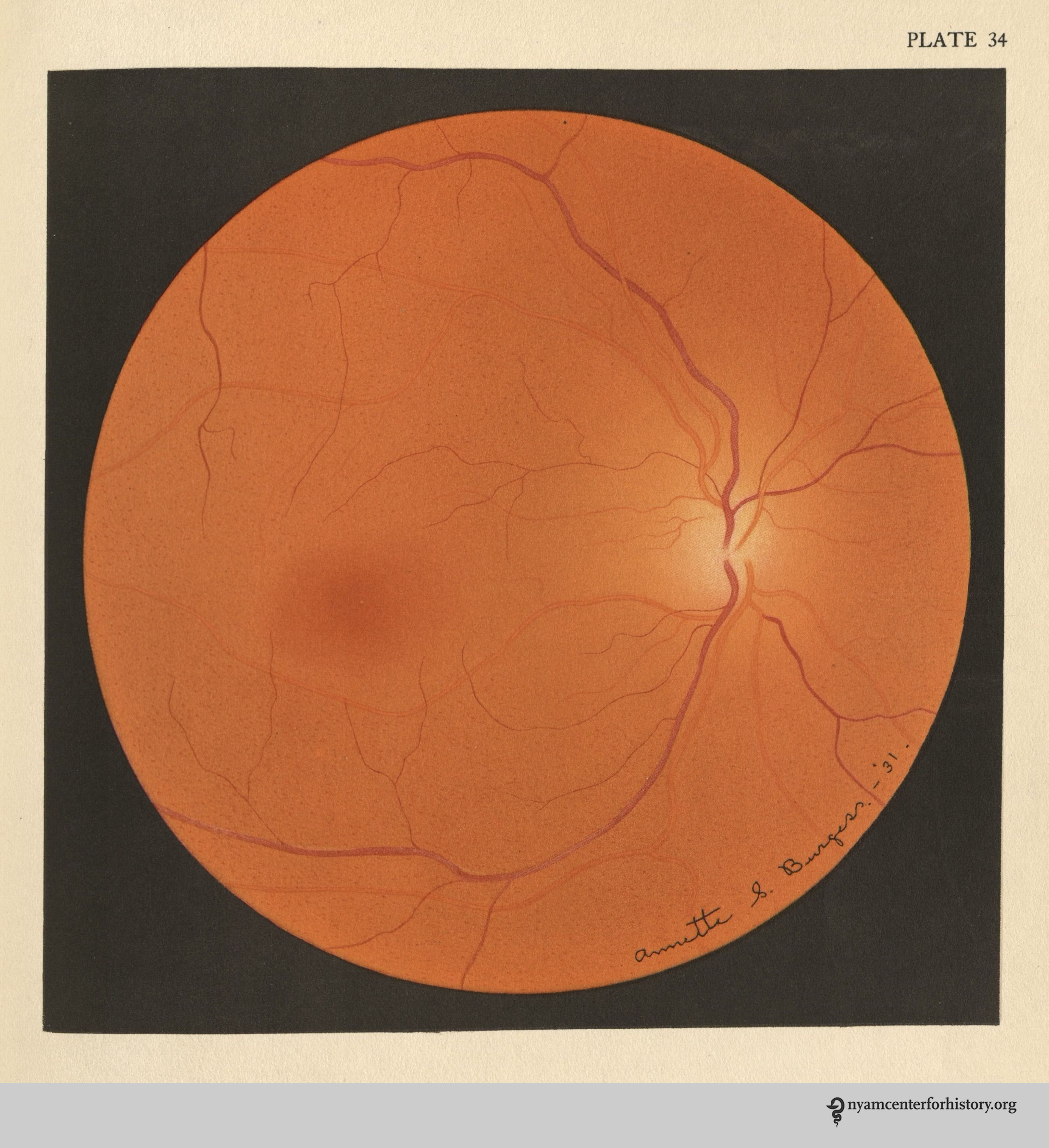
“Choroiditis, Diffuse, with Ascending Perineuritis” (plate 34) from Atlas Fundus Oculi (1934).

Burgess Pl. 34 halftones Wilmer_watermark

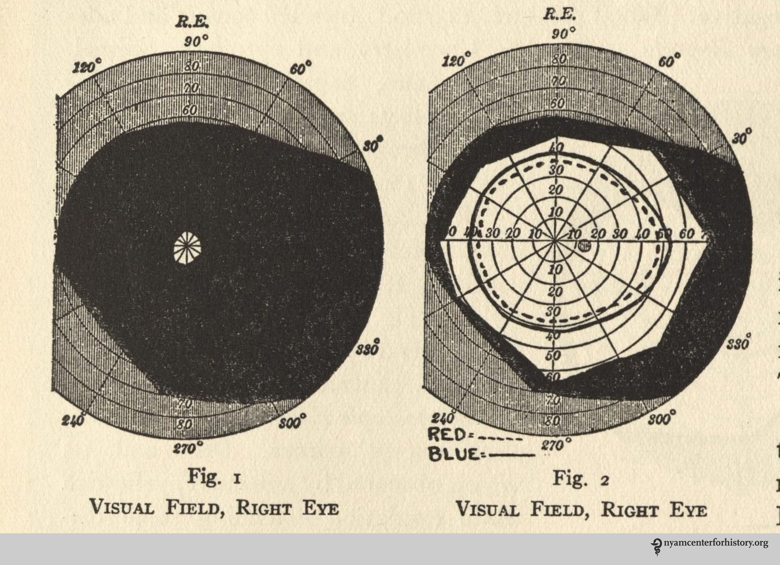
“Choroiditis, Diffuse, with Ascending Perineuritis” (plate 34) halftones from Atlas Fundus Oculi (1934).

Burgess also collaborated with Woods, providing the illustrations for Endogenous Uveitis (1956) and Endogenous Inflammations of the Uveal Tract (1961), although in both of those volumes her paintings were reproduced using photographic processes rather than lithography, and reduced in size.  While still extraordinarily beautiful, the texture found in the earlier lithographs disappears in the reproductions in these later publications.

Plates XXVII and XXVIII (left) and plates XXIX and XXX (right), from Endogenous Uveitis (1956).[4]

For decades after her death, the Department of Art as Applied to Medicine at Hopkins continued to celebrate Annette Burgess’s legacy with an award to honor excellence in ophthalmological illustration.

References:
[1] Davis RW. Annette Smith Burgess (1899-1962).  Journal of the Association of Medical Illustrators. 1963; 14:25-28.
[2] Wilmer WH. Atlas Fundus Oculi. New York: MacMillan; 1934, p. 7.
[3] Woods AC.  Obituary in “News and Comment.” Archives of Ophthalmology. 1962; 68(6): 880.
[4] Woods AC. Endogenous Uveitis. Baltimore: Wiliams & Wilkins, 1956.

At the Crossroads of Art and Medicine

By Anne Garner, Curator, Center for the History of Medicine and Public Health

Our collections have always reflected the strong relationship between medicine and visual culture. Accordingly, since its creation in 2012 our blog has frequently taken up the intersection between medicine and art as subject. Below, we link below to a few posts that explore these crucial connections.

Most recently, Caitlin Dover featured The New York Academy of Medicine’s collections of illustrated medical books on the Guggenheim’s blog in “Doctors Without Borders: Exploring Connections Between Art and Medicine.” Her findings are in part the fruit of a visit with the Academy’s Historical Collections Librarian Arlene Shaner, who showed her a selection of books and ephemera from our Drs. Barry and Bobbi Coller Rare Book Reading Room, showcasing the connection between physicians and artwork.

Robert Latou Dickinson sketch of the Rare Book Room on its opening in 1933, from the Academy's Annual Report, 1933

Robert Latou Dickinson sketch of the Rare Book Room on its opening in 1933, from the Academy’s Annual Report, 1933.

Our extensive collection of anatomical atlases demonstrates the close relationships of physicians and artists, who frequently collaborated to create works both for students of medicine and of art. These atlases show both the successes and failures of collaborations between anatomists and artists who worked together to communicate new medical knowledge. For Vesalius, the collaboration was a great success. In a guest post from 2015, our 2014–2015 Helfand Research Fellow Laura Robson discusses the way Andreas Vesalius’ great milestone work of 1543, De Humani Corporis Fabrica, relies on the synergy between plates and text, and how a later work that uses the Vesalian plates suffers when the anatomist’s text is eliminated. Another guest post by New York physician Jeffrey Levine explores the visual imagery of Vesalius’ famous frontispiece of this same work. Other writers use illustration to signal authority and knowledge. A 2015 post on Walther Ryff explores the ways that Ryff’s use of the counterfeit style in his illustrations implied eye-witness discovery.

Andreas Vesalius (1514-1564). De humani corporis fabrica libri septum. Basel: Johannes Oporinus, 1543. The most famous illustrations are the series of fourteen muscle men, progressively dissected. Some figures, such as this one, are flayed. Hanging the muscles and tendons from the body afforded greater detail, not only showing the parts, but how they fit together.

Andreas Vesalius (1514-1564). De humani corporis fabrica libri septum. Basel: Johannes Oporinus, 1543.

Our 2014 festival Art, Anatomy and the Body: Vesalius at 500 offered ample opportunity for critical thinking about the relationship between art and the body. Guest curator and visual artist Riva Lehrer describes her personal experience of the ways the body informs identity, and how that has shaped her own work as an artist in a 2014 post. A selection of images from several of our early anatomical atlases are featured in “Brains, Brawn and Beauty,” an exhibit that accompanied the festival, and are discussed here.

Finally, two posts on skeleton imagery highlight the tradition of danse macabre imagery in anatomical illustrations. Brandy Shillace’s guest post, “Naissance Macabre: Birth, Death, and Female Anatomy” examines depictions of the female body over time. For a look at the evolution of anatomical imagery with special attention to the tradition of portraying the human skeleton in vivo, visit our blog here. You’ll find a slide show hosted by Flavorwire featuring spectacular anatomical images from our collections.

Surgite mortui, et venite ad judicium (Arise, ye dead, and come to the judgment). Table 6. Click to enlarge.

Surgite mortui, et venite ad judicium (Arise, ye dead, and come to the judgment). Table 6. Click to enlarge.

Next month, the New York Academy of Medicine library will be undertaking an artistic project of our own. Capitalizing on the current coloring craze, we are starting a week-long special collections coloring celebration on social media, using the hashtag #ColorOurCollections. We’ll share images from our collections, as will friends at other institutions. We encourage you to color them, and share your colored copies on social media. Read more about how you or your institution can participate.

CamelColored

Coloring a camel from Conrad Gesner’s Historia Animalium, Liber I, 1551.

Canapé Parade

By Johanna Goldberg, Information Services Librarian

Thanksgiving means many things: spending time with family, reflecting on what you’re thankful for, looking back at American history. But the essence of the holiday can be stated in three words: food, football, and parades.

Our collection contains a whimsical pamphlet that combines two of the three (sorry, football fans): “Canapé Parade: 100 Hors d’Oeuvre Recipes,” published in 1932. (We have the fourth printing, from November of that year.)

The cover of Canapé Parade. Click to enlarge.

The cover of Canapé Parade.

The personality-filled canapés from the cover reappear throughout the pamphlet, illustrating recipes like bloater paste, Japanese crabmeat, herring, and marrons in brandy. Unfortunately, the pamphlet does not credit the illustrator.

None of the recipes specify ingredient amounts, “as the consistency and proportion of ingredients used will vary according to the individual palate.” The recipes take a semi-homemade approach, adding minimal fresh items to packaged food before spreading on crackers. The cookbook also advises that “the majority of these hors d’oeuvres also make excellent sandwich fillings to be served between thinly sliced bread at afternoon tea or buffet suppers.”

Enjoy the recipes on parade below (click to enlarge and view the gallery):

For more traditional Thanksgiving recipes, read Thanksgiving, 1914 Style.

Revisiting the Fabrica Frontispiece

Jeffrey M. Levine, MD, AGSF, author of today’s guest post, will present “Revisiting the Frontispiece: Vesalius’s Jewish Friend and the Impact of the Inquisition” with Michael Nevins, MD, at our October 18th festival, Art, Anatomy, and the Body: Vesalius 500.

Between the first edition in 1543 and the second edition in 1555, the frontispiece of Andreas Vesalius’ classic masterpiece, De humani corporis fabrica, was recut with many subtle variations in both style and content. I am thrilled to be presenting “Revisiting the Frontispiece: Vesalius’s Jewish Friend and the Impact of the Inquisition” at Art, Anatomy, and the Body: Vesalius 500 with my colleague and mentor, Dr. Michael Nevins. Together we will examine and compare the frontispieces and offer theories as to why differences appear. We propose, for example, that some changes to the second edition were in reaction to the Inquisition, which was revived by Pope Paul III.1

Today’s guest post introduces selected features of the frontispiece of the Fabrica’s first edition. This intricate and multilayered composition features the master Vesalius dissecting a young female corpse, her abdomen flayed open. They are surrounded by a multitude of spectators crowded into a three-tiered wooden scaffold built into a semicircular amphitheater of Corinthian columns. At top-center is the decorative escutcheon that bears the name of the book and the author. Above is the family coat-of-arms of Andreas Vesalius flanked by two putti, the chubby male children who were often a feature of Renaissance art, and two gargoyles. Below is the face of Jupiter, the Roman king of gods.

The frontispiece to the 1543 Fabrica in our collection.

The frontispiece to the 1543 Fabrica in our collection. Click to enlarge.

The frieze sitting atop the columns contains symbols including a bucranium, or ox skull with garlands hanging from its horns, which was the symbol of the University at Padua,2 and a winged lion representing the evangelist St. Mark, the symbol of neighboring Venice.3 The columns are flanked by two men, one naked with tense muscles and a worried look, the other relaxed and smartly dressed.

To the right of the skeleton bearing a risus sardonicas, a man in a truncated conical hat recoils as if in terror, squinting and raising his left hand in a defensive gesture. In his 1964 biography of Vesalius, Charles O’Malley identifies this figure as Vesalius’s Jewish friend, Lazaro de Fregeis, who assisted with the Hebrew nomenclature in the Fabrica.4 The only woman other than the corpse appears as a mysterious figure peeking between the columns. There are two Franciscan Monks among the spectators, neither exhibiting much interest in the dissection. Below right is a pickpocket caught in the act. On the opposite side, a leashed monkey screams in protest, and under the table two men fight over the dissecting tools.

There is much more to learn about the frontispiece of the first edition of the Fabrica, and even more when compared to the second edition. To find out more about the changes to the second edition frontispiece, and how they may have contained coded messages reflecting tensions of 16th-century Italian society, particularly in the context of the situation of European Jewry, come to our presentation at the New York Academy of Medicine’s Vesalius 500 celebration on October 18.

References

1. Historical overview of the Inquisition. 2001. Available at: http://galileo.rice.edu/lib/student_work/trial96/loftis/overview.html. Accessed September 23, 2014.

2. Padova Terme Euganee. University of Padua – Palazzo Bo. Available at: http://www.turismopadova.it/en/university-padua-palazzo-bo. Accessed September 23, 2014.

3. Imboden D. Winged Lion of St. Mark. Durant Cheryl Imboden’s Venice Visit. Available at: http://europeforvisitors.com/venice/articles/winged_lion_of_st_mark.htm. Accessed September 23, 2014.

4. O’Malley C. Andreas Vesalius of Brussels, 1514-1564. Berkeley: Univ. of California Press; 1964.

Introducing Graphic Medicine

Ian Williams and MK Czerwiec, authors of today’s guest post, co-run the website GraphicMedicine.org. They will present “Graphic Medicine and the Multiplanar Body” at our October 18th festival, Art, Anatomy, and the Body: Vesalius 500.

The 2010 Comics & Medicine gathering before Senate House.

The 2010 Comics & Medicine gathering before Senate House.

In the summer of 2010 a group of scholars, health care professionals, and comics artists gathered in Senate House, London. This brutal-looking art deco building, said to have inspired George Orwell’s “Ministry of Truth,” represented Gotham City Courts in the films Batman Begins and The Dark Knight. Those gathered, however, were not particularly interested in superheroes. They focused on graphic memoirs of illness, a modern phenomenon born of the counterculture in the 60s and 70s that has gathered momentum over the last 20 years.

Among the 75 delegates from around the world were the authors of this blog entry. The lead organizer of the conference was Ian Williams, a doctor and comics artist, creator of The Bad Doctor (2014, Myriad Editions). MK Czerwiec (pronounced sir-wick), aka Comic Nurse, has been making comics about her work in HIV/AIDS and hospice care since the late 1990s as a way of processing these caregiving experiences. We have now worked together for four years, talking and writing about the interplay between the comics and health care. We make comics, collaboratively and separately, and will give a talk on October 18th at “Art, Anatomy, and the Body: Vesalius 500” about Graphic Medicine, the field we helped pioneer.

The Bad Doctor. Cover by Ian Williams.

The Bad Doctor. Cover by Ian Williams.

MK Czerwiec teaching at Northwestern Feinberg Medical School. Still from BBC story by Katie Watson.

MK Czerwiec teaching at Northwestern Feinberg Medical School. Still from BBC story by Katie Watson.


Often when we describe Graphic Medicine, people say that comics must make an excellent educational medium for patients, especially those with poor literary skills and marginalized groups such as drug addicts, teenage mothers, or the mentally ill. While comics have certainly been used to reach these audiences, the idea behind this response is freighted with assumptions about comics, their target demographics, and the literacy skills or aesthetic proclivities of the social groups so named.

Stack of medically-themed graphic novels. Photo by Ian Williams.

Stack of medically-themed graphic novels. Photo by Ian Williams.

We regard comics as a sophisticated, rich, and adaptable system through which to explore the complex issues of health care. Our primary interest has been the use of graphic illness narratives to provide new knowledge about the illness experience and commentary on the pervading cultural conceptions of disease and health care. We are also interested in the psychological process of making comics. We have also been teaching using comics—both making them and reading them—in medical schools in the US and UK.

Binky Brown Meets The Holy Virgin Mary cover by Justin Green

Binky Brown Meets The Holy Virgin Mary cover by Justin Green

In 1972 Justin Green became the first comics artist to unburden his psychological troubles onto the page, creating Binky Brown Meets the Holy Virgin Mary. This inspired subsequent generations of artists to articulate their corporeal experiences in words and pictures, a process that Elisabeth El Refaie refers to as “pictorial embodiment.”1 More than 40 years later, the myriad comics titles that appear each year include stories of disease or trauma, known as “graphic pathographies,”2 in which the authors give highly subjective accounts of their own illnesses or caregiving experiences. The production of these works involves the repeated drawing of the author’s or subject’s body over a prolonged period, which may have interesting effects on how the artist perceives the body. The relentless decision-making process forces the artist to examine fears, suffering, anger, disgust, disappointment, and grief and distill the whole into a succinct series of sequential panels through which to transfer the narrative to the reader.

2014 Comics & Medicine poster. Art by Lydia Gregg.

2014 Comics & Medicine poster. Art by Lydia Gregg.

Since the London gathering, we have held international conferences in Chicago, Toronto, Brighton, and Baltimore. The movement is growing and what was initially viewed by some as a novelty interest is gaining respect in academia. As the nature of literacy changes, moving from the textual towards the image, comics is once again in ascendance, gaining new readers who might have previously dismissed the medium.


References

1. El Refaie, E. (2012). Autobiographical comics: Life writing in pictures. Jackson: University Press of Mississippi.

2. Green, M. J., & Myers, K. R. (2010). Graphic medicine: Use of comics in medical education and patient care. BMJ, 340, c863.

Virtual Dissection

Kriota Willberg, the author of today’s guest post, explores the intersection of body sciences with creative practice through drawing, writing, performance, and needlework. She will present at our October 18th festival, Art, Anatomy, and the Body: Vesalius 500.

Artistic and clinical examinations of the body share many of the same processes. The artist and the clinician study the body’s mass, look for irregularities in its shape and color, locate bones, joints, and muscles, take notice of the breath. They watch the body and its parts move through space, assess joint alignment, and determine if their subject’s physical parts and relationships are assembled or functioning in a desirable form. The languages for and techniques of analysis vary by discipline but the object of exploration is the same.

"Dhanurasana illustration" by Kriota Willberg

“Dhanurasana illustration” by Kriota Willberg. Click to enlarge.

My careers are grounded in the exploration of the body. There was a time when I would take a morning ballet class, teach anatomy in the afternoon, and in the evening either work a shift as a massage therapist or go to a dance rehearsal. To relax on the weekends I would draw musculoskeletal anatomy illustrations for my class handouts.

Drawing, dancing, and massage all require skills in postural assessment. As a massage therapist I also palpate deeper structures, locating them under skin, fat, and other layers of muscle. As a dancer I learned to feel my musculoskeletal structures via movement exercises that isolate muscle groups or coordinate the body as a whole. Through years of building experiential and objective understanding of the body, physical assessment has become second nature to me.

Friction “…Bundle of Fibers” by Kriota Willberg.

“Friction” by Kriota Willberg. Click to enlarge.

Anatomy entertains and delights me everywhere I go. I study the foot and ankle alignment of strangers as they climb the subway steps. I monitor my two amputee cats for the development of functional scoliosis. I measure and palpate the skin and adipose of my husband, or myself, or the cats, as we sit on the couch and watch the Bond film Tomorrow Never Dies. I comment on Pierce Brosnan’s resemblance in the film to a dissected human subject illustrated in an Albinus anatomy text from 1749.

Tomorrow Never Dies from “The Anatomy of 007” by Kriota Willberg. Click to enlarge

Tomorrow Never Dies from “The Anatomy of 007” by Kriota Willberg. Click to enlarge

The world is an anatomical wonderland. Anatomy is all around us and all we have to do is see it and feel it.

"Pictorial Anatomy of the Cute" by Kriota Willberg. Click to enlarge.

“Pictorial Anatomy of the Cute” by Kriota Willberg. Click to enlarge.

I’m not unique in my perspective of the world of anatomy. There are many people in arts, sciences, and health professions who are skilled at virtual dissection. We can look at you, through your clothing, and through your skin and fat to see the muscle and bone beneath. We share the same skills and sometimes we share the same sense of humor. But our cohort is somewhat rarified. I intend to bring more people into the knowledge and skills that will enable them to join our “club.”

I train others in methods of seeing the body as a clinical or artistic tool. As a part of this instruction, I draw the body on a body. Using a live model, I locate bones and joints, tracing bony landmarks in rinseable ink. Then I locate a muscle’s attachment sites, connect them, and “flesh out” the muscle’s contours and fiber direction.

Willberg anatomy drawing. Model: Wendy Chu

Willberg anatomy drawing. Model: Wendy Chu

Willberg anatomy drawing. Model: Wendy Chu

Willberg anatomy drawing. Model: Wendy Chu

We watch levator scapula lengthen with upward rotation of the scapula. Or the hamstring elongate to seemingly impossible length as the model moves through deep hip flexion. The upper pectoralis major shortens as the lower part lengthens when the model brings her arms overhead. After 27 years of teaching, I am still entranced by these simple movements.

At the Vesalius 500 celebration on October 18, we will look at the body with the double vision of the anatomist. Part live-drawing performance, part slide show/lecture, part conversation, we will explore the (kin)esthetic relationships of our anatomy. I’ll present a narrated slideshow of artworks from A(lbinus) to V(esalius) to enhance and define actual and fanciful relationships of our parts to our whole. A live model and I will create associations between these illustrations and the living body by tracing superficial and deep connections of muscle to movement. The presentation will include opportunities for you (the audience) to ask questions and comment on your own experiences with the study of anatomy.

See you there!

Winsome Fetal Skeletons Bearing Scythes: Monro’s Traité d’ostéologie of 1759: Guest post by Morbid Anatomy

A note from the Center for the History of Medicine & Public Health: This is the last post in Morbid Anatomy‘s guest series leading up to our Festival of Medical History and the Arts. If you’ve enjoyed these posts as much as we have, don’t despair! Tomorrow’s event holds a full day of lectures and activities from Morbid Anatomy, Lawrence Weschler, and the Center. We hope you can make it! See the full schedule here.

FrontispieceThe NYAM rare book collection holds a gorgeous copy of the first French edition of Alexander Monro’s (1697–1767) celebrated Traité d’ostéologie (or “Anatomy of Bones”). Monro was trained in London, Paris, and Leiden before going on to become the first professor of anatomy at the newly established University of Edinburgh. It was under his leadership, and that of his successors, that the school went on to become a renowned center of medical learning.

Monro originally published this book without images, thinking them unnecessary after William Cheselden’s lavishly heavily-illustrated Osteographia, or the anatomy of the bones of 1733 (more on that book at this recent post). The very fine copperplates you see here were added to the French edition by its translator, the anatomist Jean-Joseph Sue (1710–1792).

My favorite image in the book is a kind of memento mori–themed tableau morte of winsome, scythe-bearing fetal skeletons enigmatically arranged in a funereal landscape (images 1–3). I also love the frontispiece in which a group of plump putti proffer anatomical atlases and dissecting tools under the oversight of a skeletal bird (above).

This post was written by Joanna Ebenstein of the Morbid Anatomy blog, library and event series; click here to find out more. All images are my own, photographed at the New York Academy of Medicine.

A Renaissance Man at Work: Volcher Coiter’s “Externarum et internarum principalium humani corporis” of 1573: Guest post by Morbid Anatomy

Coiter2


Coiter10

One under-seen and fascinating book to be found in the NYAM rare book collection is Externarum et internarum principalium humani corporis partium tabulae published by Dutch Renaissance man Volcher Coiter (1534–1576) in 1573. Not only was Coiter renowned as an anatomist, surgeon, and physician accomplished in the fields of physiology, ornithology, and embryology; not only did he establish the study of comparative osteology and describe cerebrospinal meningitis before any of his peers; he was also an artist, and signed many of the finely drawn copper engravings in his books, including those you see here.


All images are my own, photographed at the New York Academy of Medicine, save the painted portrait of Coiter, which was sourced here. The caption, attributed to Dorothy M. Schullian, reads: “Coiter’s portrait (1575) in oils, attributed to Nicolas Neufchatel and representing him demonstrating the muscles of the arm, with the écorché he had constructed on his left and a shelf of medical classics behind him, is preserved in the Germanisches Nationalmuseum, at Nuremberg; there are later portraits at Weimar and Amsterdam.” (source for caption here)

Sources: Lessico Volcher CoiterWikipedia

This post was written by Joanna Ebenstein of the Morbid Anatomy blog, library and event series; click here to find out more.

Remmelin’s Dissectible Cosmic Anatomical Extravaganza: Guest post by Morbid Anatomy

Johan Remmelin (1583–1632) was town physician of Ulm and Plague physician of Augsberg. He was also the man behind both the concept and the original drawings (engraved by Lucas Kilian) for the ingenious moving-parts anatomical extravaganza Catoptrum microcosmicum, published in 1619, with numerous editions in many languages thereafter.  NYAM has both the 1619 and a 1639 edition in its rare book collection

title

This astounding book—in which flaps of paper can be drawn back to virtually dissect the human body—features a heady blend of the anatomical, the theological, and the metaphysical, beautifully expressing the worldview of Natural Philosophy, that precursor to science, which oversaw investigations into the human body in the early modern era. In this worldview, God and man, metaphor and the encountered world, were indivisible; the human being was the microcosm of all creation, so to understand the secrets of the human body would be to know the mind of God. Accordingly, as explained by Martin Kemp and Marina Wallace in their book Spectacular Bodies:

The purpose of anatomical images during the period from the Renaissance to the nineteenth century had as much to do with what we would call aesthetic and theological understanding as with the narrower intentions of medical illustration as now understood. . . .They were not simply instructional diagrams for the doctor technician, but statements about the nature of human beings as made by God in the context of the created world as a whole [as well as] the nature of life and death. . . .

It should not be surprising, then, that the dissectible humans herein are inextricably entwined with images of Jesus Christ (image 9,17); memento mori mottos (16) and imagery (images 12, 17); allusions to God and the angels (image 1); and even the head of the devil, serving as a kind of fig leaf covering the female sex organs in one instance (image 2). There are also numerous biblical references, including a serpent slithering through a human skull holding a branch from the tree of knowledge in its mouth (image 13), lest we forget that original sin introduced death and disease into our world in the first place; without it, we would still be luxuriating in Eden with no need for medicine, or, by extension, books such as this one. The book also contains the occasional inadvertent (?) eroticism, as the peeling back of obscuring layers brings you, in a sort of pre-modern striptease, to the unveiled sexual organs below (image 14 and 15).

If you page through all of the images below, you will get a sense of the carnivalesque exuberance and dynamism of this book; you can also virtually dissect them yourself by clicking here, or here, compliments of The Hardin Library of The University of Iowa, which was also a source for much of the factual content of this piece.

This post was written by Joanna Ebenstein of the Morbid Anatomy blog, library and event series; click here to find out more. All images are my own, photographed at the New York Academy of Medicine.

William Cowper’s Myotomia reformata: or an anatomical treatise on the muscles of the human body, Guest Post by Morbid Anatomy

Cowper01

William Cowper (c. 1666–1709) was a British surgeon and anatomist best known today for describing “Cowper’s Gland,” part of the genito-urinary system. He has also been described by at least one scholar as “the first of the surgeon-scientists of Great Britain… the first to bring the power of the experimental method to bear on practical surgical problems… [anticipating] the celebrated Hunterian school of surgery by more than half a century.” (In the last half of the 18th century, famous London surgeon John Hunter made his reputation in part by advocating for the scientific method in medicine.)

Cowper02

Cowper’s book Myotomia reformata: or an anatomical treatise on the muscles of the human body, first published in 1694 (NYAM’s copy is from 1724), is filled with grimly literal anatomical and dissection-themed initial capital letters, and charming, if somewhat rough, illustrations.

This post was written by Joanna Ebenstein of the Morbid Anatomy blog, library and event series; click here to find out more.