Get Crafty at the Museum Mile Festival on June 14

By Emily Miranker, Project Coordinator

When my office is perfumed by the smell of crayons and stocked with boxes of jumbo-sized sidewalk chalk, I know its Museum Mile Festival time. This year’s Museum Mile Festival takes place on Tuesday, June 14 from 6:00-9:00 pm, rain or shine.

Museum Mile (New York City’s Fifth Avenue from 82nd to 105th Street, which is technically three blocks longer than a mile) is one of the densest cultural stretches in the world.1 For the last 38 years, Fifth Avenue closes to traffic for a few hours on an early June evening. The eight major museums and their neighbors–that’s us!–throw open their doors and spill out onto the street in a block party.

Museum Mile at the New York Academy of Medicine. Courtesy of the Academy's Communications Office.

Museum Mile at the New York Academy of Medicine. Courtesy of the Academy’s Communications Office.

The first festival was held in 1979, the brainchild of the Museum Mile Association, to increase cultural audiences and garner support for the arts in time of great fiscal crisis in the city. The festival has since brought many New Yorkers and tourists to upper Fifth Avenue for the first time, and total attendance over the years has surpassed one million visitors.

Besides free admission to the museums along the mile, street performers, chalk drawing, live bands, balloons, and family-friendly activities abound. Dedicated to improving the health and well-being of people living in cities, the Academy has partners from the East Harlem Asthma Center of Excellence and Shape Up NYC joining us for the evening.

Getting physical with our community partners at Museum Mile. Courtesy of the Academy's Communications Office.

Getting physical with our community partners at Museum Mile. Courtesy of the Academy’s Communications Office.

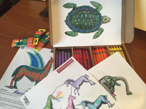
The Library has planned some special crafts for the festival. We have the perennial favorite: coloring pages based on images from our collections. Feel free to download your own pages any time from #ColorOurCollections online.

Coloring sheets fro the New York Academy of Medicine Library. Photo: Emily Miranker.

Coloring sheets from the New York Academy of Medicine Library. Photo: Emily Miranker.

Among the treasures of our collection are the anatomical flap books. These are detailed anatomical illustrations superimposed so that lifting the sheets reveals the anatomy and systems of the body as they would appear during dissection. We created a simple DIY version of a flapbook inspired by these remarkable figures from the 1559 edition of Geminus’ Compendiosa totius anatomiae delineatio, aere exarata. The sheets are quite delicate, so it’s rare to see intact versions like this 400 years after they were made. Make your own flapbook with us during the festival.

Male flap anatomy from The Academy's copy of the 1559 English edition of Geminus’ Compendiosa.

Male flap anatomy from The Academy’s copy of the 1559 English edition of Geminus’ Compendiosa.

Female flap anatomy from The Academy's copy of the 1559 English edition of Geminus’ Compendiosa.

Female flap anatomy from The Academy’s copy of the 1559 English edition of Geminus’ Compendiosa.

Make this flap anatomy craft with us at Museum Mile! Photo: Emily Miranker.

Make this flap anatomy craft with us at Museum Mile! Photo: Emily Miranker.

And there’s nothing like using your own body to create art—finger print art!2

Make fingerprint art with us at Museum Mile! Photo: Emily Miranker.

Make fingerprint art with us at Museum Mile! Photo and artwork: Emily Miranker.

We look forward to seeing you at Museum Mile!

References

1. “Museums on the Mile.” Internet Archive Wayback Machine (June 2011). Accessed June 3, 2016. https://web.archive.org/web/20120101013336/http://www.museummilefestival.org/museums/

2. “Fingerprint Fun.” Bookmaking with Kids (June 2010). Accessed June 6, 2016. http://www.bookmakingwithkids.com/?p=1826

The Foresight of Trans-vision: An Innovative Anatomy of the Eye

By Anne Garner, Curator of Rare Books and Manuscripts

Early European anatomical lift-the-flap books made use of technologies available during the 16th century: woodcut and engraving, combined with manual cutting and pasting.1 Flap anatomies like Geminus’ Compendiosa (1559) allowed readers to peel away the layers of the body to reveal different organs, but these flaps were made of paper and opaque, and didn’t allow the reader to view the strata of the body simultaneously.

Female flap anatomy from The Academy's copy of the 1559 English edition of Geminus’ Compendiosa.

Female flap anatomy from The Academy’s copy of the 1559 English edition of Geminus’ Compendiosa.

Fast forward four centuries, when an innovation in printing technology let readers take a deep dive through the layers of the body all at once.

Anatomical illustrators used transparencies to show the layers of the body as early as the 1920s. J.E. Cheesman published Baillière’s Synthetic Anatomy, a series of 14 booklets, in London from 1926 to 1936. The series used a set of glassine sheets to show what lay beneath the surface of the skin.

Forearm in Cheesman, Baillière's Synthetic anatomy, 1926-1936.

Forearm in Cheesman, Baillière’s Synthetic Anatomy, 1926-1936. Click to enlarge.

Hand in Cheesman, Baillière's Synthetic anatomy, 1926-1936.

Hand in Cheesman, Baillière’s Synthetic Anatomy, 1926-1936. Click to enlarge.

In 1942, Richard Lasker patented a new printing process for Milprint Inc., a Milwaukee-based company. He called this new method trans-vision. Trans-vision allowed for the printing of images on the inner surfaces of folded sheets of transparent acetate. These sheets could then be piled on top of one another so that they overlapped, enabling a multi-layered view with the top sheets depicting the most superficial layers of an object and the bottom sheets the deepest level.2

The patent application for Lasker’s trans-vision process used a cutaway illustration of a mattress, with different layers of acetate offering views of the mattress’ filling.3 Trans-vision’s medical applications proved significantly more useful: it made possible the representation of complex anatomical relationships to health professionals and public audiences alike.

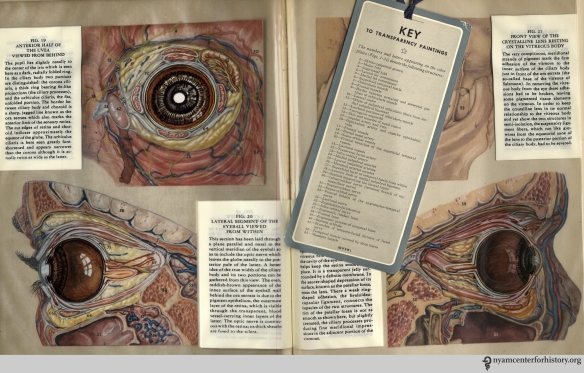
In 1943, Peter C. Kronfeld, an ophthalmology professor at the University of Illinois, published The Human Eye in Anatomical Transparencies. The book contains 34 color anatomical paintings printed using trans-vision.4 Each page offers a frontal and temporal view of the eye and its area, with transparent layers that can be peeled away by turning the pages. The paintings are printed on the inner side of the acetate to minimize damage from handling. The parts of the eye are numbered. Readers can use a bookmark key laid-in to identify the different parts by name.5

Figure I in Kronfeld, The Human Eye in Anatomical Transparencies, 1943. Click to enlarge.

Figure 1 in Kronfeld, The Human Eye in Anatomical Transparencies, 1943. Click to enlarge.

Kronfeld describes the project in The Human Eye’s preface:

It was at once obvious that the eye could advantageously be represented by this means, for it is a three-dimensional object in which great structural intricacy is combined with relatively small size. Ordinary drawings of its separate parts tend to isolate them too much from each other in the mind of the observer….The text has been so organized as to present not only a systematic account of ocular anatomy—taking up the various structures in a functionally logical order—but also a topographic treatment of the anatomy…which necessarily reveal the structures layer by layer in an order determined somewhat by the layers of dissection techniques.6

The paintings were made at twice the actual size by Gladys McHugh, an illustrator at the University of Chicago. The pioneering medical illustrator Max Brödel had been McHugh’s teacher and mentor at Johns Hopkins University, where she studied. It was Brödel who influenced McHugh to make her own dissections.

Figure II in Kronfeld, The Human Eye in Anatomical Transparencies, 1943. Click to enlarge.

Figure 2 in Kronfeld, The Human Eye in Anatomical Transparencies, 1943. Click to enlarge.

McHugh augmented her dissections of human eyes with specimens from pigs and monkeys. She describes her extensive dissection work in her introduction:

Over a course of time I obtained from baby autopsies ten good cases, making a total of twenty eyes and orbits. These I dissected layer by layer, making color notes and drawings from the fresh specimens. To develop a technique for separating the layers of the eyeball as intact semispheres, pigs’ eyes were employed. Also, to supplement my observation of the muscles and other structures not fully developed in the infant, monkey orbits were dissected.

As Professor Shelley Wall has argued, turning the pages in The Human Eye mimics the dissection process. As the layers on the recto side of the book are more deeply revealed with each page turn, the layers build on the opposite verso, allowing for the eye’s reconstruction.7 With The Human Eye, the union of format, technology, and material is harmoniously in sync.

Figues 3-6 in Kronfeld, The Human Eye in Anatomical Transparencies, 1943. Click to enlarge.

Figues 3-6 in Kronfeld, The Human Eye in Anatomical Transparencies, 1943. Click to enlarge.

Figure 14 in Kronfeld, The Human Eye in Anatomical Transparencies, 1943. Click to enlarge.

Figure 14 in Kronfeld, The Human Eye in Anatomical Transparencies, 1943. Click to enlarge.

Figures 19-22 in Kronfeld, The Human Eye in Anatomical Transparencies, 1943. Click to enlarge.

Figures 19-22 with bookmark key in Kronfeld, The Human Eye in Anatomical Transparencies, 1943. Click to enlarge.

By 1958, the book had enjoyed five editions.8 Students and educators embraced the text and its ingenious illustrations. In 1946, the trans-vision process was applied again to McHugh’s paintings in for The Human Ear in Anatomical Transparencies. Initially conceived as a wartime project useful to the aviation industry, the book’s value, as with The Human Eye, was in its power to demonstrate to both lay and specialized audiences the inner workings of organs not easily seen.

The finest examples of trans-vision printing occurred when the coffers of the pharmaceutical companies who published them were at their fullest. After The Human Eye and The Human Ear, medical illustrator Ernest Beck used trans-vision technology to produce more than 30 anatomical transparency projects published by Milprint for encyclopedias, pharmaceutical companies and other commercial concerns.9 A decade after Kronfeld, Ciba Pharmaceuticals published a 13-volume collection of anatomical illustrations using anatomical transparencies between 1953 and 1989. The illustrator of these, was a native New Yorker, fellow of the Academy, and former member of the Art Students League of New York. His name was Frank Netter.10

References

1. For more on this, see Andrea Carlino’s excellent Paper bodies: A catalogue of anatomical fugitive sheets, 1538-1687.

2. Wall, Shelley. “Mid-twentieth-century anatomical transparencies and the depiction of three-dimensional form.” Clinical Anatomy 23 (2010) 915-921. Accessed December 2015.

3. Wall, 917.

4. David Templeman, review of The Human Eye in Anatomical Transparencies by Peter C. Kronfeld. Optometry & Vision Science, 35 7 (1958). 388.

5. Wall, 919.

6. Kronfeld, Peter. The Human Eye In Anatomical Transparencies. Rochester, New York: Bausch & Lomb Press, 1943. iii.

7. Wall, 919.

8. David Templeman, review of The Human Eye in Anatomical Transparencies by Peter C. Kronfeld. Optometry & Vision Science, 35 7 (1958). 388-389.

9. Brierley, Meghan. Dialogue and Dissemination: The Social Practices of Medical Illustrators in the Pharmaceutical Context. Dissertation, University of Alberta. 2013. Accessed online May 31, 1916.

10. Frank Netter was the most prolific medical illustrator of the twentieth-century. During his more than sixty-years as a medical illustrator he produced more than 4,000 illustrations of the entire anatomic and pathologic character of the human body, system by system. His comprehensive and detailed illustrations, published in The Ciba Collection of Medical Illustrations, Clinical Symposia, and The Atlas of Human Anatomy educated generations of medical professionals and students all over the world.

20th-Century Teeth: Dentistry at the Turn of the Century

By Johanna Goldberg, Information Services Librarian

This is part of an intermittent series of blogs featuring advertisements from medical journals. You can find the entire series here.

“How did you learn to be a dentist? Did you go to a college?”

“I went along with a fellow who came to the mine once. My mother sent me. We used to go from one camp to another. I sharpened his excavators for him, and put up his notices in the towns–stuck them up in the post-offices and on the doors of the Odd Fellows’ halls. He had a wagon.”

“But didn’t you never go to a college?”

“Huh? What? College? No, I never went. I learned from the fellow.”

Trina rolled down her sleeves. She was a little paler than usual. She fastened the buttons into the cuffs and said:

“But do you know you can’t practise unless you’re graduated from a college? You haven’t the right to call yourself, ‘doctor.'”1

In Frank Norris’ 1899 novel McTeague: A Story of San Francisco—better known for its depiction of greed than the professionalization of dentistry—the title character loses his 12-year-old dental practice after California requires practitioners to hold a degree in the field. The timing couldn’t be worse for McTeague: he’d only just fulfilled a long-held dream, obtaining and hanging an enormous golden tooth outside his dental parlor.

McTeague’s fictionalized struggle was based in reality: until the mid to late 1800s, dentistry in the United States was not a regulated profession. Alabama became the first state to regulate dentists in 1841, and other states followed suit through the end of the century.2 In 1885, California passed a law requiring practicing dentists to register with a board, which could call up registrants for examination. Diplomas from a licensed dentistry school—the University of California College of Dentistry opened in San Francisco in 1882—also qualified registered dentists to practice. In 1901, a new law made practicing dentistry in California even more restrictive, part of a nationwide move to tighter regulation.3,4 In the novel as it would have been in real life, McTeague’s practice was toast.

Advertisements in dental journals from the era depict the trend toward professionalization, along with other technological advances. In 1840, the Baltimore College of Dental Surgery opened its doors as the first dental school in the world; by 1895, it had some local competition, including the Dental Department of the Baltimore Medical College.4 This school advertised heavily in journals like the American Journal of Dental Science.

Ad for the Dental Department of the Baltimore Medical College in the American Journal of Dental Science, vol. 33, no. 10, February 1900.

Ad for the Dental Department of the Baltimore Medical College in the American Journal of Dental Science, vol. 33, no. 10, February 1900.

Intriguingly, not only dental schools advertised in dentistry journals: The February 1901 volume of Dental Hints includes an ad encouraging dentists to take up a correspondence course in optometry, “on account of the intimate relationship between the eye and the teeth.” Huh?

Advertisement for the Philadelphia Optical College in Dental Hints, vol. 3, no. 2, February 1901.

Advertisement for the Philadelphia Optical College in Dental Hints, vol. 3, no. 2, February 1901.

Dental journal advertisements also reflect anesthetic advances. William Morton, a dentist, performed the first public demonstration of ether as a surgical anesthetic in 1846.2 A similar demonstration of nitrous oxide in 1845 did not go so well: dentist Horace Wells extracted a tooth before administering the proper dosage, and the patient cried out in pain. The drug was tabled for about 20 years; by 1869, it was commonly used either on its own or in conjunction with ether for dental procedures.2,5 Dental surgeries held less risk than other medical procedures, as they were commonly performed either in the patient’s or dentist’s home, locations less teeming with deadly microbes than operating theaters. After advances in antiseptic surgery by people like Joseph Lister, dental surgery became even safer—and Dr. Joseph Lawrence named an antiseptic mouthwash in his honor.5,6

Codman & Shurtleff's Inhaler for Gas or Ether advertisement in Dental and Oral Science Magazine, vol. 1, no. 2, May 1878.

Codman & Shurtleff’s Inhaler for Gas or Ether advertisement in Dental and Oral Science Magazine, vol. 1, no. 2, May 1878.

Listerine advertisement in the American Journal of Dental Science, vol. 33, no. 10, February 1900.

Listerine advertisement in the American Journal of Dental Science, vol. 33, no. 10, February 1900.

Local anesthetics also entered the market around the turn of the century. Some, like Mylocal, contained cocaine—though in the case of Mylocal, that cocaine was to be added by the practitioner prior to use. Perhaps unsurprisingly, the amount of cocaine used in local anesthetics was often poorly controlled, with sometimes dire results.5 Another local anesthetic, Eureka, proudly advertised that it “[avoids] that most dangerous drug that is known to the profession as COCAINE.” A third, Wilson’s Local Anaesthetic, notes that it is “non-secret and positively guaranteed.” Unfortunately, its ads don’t state what these non-secret ingredients are.

Advertisement for Mylocal anaesthetic in the American Journal of Dental Science, vol. 39, no. 1, January 1908.

Advertisement for Mylocal anaesthetic in the American Journal of Dental Science, vol. 39, no. 1, January 1908.

Advertisement for Eureka Local Anaesthetic in Dental Hints, vol. 3, no. 2, February 1901.

Advertisement for Eureka Local Anaesthetic in Dental Hints, vol. 3, no. 2, February 1901.

Advertisement for Wilson's Local Anesthetic in Dental Clippings, vol. 3, no. 6, April 1901.

Advertisement for Wilson’s Local Anesthetic in Dental Clippings, vol. 3, no. 6, April 1901.

Other turn-of-the-century advances include the development of tube toothpaste in the 1880s (previously, toothpaste had only been available in powdered form); awareness of microbial causes of tooth decay, leading to the promotion of flossing and brushing in the 1890s; and the use of gold foil as a cavity filling in the 1850s.2 The ads below reflect these advances and others, and were selected to show the relatively pain-free side of dentistry.

R. S. Williams Toothbrushes advertisement in Dental and Oral Science Magazine, vol. 1, no. 2, May 1878

R. S. Williams Toothbrushes advertisement in Dental and Oral Science Magazine, vol. 1, no. 2, May 1878.

Ney's Gold Plates advertisement in the American Journal of Dental Science, vol. 33, no. 1, May 1899.

Ney’s Gold Plates advertisement in the American Journal of Dental Science, vol. 33, no. 1, May 1899.

Dental Floss Silk advertisement in advertisements in the American Journal of Dental Science, vol. 33, no. 10, February 1900.

Dental Floss Silk advertisement in advertisements in the American Journal of Dental Science, vol. 33, no. 10, February 1900.

McConnell Dental Chair advertisement in Dental Hints, vol. 3, no. 4, April 1901.

McConnell Dental Chair advertisement in Dental Hints, vol. 3, no. 4, April 1901.

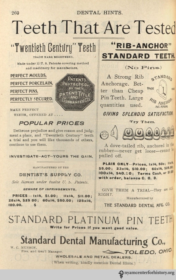
Standard Dental Manufacturing Co. advertisement in Dental Hints, vol. 3, no. 5, May 1901.

Standard Dental Manufacturing Co. advertisement in Dental Hints, vol. 3, no. 5, May 1901.

Dentacura toothpaste advertisement in Dental Hints, vol. 3, no. 11, November 1901.

Dentacura toothpaste advertisement in Dental Hints, vol. 3, no. 11, November 1901.

Munson's Standard Teeth advertisement in Dental Hints, vol. 3, no. 12, December 1901.

Munson’s Standard Teeth advertisement in Dental Hints, vol. 3, no. 12, December 1901.

Prophylactic Toothbrush advertisement in Dental Summary, vol. 22, no. 7, July 1902.

Prophylactic Toothbrush advertisement in Dental Summary, vol. 22, no. 7, July 1902.

Antikamnia and Odontoline advertisements in advertisements in the American Journal of Dental Science, vol. 39, no. 4, April 1908.

Antikamnia and Odontoline advertisements in advertisements in the American Journal of Dental Science, vol. 39, no. 4, April 1908.

Baker Coat Co. and Keeton Gold advertisements in the American Journal of Dental Science, vol. 39, no. 4, April 1908.

Baker Coat Co. and Keeton Gold advertisements in the American Journal of Dental Science, vol. 39, no. 4, April 1908.

Bowl Spittoon advertisement in the American Journal of Dental Science, vol. 39, no. 4, April 1908.

Bowl Spittoon advertisement in the American Journal of Dental Science, vol. 39, no. 4, April 1908.

References

1. Norris F. McTeague.; 1899. Available at: http://www.gutenberg.org/files/165/165-h/165-h.htm. Accessed May 9, 2016.

2. History of Dentistry Timeline. Available at: http://www.ada.org/en/about-the-ada/ada-history-and-presidents-of-the-ada/ada-history-of-dentistry-timeline. Accessed May 9, 2016.

3. Newkirk G. California. In: Koch CRE, ed. History of dental surgery: Dental laws and legislation, dental societies and dental jurisprudence, Vol. III. Fort Wayne, Ind.: National art publishing Company; 1910:755–756. Available at: https://books.google.com/books?id=9iE-AQAAMAAJ&pgis=1. Accessed May 9, 2016.

4. Schulein TM. A chronology of dental education in the United States. J Hist Dent. 2004;52(3):97–108.

5. Enever G. History of dental anaesthesia. In: Shaw I, Kumar C, Dodds C, eds. Oxford Textbook of Anaesthesia for Oral and Maxillofacial Surgery. Oxford: Oxford University Press. doi:10.1093/med/9780199564217.001.0001.

6. From Surgery Antiseptic to Modern Mouthwash | LISTERINE®. Available at: http://www.listerine.com/about. Accessed May 10, 2016.

Many Anatomy Lessons at the New York Academy of Medicine

Kriota Willberg, the author of today’s guest post, explores the intersection of body sciences with creative practice through drawing, writing, performance, and needlework. She is offering the workshop “Visualizing and Drawing Anatomy” beginning June 6 at the Academy. Register online.

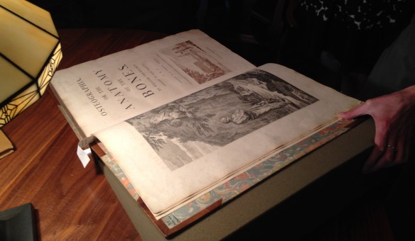
Cheselden's Osteographia, 1733, opened to the title page and frontispiece.

Cheselden’s Osteographia, 1733, opened to the title page and frontispiece.

Different Disciplines, Same Body

I teach musculoskeletal anatomy to artists, dancers, and massage therapists. In my classes the students study the same raw material, and the set of skills each group acquires can be roughly organized around three distinct areas: representation of the body, kinesiology (the study of movement), and palpation (feeling the body).

As an anatomy teacher I am constantly on the prowl for images of the body that visually reinforce the information my students are learning. The Internet has become my most utilized source for visual teaching tools. It is full of anatomy virtual galleries, e-books, and apps. 3D media make it ever easier to understand muscle layering, attachment sites, fiber direction, and more.

In spite of the overwhelming volume of quality online cutting-edge anatomical imagery, I find myself drawn to historical 2D printed representations of the body and its components, once the cutting-edge educational technology of their respective centuries. Their precision, character, size, and even smell enhance my engagement with anatomical study. Many of these images emphasize the same principles as the apps replacing them centuries later.

The Essential Structure Of The Body

Different artists prefer different methods of rendering bodies in sketches. One method is to organize the body by its masses, outlining its surface to depict its bulk. Another method is to draw a stick figure, organizing body volume around inner scaffolding.

Plate XXXIII in Cheselden, Osteographia, 1733.

Plate XXXIII in Cheselden, Osteographia, 1733.

And what is a skeleton but an elaborate stick figure? William Cheselden’s Osteographia (1733) presents elegant representations of human and animal skeletons in action. These images remind us that bones are rigid and their joints are shaped to perform very specific actions. The cumulative position of the bones and joints gives the figure motion. In Cheselden’s world of skeletons, dogs and cats fight, a bird eats a fish, a man kneels in prayer, and a child holds up an adult’s humerus (upper arm bone) to give us a sense of scale while creating a rather creepy theatrical moment.

Muscle Layering

3D apps and other imaging programs facilitate the exploration of the body’s depth. One of the challenges of artists and massage therapists studying anatomy is transitioning information from the 2D image of the page into the 3D body of a sculpture or patient.

Planche 11 in Salvage, Anatomie du gladiateur combattant, 1812.

Planche 11 in Salvage, Anatomie du gladiateur combattant, 1812.

Salvage’s Anatomie du gladiateur combattant: applicable aux beaux artes… (1812) is a 2D examination of the 3D Borghese Gladiator. Salvage, an artist and military doctor, dissected cadavers and positioned them to mimic the action depicted in the statue. His highly detailed images depict muscle layering of a body in motion. The viewer can examine many layers of the anatomized body in action from multiple directions, rendered in exquisite detail. Salvage retains the outline of the body in its pose to keep the viewer oriented as he works from superficial to deeper structures.

Tab. VIII in Albinus, Tabulae sceleti et musculorum corporis humani, 1749.

Tabula VIII in Albinus, Tabulae sceleti et musculorum corporis humani, 1749 edition.

Bernhard Siegried Albinus worked with artist Jan Wandelaar to publish Tabulae sceleti et musculorum corporis humani (1749). Over their 20-year collaboration, they devised new methods for rendering the dissected body more accurately.  The finely detailed illustrations and large size of the book invite the reader to scrutinize the dissected layers of the body in all their detail. Although there is no superficial body outline, the cadaver’s consistent position helps to keep the reader oriented. On the other hand, cherubs and a rhinoceros in the backgrounds are incredibly distracting!

Fiber Direction

Familiarity with a muscle’s fiber direction can make it easier to palpate and can indicate the muscle’s line of pull (direction of action).

Figure in Berengario, Anatomia Carpi Isagoge breves, 1535.

Figure in Berengario, Anatomia Carpi Isagoge breves, 1535.

The images of Jacopo Berengario da Carpi’s Anatomia Carpi Isagoge breves, perlucide ac uberime, in anatomiam humani corporis… (1535) powerfully emphasize the fiber direction of the muscles of the waist. This picture in particular radiates the significance of our “core muscles.” Here, the external oblique muscles have been peeled away to show the lines of the internal obliques running from low lateral to high medial attachments. The continuance of this line is indicated in the central area of the abdomen. It perfectly illustrates the muscle’s direction of pull on its flattened tendon inserting at the midline of the trunk.

The Internal Body Interacting with the External World

One of the most important lessons of anatomy is that it is always with us. Gluteus maximus and quadriceps muscles climb the stairs when the elevator is broken. Trapezius burns with the effort of carrying a heavy shoulder bag. Heck, that drumstick you had for lunch was a chicken’s gastrocnemius (calf) muscle.

Tab. XII in Speigel, De humani corporis fabrica libri decem, 1627.

Tab. XII in Speigel, De humani corporis fabrica libri decem, 1627.

Anatomists from Albinus to Vesalius depict the anatomized body in a non-clinical environment. One of my favorites is Adriaan van de Spiegel and Giulio Casseri’s De humani corporis fabrica libri decem (1627). In this book, dissected cadavers are depicted out of doors and clearly having a good time. They demurely hold their skin or superficial musculature aside to reveal deeper structures. Some of them are downright flirtatious, reminding us that these anatomized bodies are and were people.

Kriota Willberg's self portrait. Courtesy of the artist.

Kriota Willberg’s self portrait. Courtesy of the artist.

I am so enamored of van de Spiegel and Casseri that I recreated page 24 of their book as a self-portrait. After my abdominal surgery, the image of this cadaver revealing his trunk musculature resonated with me. In my portrait I assume the same pose, but if you look closely you will see stitch marks tracing up my midline. I situate myself in a “field” of women performing a Pilates exercise that challenges abdominal musculature. And of course, I drew it in Photoshop.

Narrative, Health, and Social Justice: Stories of the Body

Annie Robinson, today’s guest blogger, holds a Master of Science in Narrative Medicine from Columbia University. As an eating disorder recovery coach, wellness educator, and workshop and retreat leader, Annie uses story to facilitate healing, self-reflection, and narrative competence. She will lead a Health and Social Justice Reading Group at the Academy six Wednesday afternoons from June 22 to July 27. Find out more and register online.

In middle school I developed a severe eating disorder that persisted into my mid-twenties. I have told this story in so many ways over the years. Initially, I subscribed to the common narrative of disorder-as-enemy: “I am battling an eating disorder.” But ultimately, this story did not serve my healing. It made me feel like I was at the mercy of my symptoms. Perceiving my eating disorder as a demon that I needed to fight against positions me as an enemy of myself, insofar as the eating disorder is inherently a part of me.

So I tried out a new story. What if my eating disorder is a wounded part trying to protect me from pain? It offers temporarily helpful—though ultimately ineffective—strategies to meet my needs for comfort and safety. It is young and naive, frantic and scared. It needs to be loved and listened to, not condemned and silenced. I took on the role of mother caring for a feisty, frightened child who needed firm but kind parenting. My mothering self and my eating disorder self engaged in frequent dialogues, both out loud and in writing, to rework the stories I’d been living for so long.

To truly enter recovery, I had to not only rewrite the story of my eating disorder, but to share it with others. As author instead of victim, I am freed from the secretiveness and shame that eating disorders thrive on. And by sharing my story, I make myself vulnerable, which allows me to connect authentically with others.

Using poetry for self-reflection and healing

Using poetry for self-reflection and healing

Renowned researcher Brené Brown studies the correlation between exposing shame, embracing vulnerability, and wellbeing. Her research also examines how the stories we tell about ourselves possess tremendous power—either to trap us, or to instigate radical change. She postulates: “When we deny the story, it defines us. When we own the story, we can write a brave new ending.”

By changing my story, I changed my behaviors, and by changing my behaviors, I changed my life. While stories can disempower, they can also generate agency. They can ascribe blame, or bestow forgiveness. At their best, I believe stories are some of our greatest tools for healing both individual pain and social injustices.

I realized the potency of language not only in recovery, but also in my role as a doula—someone trained to support individuals as they navigate pregnancy, abortion, birth, and fetal loss. In this role, I have witnessed how issues of social justice are deeply entwined with bodily experiences.

Maria, a quiet 16-year-old Hispanic girl living a foster care home for pregnant teens, was 36 weeks along when I met her. For the majority of her labor at a large hospital in the city, no one looked her in the eye. Family-less, jobless, degree-less, and soon to be responsible for a newborn, she seemed too much for her providers to bear. Her labor was blessedly short, her birth smooth, and her beautiful baby boy healthy. But no one showed up to celebrate with her. Though she spoke no words of disappointment, tears welled in her downcast eyes as she smiled down at her tiny child.

Pooja, a vibrant 30-year-old Bengali woman in her second trimester of pregnancy, came into my care at a public hospital. She had just learned that if she carried her pregnancy to term, her baby would be born with severe disabilities. She had no choice but to terminate the pregnancy, because she and her husband could not financially accommodate a child with expensive chronic medical needs. During the termination procedure, she wailed and dug her nails into my hand as I held fast, whispering soothing words in her ear. As her cries escalated, the doctors spoke louder to her in English (a language she barely understood) about what steps they were taking, as if they could extinguish her deep suffering with their voices and expertise.

The stories of Maria and Pooja, along with those of dozens of other women I have served, reflect how social injustice is so often based in the body. Their distinct cultural conditions, social vulnerabilities, and economic disparities all influenced the care they received.

In the world of medicine, the term “social justice” refers to the differences in how people experience health conditions and interface with the healthcare system. It is imperative to consider the roles these factors potentially play in the story of someone’s health experience, leading to inequities in resources, unique linguistic and cultural reference points, and distinct vulnerabilities and disadvantages.

While serving as a doula, I also was a graduate student at Columbia University studying an innovative discipline called narrative medicine. This approach endeavors to train clinicians to deeply hear and respond to their patients’ stories, not just their symptoms. Narrative medicine provides a way for patients and providers to co-create humanized stories of illness and embodied experiences, and offers strategies for studying the meaning of these experiences through telling, reading, and writing about them. I came to appreciate body-based stories as deeply vulnerable ones, as they concern both our physical selves as well as the parts of our identity that transcend biology.

I will be facilitating a six-week course at the New York Academy of Medicine from June 22-July 27, 2016 on how language can serve as a mechanism for social justice in health (register online). We will use narrative practices such as reading fiction and nonfiction texts, having group discussions, and writing self-reflectively for an in-depth exploration of how language influences our experiences of our bodies and can serve as a mechanism for enacting social justice in healthcare.

Workshop on stories and self-care led by Annie Robinson, November 2015

Workshop on stories and self-care led by Annie Robinson, November 2015.

We will use narrative depictions to unpack how health, illness, and disability are issues of social justice. How do social justice and health relate to gender, sexuality, race, trauma, caregiving, privilege, disability, age, class, and geography? How can creative expressions of embodied experiences facilitate self-realization and healing? Whose voices are most often heard, and whose are not? Where do private matters of health, illness, and disability fit in the public arena? What are the effects of how they are politicized, for better and for worse? And how can social, cultural, and political change that benefits embodied experiences be instigated by individuals?

Sources will include (among others): poems by physician-poet Rafael Campo, stories by Sherman Alexie, essays by Eve Ensler, excerpts from Illness as Metaphor by Susan Sontag, first-person perspectives about gendered embodiment from Minding the Body, and pieces from Leslie Jamison’s The Empathy Exams.

Please join us to explore what social justice, story, and embodiment mean to you!

Questions? Please email culturalevents@nyam.org.

Have You Heard of the Lincoln Collective?

Today’s guest blogger, Merlin Chowkwanyun, is an assistant professor of sociomedical sciences at Columbia University’s Mailman School of Public Health. He will present “The Lincoln Collective: The World of New York City Health Activism in the 1970s” at the Academy on May 24. Learn more and register.

I’m really looking forward to visiting the New York Academy of Medicine next week, in no small part because the health activism I’m going to discuss took place in New York City itself. My talk will focus on a couple dozen physicians, fresh out of medical school, who decided to do their residencies at Lincoln Hospital in the South Bronx in the 1970s.

They arrived in the summer of 1970 and called themselves “the Lincoln Collective,” hoping to form a critical mass of politically conscious physicians who could effect change in one institution, and in the process, provide a model for other activists across the country to follow. In its recruitment pamphlet, the Collective’s founders wrote that they intended “to become part of the solution rather than part of the problem” and “affirm[ed] that we are in training to serve the community, and that we are committed to dealing with the problems of the urban ghetto community in a long-run way.” That commitment entailed not just ephemeral service projects that lasted a few weeks, but finding ways to facilitate more permanent community input into healthcare facilities’ operations.

Cover of a Lincoln Collective Recruitment pamphlet.

Cover of a Lincoln Collective Recruitment pamphlet.

Lincoln epitomized the overtaxed, under-resourced urban hospital. One official document described it as “a hopelessly inefficient and inadequate building” with “dirt and grime and general dilapidation [that] make it a completely improper place to care for the sick…” And locals had nicknamed it “The Butcher Shop.” By conventional standards, then, Lincoln was not exactly a desirable or prestigious choice for your typical medical graduate at this time. So what was it that set the Lincoln Collective’s members apart? Who were these people? And where did their values come from? What were they hoping to get by converging on one of the most dilapidated hospitals in one of the most resource-deprived areas of the United States? And most important of all, what did it all mean in the end, when the Lincoln Collective came to a close in the mid-1970s?

To answer these questions, I’ll place the Lincoln episode in a wider story about changes that wracked the healthcare sector during the 1960s and 1970s. Many Collective members had been involved in student organizing on medical campuses, not exactly known, then and now, as cauldrons of political foment. Others had come from community organizing. And some were not particularly political and simply looking for a place to serve the most indigent and medically deprived. They came to Lincoln when the health field was undergoing what I have called a “governance revolution”—multi-pronged efforts throughout the era to decrease hierarchy within medicine and increase the participation of professionals in healthcare governance.

Article on medical student unrest in Medical World News, Oct. 13, 1967, pp. 63–67.

Article on medical student unrest in Medical World News, Oct. 13, 1967, pp. 63–67.

The Collective arrived at a time of tumult around the hospital itself. Groups like the Black Panthers and the Young Lords had made healthcare equality a major tenet of their organizing. At times, the Collective’s relationship with these groups was cooperative and fruitful, at other times, tense and ambiguous. Much of that depended on Collective members’ individual ideological inclinations, which were hardly uniform throughout the group. Tensions undergirded the encounter between mostly white physicians and mostly non-white, non-professional activists, and I’ll explore these challenges throughout the talk.

Pamphlet of Health Revolutionary Unity Movement, a health-oriented adjunct of the Young Lords that also organized around Lincoln.

Pamphlet of Health Revolutionary Unity Movement, a health-oriented adjunct of the Young Lords that also organized around Lincoln.

I’ve been thinking about the Lincoln Collective for more than a decade now. The title of my talk is an utterance I heard repeatedly when I was a college student in New York City studying activist movements in public health and medicine. “Have you heard of the Lincoln Collective?” people would ask. Some who posed the question were in it (and some claimed to be but, I’d later discover, were not). When I went off to graduate school, I put the story aside for a long time. At the confused age of 22, I didn’t feel I had the political maturity to really write about some pretty politically fraught and emotional events. Now, with more distance, I’ve returned to it.

We’re now in an era when people in the health sector—in the wake of a wave of police brutality and the Flint disaster—are asking themselves serious questions about the role political activism should play in their work. Turning back the clock and looking at a group of health activists from 50 years ago is a way of moving that conversation forward.

Edward Jenner and the Development of the Smallpox Vaccine

By Rebecca Filner, Head of Cataloging

Smallpox was one of the leading causes of death in 18th-century Europe, killing about 400,000 people annually and leaving many more disfigured or blind. Epidemics in major cities routinely killed 20% or more of those infected, and this case-fatality rate rose to 80% for infants.

Variolation, a process through which people were deliberately infected with mild strains of smallpox in an effort to shield them from catching a more virulent form of the disease, was first used in England by Lady Mary Wortley Montagu in 1721 and became increasingly common later in the 18th century. However, 0.5 to 3% of variolated patients still died of smallpox, and there was always the risk that a variolated person would infect others with the disease.1

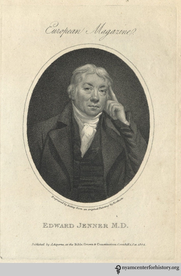
Engraving of Edward Jenner by William Ridley from an original painting by James Northcote.

Engraving of Edward Jenner by William Ridley from an original painting by James Northcote.

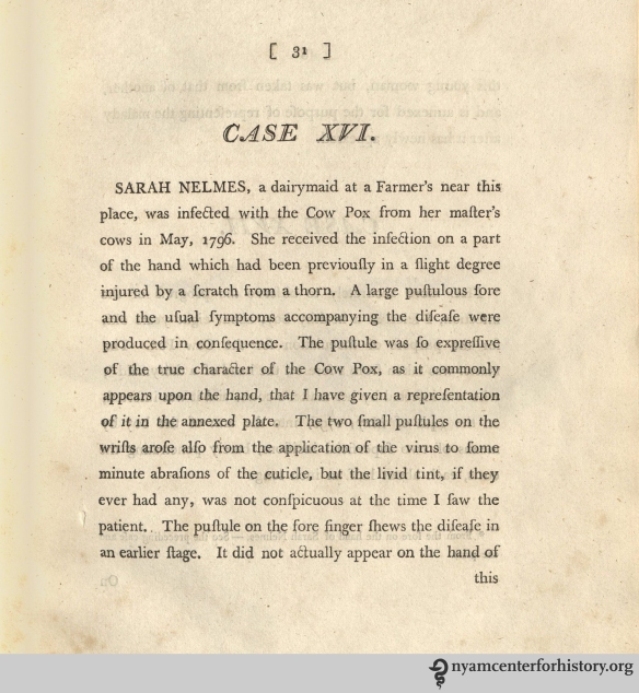
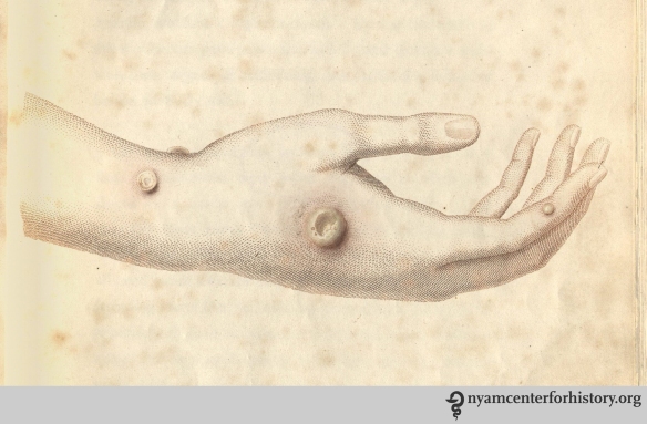
Enter Edward Jenner, an English doctor working in Gloucestershire in the late 18th century. Jenner was interested in testing the commonly held rural belief that dairy maids exposed to cowpox were no longer susceptible to the disease.2 He first heard this theory in 1770,3 and he began compiling case studies to test it in the 1780s and 1790s. On May 14, 1796, Jenner performed the first documented cowpox inoculation, taking a sample from a cowpox pock on the hand of a dairy maid named Sarah Nelmes and deliberately inserting the cowpox material into the arm of James Phipps, an eight-year-old boy. Phipps developed a cowpox lesion that healed within two weeks. When Jenner later exposed Phipps to smallpox through variolation, the boy was immune to the disease. Jenner was so confident that cowpox infection was a safe and effective method to prevent smallpox that he inoculated his infant son.

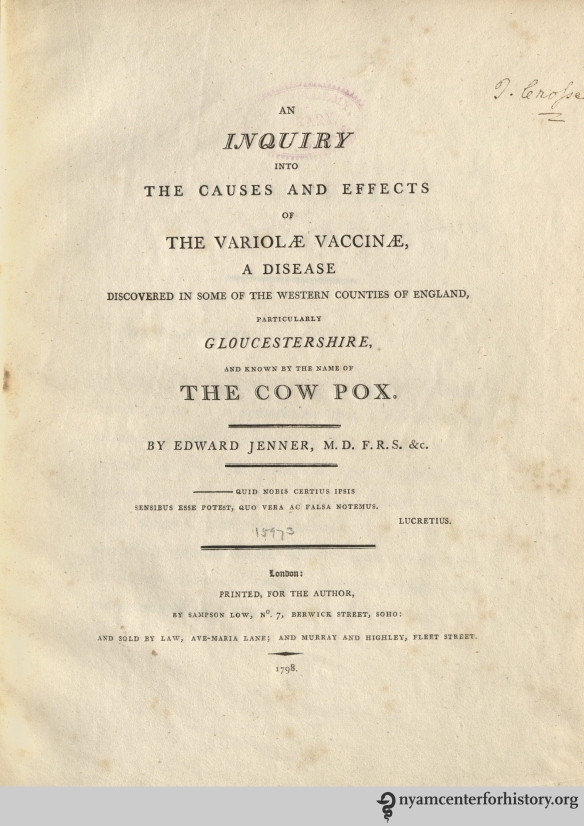
Jenner wrote a paper about his findings and submitted it to the Royal Society in 1797. The paper was not accepted for publication, however, because the reviewers found it had insufficient data. Undeterred, Jenner conducted additional trials and in 1798 published An Inquiry into the Causes and Effects of the Variolae Vaccinae, a Disease Discovered in Some of the Western Counties of England, Particularly Gloucestershire, and Known by the Name of the Cow Pox. He wrote three additional books on the same subject by 1801. Shown below is the title page from the first edition of the Inquiry, Jenner’s description of Sarah Nelmes’ case, and a detail from one of the color plates showing the cowpox on Nelmes’ hand.

The title page of the first edition of Jenner’s Inquiry, 1798.

The title page of the first edition of Jenner’s Inquiry, 1798.

Jenner’s description of Sarah Nelmes’ cowpox infection in Inquiry, 1798.

Jenner’s description of Sarah Nelmes’ cowpox infection in Inquiry, 1798.

Color engraving of Sarah Nelmes’ cowpox pocks from the first edition of Jenner’s Inquiry.

Color engraving of Sarah Nelmes’ cowpox pocks from the first edition of Jenner’s Inquiry.

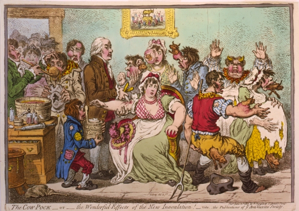
Early reactions to the Inquiry from the medical community and the general public were mixed. A cartoon by the famous caricaturist James Gillray, shown below, plays on the fear that using animal matter on people would cause the patients to assume animal characteristics. Members of the medical community, especially those with investments in the practice of variolation, tried to discredit Jenner’s discovery or stake their own claim to it. Jenner had especially acrimonious feuds with two London physicians, Dr. George Pearson and Dr. William Woodville. Pearson even gave evidence against Jenner’s 1802 petition to the House of Commons for recognition of his work on vaccination.

James Gillray’s “The Cow Pock – or – the Wonderful Effects of the New Inoculation,” published June 12, 1802, by H. Humphrey, St. James’s Street. Image courtesy of the Library of Congress.

James Gillray’s “The Cow Pock – or – the Wonderful Effects of the New Inoculation,” published June 12, 1802, by H. Humphrey, St. James’s Street. Image courtesy of the Library of Congress.

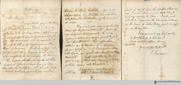
The New York Academy of Medicine holds a number of Edward Jenner’s autograph letters. In them, he rails against the misguided opposition to his discovery. On June 30, 1806, he writes to the Rev. Mr. Joyce:

“How wonderful that this horrid pestilence should at this time even have an existence in our island . . . A Physician from Copenhagen call’d on me today, and express’d his astonishment at an opposition to vaccine Inoculation, as by that means only the smallpox was completely extinguish’d in that City.”

Writing to a Mr. Phillips on Jan. 16, 1807, Jenner notes that the College of Physicians:

“[Has] not yet finished this Inquiry, which will, when completed, be laid before the House [of Commons]. This Inquiry will lay all those troublesome ghosts which have so long haunted the Metropolis with their ox-faces, & dismal hootings against Vaccination. However, tis all for the best – you may depend upon it the new Investigation will prove the touchstone of the vaccine discovery.”

Jenner also offers advice about his smallpox vaccine:

“A word more respecting your little one. Altho’ I should be happy to shield it myself from the speckled Monster, yet I would advise you not long to risk my coming to Town. I will just add that I consider the Vaccine Lancet in the hand of [Dr.] John Ring, just as safe as in my own.”4

In an autograph letter signed to Mr. Phillips, dated Jan. 16, 1807, Edmund Jenner discusses the ongoing vaccination controversy and offers advice for vaccinating Phillips’ new baby.

In an autograph letter signed to Mr. Phillips, dated Jan. 16, 1807, Edmund Jenner discusses the ongoing vaccination controversy and offers advice for vaccinating Phillips’ new baby. Click to enlarge.

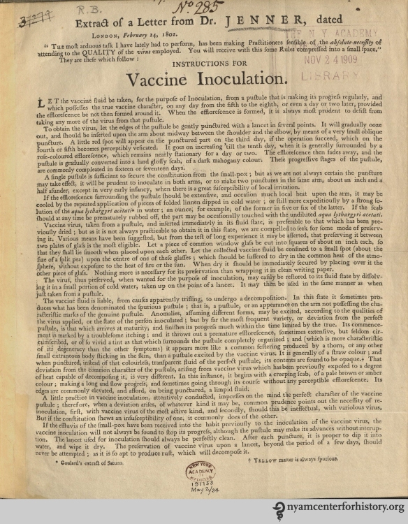
Despite some people’s doubts about the safety and efficacy of Jenner’s smallpox vaccine, there was great demand for cowpox samples to conduct vaccinations in England and abroad. Jenner and other practitioners in England sent dried cowpox specimens sandwiched between glass to Europe and the United States. The hand-colored drawing below presumably accompanied cowpox samples sent from England to America; the drawing shows the difference between cowpox pustules (on the left) and smallpox pustules (on the right) at 6, 8, 10, 12, 14, and 18-20 days after infection. Also shown below is an example of printed “Instructions for Vaccine Inoculation” from an “Extract of a Letter from Dr. Jenner, dated London, February 24, 1802.”

The New York Academy of Medicine holds this hand-colored drawing that shows the difference between cowpox and smallpox pustules at various stages of infection.

The New York Academy of Medicine holds this hand-colored drawing that shows the difference between cowpox and smallpox pustules at various stages of infection.

This broadside shows what kind of instructions were available for people interested in administering the smallpox vaccine.

This broadside shows what kind of instructions were available for people interested in administering the smallpox vaccine.

The smallpox vaccine became more common during the 19th century, but smallpox epidemics continued to occur, at least partly because people did not yet fully appreciate the need for re-vaccination. In the 20th century, smallpox continued to plague third-world countries. Finally, a global eradication campaign organized by the World Health Organization (WHO) in 1967 succeeded in eliminating smallpox. The last naturally occurring case was in Somalia in 1977.5 Thanks to Edmund Jenner’s research and his efforts to promote smallpox vaccination, one of the world’s most feared diseases is now a historical curiosity instead of an ongoing deadly threat.

References

1. These statistics are from Abbas M. Behbehani’s “The Smallpox Story: Life and Death of an Old Disease” in Microbiology and Molecular Biology Reviews, v. 47, no. 4 (Dec. 1983), p. 455-509. Available online at http://mmbr.asm.org/content/47/4/455.long See also Stefan Riedel’s “Edward Jenner and the history of smallpox and vaccination” in Baylor University Medical Center Proceedings, v. 18, no. 1 (Jan. 2005), p. 21-25. Available online at https://www.ncbi.nlm.nih.gov/pmc/articles/PMC1200696/

2. The Jenner Museum gives a good summary of Jenner’s interest in cowpox as a way to prevent smallpox infection. See http://www.jennermuseum.com/vaccination.html

3. Behbehani, p. 468.

4. The quotations in this paragraph are from MS 1155 (Edmund Jenner, autograph letter signed: London, to the Revd. Mr. Joyce, 1806 June 30; first quote) and MS 59 (Edmund Jenner, autograph letter signed: Cheltenham, to [Mr. Phillips], 1807 Jan. 16; second two quotes.)

5. See the World Health Organization’s page on smallpox, available online at http://www.who.int/csr/disease/smallpox/en/

Sitadevi’s Sutra

By Emily Miranker, Team Administrator/Project Coordinator

In 1934, Sitadevi Yogendra (1912–2008) published Yoga: Physical Education for Women, the first book on yoga for women by a woman.1 Married at age 15 to Shri Yogendraji, founder of The Yoga Institute in Mumbai, they became what Sitadevi described as “the first yogi couple.”2 Her book enjoyed three editions in less than 10 years and has been translated into several languages. It leads the reader through a course of exercises and postures specially geared towards women, recognizing that the prevailing techniques of the teachers of her day were “based upon the physiopsychic needs of Man.”3

First up in the routine are the corrective prayer poses. These instill proper posture in the body, something difficult to maintain under the “imposition of unnatural living under modern conditions”4— and this was before we slouched at computers all day and cramped our fingers with constant texting.

Figures 2 and 3 in, Yogendra, Yoga: Physical Education for Women,” 1947.

Figures 2 and 3 in Yogendra, Yoga: Physical Education for Women, 1947.

With your posture thus improved, the next poses maintain or even increase your height. The common triangle pose (trikonasana) is among those recommended. It’s a spine-stretching equilateral triangle shape in contrast to the flashier right-triangle that frequently adorns today’s Western fitness magazine covers.

Figure 7 in in Yogendra, Yoga: Physical Education for Women, 1947.

Figure 7 in in Yogendra, Yoga: Physical Education for Women, 1947.

Right triangle pose. Yoga.com. https://yoga.com/pose/right-triangle-pose Accessed April 28, 2016.

Right triangle pose. Yoga.com. Accessed April 28, 2016.

Sitadevi details exercises for the trunk to develop core strength and tone, and poses to keep the sex organs healthy. She considered it the “duty of every woman to safeguard her health”5 as the bearers of children. She concludes with poses for the spine, which she found good for the nervous system and mental equity (samatvam).

She provides a table of the entire sequence, which should take just 30 minute to run through. “When practiced with precision and regularity, the hygienic results of these exercises are sure to become manifest in a few months. This, in turn, would inspire the essential faith and enthusiasm for their continued practice throughout the lifetime.”6

Guidetable for a yoga sequence in Yogendra, Yoga: Physical Education for Women, 1947.

Guidetable for a yoga sequence in Yogendra, Yoga: Physical Education for Women, 1947.

Sitadevi’s book, along with other publications of The Yoga Institute, were microfilmed and included in the Crypt of Civilization,7 which isn’t a videogame but rather a time capsule housed at Oglethorpe University in Atlanta, GA. Consider coming by our library to read up on Sitadevi and master her healthful poses to cultivate longevity so you’ll be around for the Crypt’s opening … in May of 8113.

References

1. “Mother Sita Devi Yogendra: A Brief Profile.” The Yoga Institute (May 29, 2013). Accessed May 3, 2016.

2. Mohanty, Sweta. “Fit to Lead.” DNA India (May 2007).Accessed April 28, 2016. http://www.dnaindia.com/lifestyle/grandeur-fit-to-lead-1099213

3. Yogendra, Sitadevi. Yoga: Physical Education for Women. Bombay: The Yoga Institute, 1947: 11.

4. Yogendra, Sitadevi. Yoga: Physical Education for Women. Bombay: The Yoga Institute, 1947: 27.

5. Yogendra, Sitadevi. Yoga: Physical Education for Women. Bombay: The Yoga Institute, 1947: 29.

6. Yogendra, Sitadevi. Yoga: Physical Education for Women. Bombay: The Yoga Institute, 1947: 127.

7. “Crypt of Civilization,” Oglethorpe University. Accessed April 28, 2016.  http://crypt.oglethorpe.edu/

Young Man Freud

By Paul Theerman, Associate Director, Center for the History of Medicine and Public Health

The best-known photograph of Sigmund Freud (1856–1939) is forbidding: cigar in hand, he appears grim-faced and imperious. The image bespeaks his complete confidence in the truth of his psychoanalytic theories, indeed in the whole venture of psychoanalysis, a field he created and, at least in the American sphere, a field that held sway in psychiatric treatment through the first half of the 20th century.

Freud circa 1921. In Ernst L. Freud, Lucie Freud, and Ilse Grubrich-Simitis, eds., Sigmund Freud : his life in pictures and words, 1978, reprint 1998.

Freud circa 1921. In Ernst L. Freud, Lucie Freud, and Ilse Grubrich-Simitis, eds., Sigmund Freud: His life in pictures and words, 1978, reprint 1998.

But that success was in the last half of his life. In honor of Freud’s 160th birthday, May 6, we wanted to present pictures of young Freud before his breakthrough works of the late 1890s, pictures of a man on the make in the intellectual culture of Vienna.

First, pictures of Freud with father Jakob and mother Amelie, when he was age 8 and 16, respectively, in Vienna. Born in Freiberg, Moravia, within the Austro-Hungarian Empire, Freud moved with his family to Vienna within a year. That city would remain his home until he moved to London in 1938, after Austria’s annexation by Nazi Germany.

Freud with his father Jakob in 1864. In The Freud centenary exhibit of the American Psychoanalytic Association, 1956.

Freud with his father Jakob in 1864. In The Freud centenary exhibit of the American Psychoanalytic Association, 1956.

Freud with his mother Amalie, circa 1872. In The Freud centenary exhibit of the American Psychoanalytic Association, 1956.

Freud with his mother Amalie, circa 1872. In The Freud centenary exhibit of the American Psychoanalytic Association, 1956.

After excelling in high school, the Leopoldstädter Kommunal-Realgymnasium, Freud entered the University of Vienna at 17 and graduated with his medical degree eight years later, in 1881. Interested in neurology, he hoped for a career in academic medicine.

Here, Freud in a wedding photograph with Martha Bernays in 1886, age 27. At this point, he had been out of medical school for five years, had begun his career at Vienna General Hospital (Allgemeines Krankenhaus der Stadt Wien), and had spent five months in Paris studying with the great French neurologist Jean-Martin Charcot, whose work on hypnosis would prove revelatory.

And finally, a portrait of Freud in 1891, five years after starting his private practice, where using hypnosis and free association he began to develop the new discipline of psychoanalysis. His works, Studies in Hysteria (1895) and The Interpretation of Dreams (1900), made his reputation. The second half of his life was spent elaborating and defending his ideas within the medical profession and in broader intellectual life.

Child Health Around the Maypole

By Johanna Goldberg, Information Services Librarian

From 1924 through 1960, May 1 marked the celebration of Child Health Day, as described in the pamphlet The Goal of May Day: A Year-Round Program:

May Day as Child Health Day holds within it the power of a great vision. Its goal is to focus the interest of the nation upon perfected childhood—with the hope of a start in life free, sound and richly potential for every child….

This day has been given to the country to become, like the Maypole, a central rallying point for all the diverse activities concerned with the welfare of children….1

"The summer round-up, with the State Congress of Parents and Teachers and the State Bureau of Maternity and Infancy cooperating in Oklahoma City, Oklahoma. In The Goal of May Day, 1928.

“The summer round-up, with the State Congress of Parents and Teachers and the State Bureau of Maternity and Infancy cooperating in Oklahoma City, Oklahoma. In The Goal of May Day, 1928.

The American Child Health Association (ACHA)—an organization founded by Herbert Hoover in 1923 with the merging of the American Child Hygiene Association and the Child Health Organization—began Child Health Day in 1924. The ACHA was inspired by the success of National Baby Week, an observance that spread awareness of infant care to millions by 1919 (clearly, awareness days and weeks are not a new phenomenon).1,2 The ACHA was also motivated by Congressional inaction (also not a new phenomenon); President Wilson had called for a child health program in 1919, to no avail.

Cover of The Goal of May Day, 1928.

Cover of The Goal of May Day, 1928.

Aida de Acosta Breckinridge, wife of President Wilson’s Assistant Secretary of War Henry Breckinridge (and intriguingly, the first woman to pilot an aircraft solo), thought up May Day as Child Health Day and ran with the idea. Through her efforts, three million department stores nationwide handed out booklets on child health. Magazines like Women’s World and Literary Digest promoted the day.2 In 1928, President Calvin Coolidge officially declared Child Health Day a national celebration. It remains one today, though in 1960, Child Health Day moved to the first Monday in October.3

By 1928, when the ACHA released The Goal of May Day, the organization viewed May Day-Child Health Day as a time to celebrate the past year’s child welfare successes and plan for the year ahead. As the pamphlet emphasizes, May Day-Child Health Day activities occurred thanks to the efforts of community groups and local governments rather than through centralized ACHA planning, “each [group] coloring [May Day] with its own interpretation and using it according to its needs.”1 The Goal of May Day provides these organizations tips and lists of further resources to plan events and to improve child health year-round.

"A child health clinic in a church." In The Goal of May Day, 1928.

“A child health clinic in a church.” In The Goal of May Day, 1928.

The challenge was significant: the pamphlet states that 18,000 mothers died in childbirth in the United States each year. In 1924, cites the pamphlet, “the stillbirth rate was 3.9 per 100 live births.” While infant deaths from diarrhea and enteritis were down by 1928, those from congenital malformation, birth injuries, and premature birth had risen. And between infancy and school age, fifty percent of deaths came from diphtheria, recently preventable by vaccine.1

Making them safe from the great menace - diphtheria. In The Goal of May Day, 1928.

“Making them safe from the great menace – diphtheria.” In The Goal of May Day, 1928.

On a larger scale, the pamphlet offers a community health inventory to spur local government to improve child health, with questions ranging from “Have you a safe water supply?” to “Is there a tuberculosis clinic?” to “Is there an organized course of study for the education of the school child in health?”

"The parochial school had its health float on May Day." In The Goal of May Day, 1928.

“The parochial school had its health float on May Day.” In The Goal of May Day, 1928.

For community groups, the pamphlet recommends consulting with national organizations—the Girl Reserves, Boy Scouts, Jewish Welfare Board, National Catholic Welfare Conference, Child Study Association, and more—to plan programs and events like home demonstrations, distribution of health-related literature, community clean ups, health dramatizations, athletics, and exhibits.1

"4-H girls club learn from the home demonstration agents." In The Goal of May Day, 1928.

“4-H girls club learn from the home demonstration agents.” In The Goal of May Day, 1928.

May Day - Child Health Day, on the school playgrounds at Rapid City, South Dakota. In The Goal of May Day, 1928.

May Day – Child Health Day, on the school playgrounds at Rapid City, South Dakota. In The Goal of May Day, 1928.

The American Child Health Association closed in 1935. During its 12 years of existence, it raised about $5 million for child-focused community services.2 And the observance of Child Health Day continues some 92 years after it began, though no longer around a Maypole.

References
1. The Goal of May Day: A Year-round Community Child Health Program. New York: American Child Health Association; 1928.

2. Lee RA. From Snake Oil to Medicine: Pioneering Public Health. Westport, Conn.: Greenwood Publishing Group; 2007.

3. Health Resources and Services Administration. Child Health Day History. Available at: http://mchb.hrsa.gov/childhealthday/history.html. Accessed April 27, 2016.