Sir William Osler: A Bibliophilic Benefactor

Osler_watermark

Photograph of William Osler. Osler, W., & Pollard, A. W. (1923). Incunabula medica: A study of the earliest printed medical books 1467–1480. Oxford: Bibliographical Society. NYAM Collection. 

December 29, 2019, marks the centenary of the death of Sir William Osler (1849–1919), arguably the most important and most loved physician of his era. Osler received his medical degree from MGill University in 1872, and joined the medical faculty there in 1874. A decade later he moved to Philadelphia to chair the department of Clinical Medicine at the University of Pennsylvania, and in 1889 he was one of the founders of the Johns Hopkins Hospital, serving as its first Physician-in-Chief and as the first professor of medicine at the newly opened medical school. In 1905, he left the United States to become the Regis Professor of Medicine at Oxford, a position he held for the rest of his life. An accomplished teacher of clinical medicine, Osler established the medical residency program at Hopkins and made sure that students had ample opportunity to interact with patients at the bedside. His textbook, The Principles and Practice of Medicine, first published in 1892, appeared in multiple editions and was the standard textbook of internal medicine for decades. (National Library of Medicine, 2013).

Osler was also an extraordinary collector and lover of books, and in addition to amassing the collection that became the Osler Library of the History of Medicine at McGill University, he bestowed gifts on both his friends and on institutions. The Library of The New York Academy of Medicine has him to thank for two of its most treasured items.

Late in February of 1906, Osler sent a postcard to Walter Belknap James (1858–1927), along with a copy of William Harvey’s 1628 De motu cordis, the text in which Harvey describes the circulatory system and the motion of the heart and the blood. Harvey’s work, probably the most important text in the history of physiology, was notoriously difficult to find. In the Bibliotheca Osleriana, Osler recounts his hunt for a copy of the book:

Feb. 17, 1906; I had been looking for a copy for nearly ten years.  Pickering and Chatto sent one to-day, which they had bought for £30 at the sale of Dr. Pettigrew’s library. Though a poor copy, measuring only 7 3/8 x 5 3/8 inches, I took it.  Feb. 19, two days later, they sent me another (this one) from the library of Milne Edwards… I took it too, and passed on the other to Dr. Walter James who gave it to the Library of the Academy of Medicine, New York. (Osler, Francis, Hill, & Malloch, 1929, p. 4)

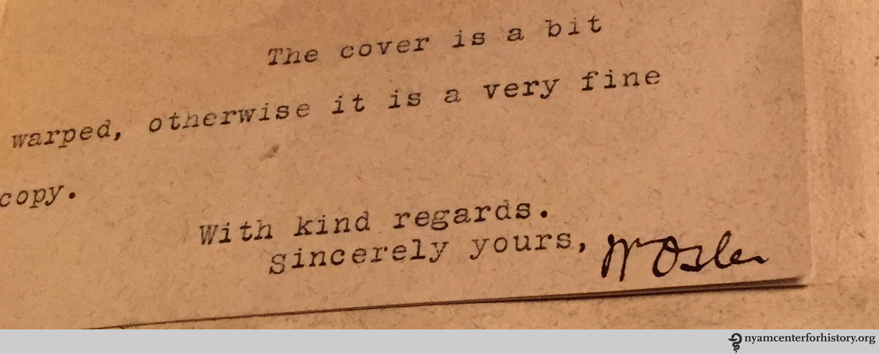
As can be seen in the image of the postcard below, Osler marketed this copy to James rather differently:

Dear James, That is a nice de Moto Cordis is it not? I had it & another copy here last week to look over and take my pick. There has not been another copy offered in England since 1895 when an imperfect copy was sold at Sotheby's for 10 guineas. Then these two turned up. My copy is from Milne Edwards library in Paris. It is an excessively rare book. Rosenthal tells me he has not had a copy offered in Germany for years. Yours sincerely, Wm Osler

Postcard to Walter Belknap James from William Osler, February 1906. NYAM Collection.

Good copy or not, the gift of the Harvey definitely enhanced the Library’s holdings, and was joined later in the 20th century by a second copy of the 1628 edition when Robert Levy gave his library of books by and about William Harvey to the Academy Library.

Harvey_1628_watermark

Title page. Harvey, W. (1628). Exercitatio anatomica de motu cordis et sanguinis in animalibus Guilielmi Harvei. Francofurti: Sumptibus G. Fitzeri. NYAM Collection.

In 1909, Osler again made a gift to the Academy’s collections. On June 16th, Osler sent Laura Smith, who worked in the library, a note relaying the following information: “Will you please tell your Superior, Mr. B [John Browne, the Academy’s librarian] that I hope to send him the Vesalius first edition this week.”

WOtoLauraSmith_96.16.1909_watermark

Letter from William Osler to Laura Smith, June 16th, 1909. NYAM Collection.

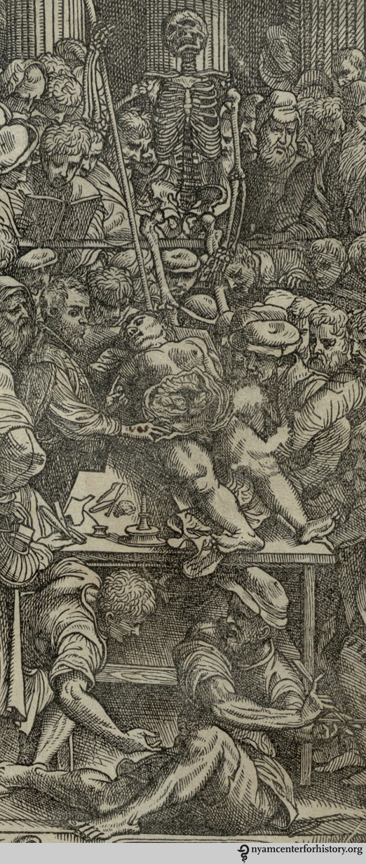
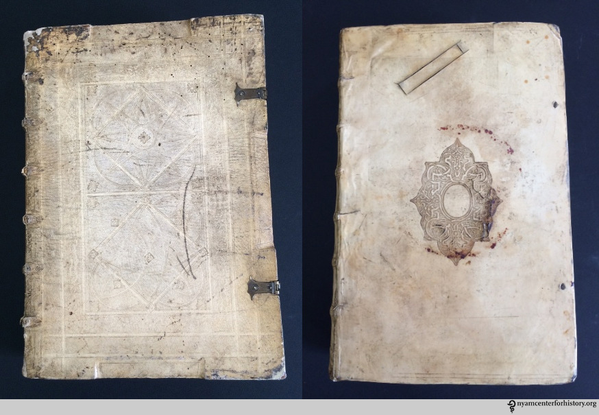
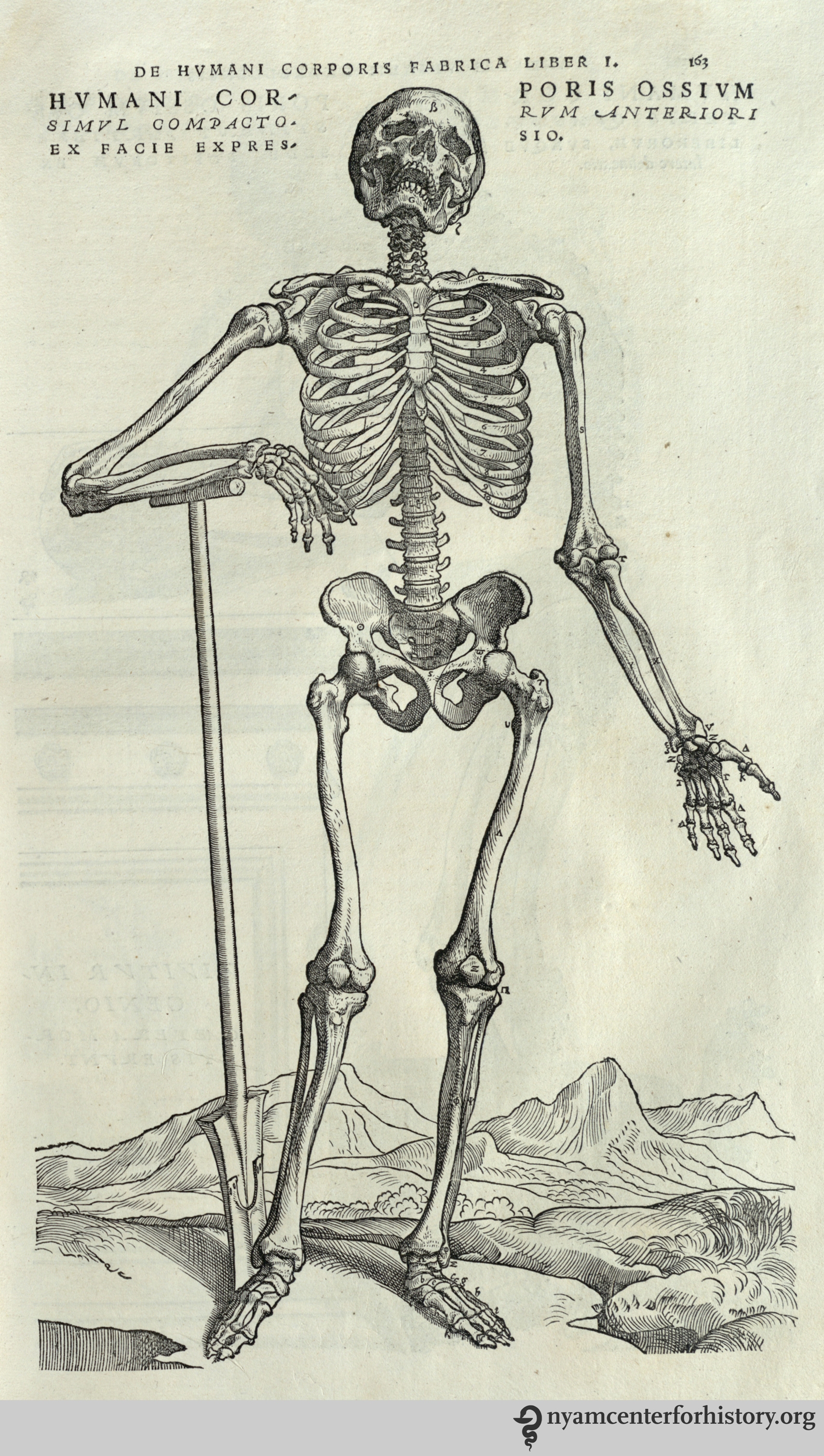
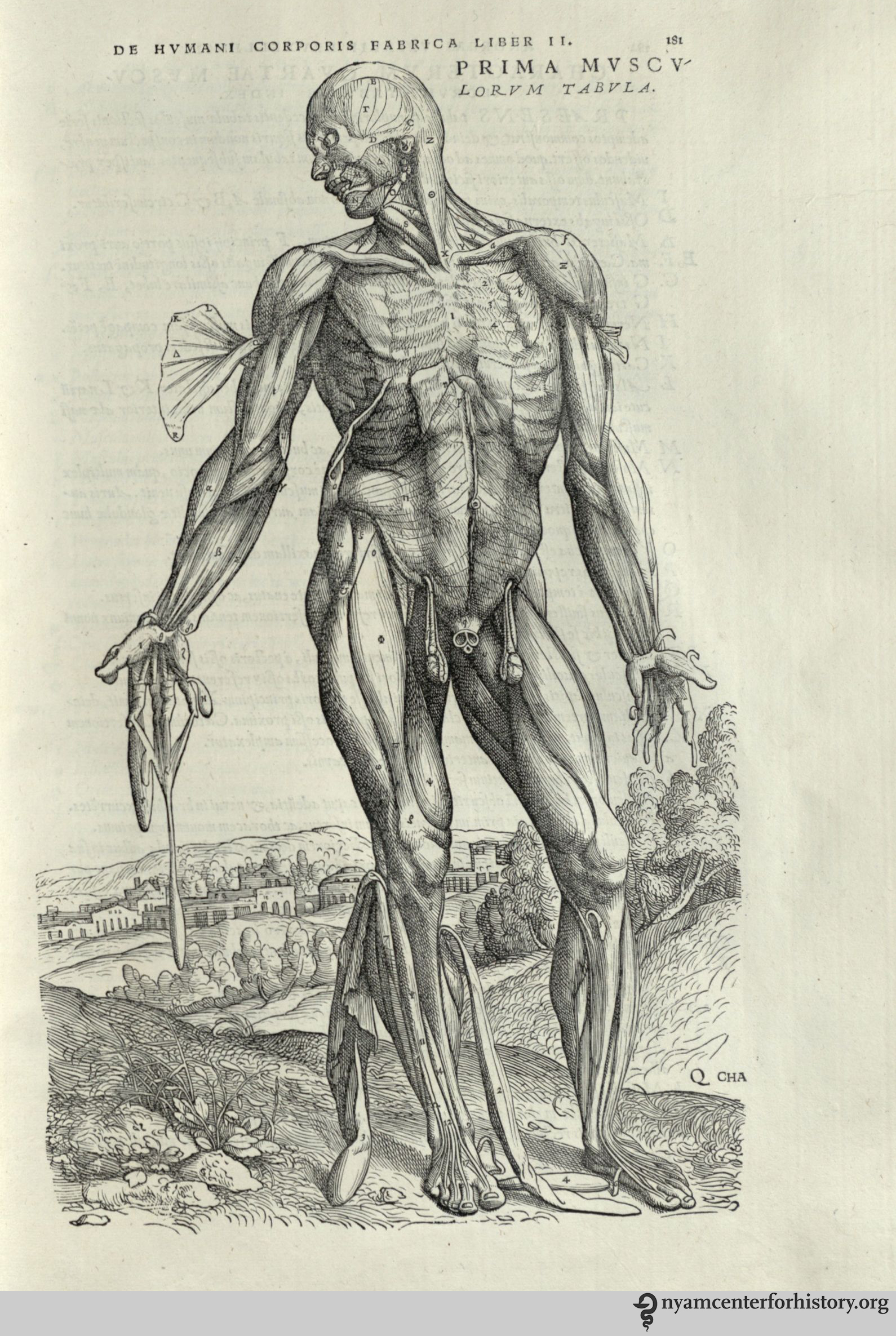
Osler had recently given a second copy of the 1543 edition of De humani corporis fabrica, Andreas Vesalius’ groundbreaking work on anatomy, to McGill, and decided that their other copy should make its way to the Academy, even going so far to say in his letter to Miss Smith that while Miss Charlton (of McGill) was “crying hard about it,” Osler was “obdurate and she was not good enough to be allowed 2 copies of so great a work” (personal communication, June 16th, 1909).

In the Bibliotheca Osleriana, Osler writes that he had in his possession at one time or another six copies of the Fabrica, also giving them as gifts to the Boston Medical Library Association; the Medical and Chirurgical Faculty, Baltimore; the Medical Department at the University of Missouri; and to his friend Llewelys Barker, who was professor of anatomy at the University of Chicago, as a wedding present. (Osler, Francis, Hill, & Malloch, 1929).

The Library’s copy still displays the inscription Osler wrote on the free endpaper of this copy when he gave it to McGill in 1903, “The original edition of the greatest medical work ever printed, the one from which modern medicine dates its beginning. W. O.”

Vesalius1543_WOinscription_watermark

Osler’s inscription on endpaper in De humani corporis fabrica (1543). NYAM Collection.

Our copy also retains the bookplates that track its movement from McGill to New York:

Vesalius_WObookplates_watermark

Bookplates in the 1543 edition of De humani corporis fabrica. NYAM Collection.

The Academy soon acquired two other copies of the 1543 Vesalius, one from the Edward Clark Streeter Collection and the other from Dr. Samuel Lambert, as well as two copies of the 1555 second edition. In fact, editions of Vesalius and related works soon became a major research strength of the collection, continue to be heavily used by readers, and are frequently shared with visiting groups and classes.

As 2019 draws to a close, the Library is grateful to its many friends and donors, who, following the spirit of Sir William Osler, continue to enrich our collections today. One hundred years later, the memory of Osler’s generosity reminds us that these books still matter.  Generations of earlier readers held the Osler copies of the Harvey and Vesalius in their hands over the course of hundreds of years before they finally landed on our shelves. It is a privilege to be able to continue to share them.

 References

National Library of Medicine. (2013). William Osler: Biographical overview. Retrieved from https://profiles.nlm.nih.gov/spotlight/gf/feature/biographical-overview

Osler, W., Francis, W. W., Hill, R. H., & Malloch, A. (1929). Bibliotheca Osleriana: A catalogue of books illustrating the history of medicine and science. Oxford: At the Clarendon Press.


A Visit to the Drs. Barry and Bobbi Coller Rare Book Reading Room

Dr. Patrick Brunner, the author of today’s guest post, is Instructor in Clinical Investigation at The Rockefeller University.

On July 26 2016, a group of young physician-scientists from The Rockefeller University visited the Drs. Barry and Bobbi Coller Rare Book Reading Room at the New York Academy of Medicine. As part of the Clinical Scholars curriculum, led by Dr. Barry Coller and Dr. Sarah Schlesinger, these researchers regularly meet for educational tutorials, and the excursion to the Rare Book Room has clearly been one of the highlights of this past semester.

Arlene Shaner, the curator of this exceptional collection, presented seminal works to the group, and her deep insight and passion for the history of medicine made the excursion a unique experience. Ms. Shaner started the tour with the presentation of one of the most outstanding works of Western medicine – Andreas Vesalius’ opus magnum “De humani corporis fabrica libri septem” (On the fabric of the human body in seven books) from 1543. Ms. Shaner comprehensively and clearly outlined the historical context in which this book had been published, and fascinated her audience with a display of the book’s iconic woodprints. This artwork, which everyone in the room had seen in numerous reproductions, now laid open in its original form – showing the famous muscle man posing in front of an Italian landscape, and the skeleton, leaning on a spade, gazing towards the sky.

vesaliusgravedigger_watermark

Skeleton from the 1543 Fabrica. Click to enlarge.

A letter, sent from Oxford, dated July 7th, 1909, had been incorporated into the book as an inscription. From this letter one can learn that Sir William Osler himself donated the book to the New York Academy of Medicine. Ms. Shaner clearly knows each and every inch of this version of Vesalius work, one of three copies that the New York Academy of Medicine holds.

osler_detail_watermark Inscription by Sir William Osler found in our 1543 Fabrica. Click to enlarge.

Vesalius’ Fabrica has undoubtedly been one of the most influential books on human anatomy, overthrowing the observations and influences of the Greek physician Galen, which had been uncontested by Western medical science for more than 1300 years. And it was not until 1628 that another seminal work, which had also been put on display for the evening, William Harvey’s treatise “Exercitatio anatomica de motu cordis et sanguinis in animalibus” (On the motion of the heart and blood in animals), established that blood circulates in a closed system, and that the heart acts as a pump – a manuscript considered by many scholars to be the single most important publication in the history of physiology.

The visitors from The Rockefeller University were greatly impressed by the richness of this library – especially as they learned that all the books are available for review through the library’s archives, be it the “Anatomia hepatis” (The anatomy of the liver) by Francis Glisson, or the first atlas of skin diseases by the dermatological founding father Ferdinand von Hebra.

Arlene Shaner also presented Bernhard Siegfried Albinus’ “Tabulae sceleti et musculorum corporis humani” (Tables of the skeleton and muscles of the human body), first published in Leiden in 1747, which not only depicts anatomical studies in a monumental fashion, but presents the models within elaborate and artful surroundings – overall, an impressive testimonial of its time.

The climax of the visit was the display of a very special gift donated to the New York Academy by Sir Alexander Fleming – a capsule containing a colony of Penicillium, taken from the original culture that produced one of the world’s first antibiotics for medical use. And it has only been about 70 years since this medication became available!

fleminggrayobverse_watermarkCapsule containing a colony of Penicillium, donated by Sir Alexander Fleming. Click to enlarge.

Seeing all these treasures that irreversibly changed the world, and learning about the stories behind them in the context of both medical and art history, was a unique, and almost sensual, learning experience for the visitors, and Ms. Shaner’s never-ending expertise helped everyone in the room to deeply dive into history.

Aldous Huxley once said: “The charm of history and its enigmatic lesson consist in the fact that, from age to age, nothing changes and yet everything is completely different.”  Understanding the challenges that these authors face during their life times, which may not have been quite so different from the ones that we face today, while, at the same time witnessing the dramatic changes that have been instigated by their works, was a true inspiration. The afternoon passed quickly, and everyone agreed that they wanted to come back and further explore this treasury in the middle of New York City.

Counterfeiting Bodies: Examining the Work of Walther Ryff

By Anne Garner, Curator, Center for the History of Medicine and Public Health

The surgeon Walther Hermann Ryff worked in Strasbourg in the early 16th century. A prolific author, he wrote as many as 65 works on diverse subjects, including architecture, poisons, cookbooks, herbal remedies, obstetrics and mathematics.1 The author’s best known work, Des aller furtrefflichsten, hoechsten und adelichsten Gschoepffs aller Creaturen, was published in Strasbourg in 1541, just two years before the publication of Vesalius’ ground-breaking Fabrica.

Plate 1 of Ryff's Des aller furtrefflichsten, hoechsten und adelichsten Gschoepffs aller Creaturen (1541)

Plate 1 of Ryff’s Des aller furtrefflichsten, hoechsten und adelichsten Gschoepffs aller Creaturen (1541). Click to enlarge.

The text compiles Ryff’s lectures in anatomy and physiology and 42 beautifully hand-colored woodcuts, compiled from a number of Renaissance sources. These images, mostly of bodies or partially dissected bodies, offer what scholar Alexander Marr describes as an immediate “rhetoric of authenticity.”2 Depicted in the counterfeit style, a type of representation common in the 16th century in Northern Europe, the illustrations in this book would have implied first-hand knowledge and discovery. The captions for plates produced in this style used the word “counterfeit” (above, contrafactur) to assert their accuracy as true representations. In this way, Ryff’s book positioned itself as a credible description of anatomy (though its illustrations were far from anatomically precise).

Little is known about Ryff’s training. He seems to have studied pharmacy in Basel, and absorbed much of his considerable medical knowledge by travelling through Europe. He was a successful author, frequently sought after by publishers. Among his peer group of writers, however, he would not have won any popularity contests. To the Swiss scientist Albrecht von Haller, he was a “compiler and polygraph of dubious morals,” and to Vesalius, simply, “the Strasbourg plagiarist.” Leonard Fuchs, the great botanist, whose work was reprinted in Ryff’s name twice, called him an “extremely outrageous, reckless, fraudulent writer.”3 The grounds for their complaints are easily recognizable by examining this volume, which lifts images from Vesalius’s Tabulae Sex (1538), from Eucharius Rösslin’s Der Rosengarten (1513), and from the anatomies of Johannes Dryander (1536), Jacapo Berengario da Carpi (1522), and Lorenz Fries (1518).

Plate 2 Ryff's Des aller furtrefflichsten, hoechsten und adelichsten Gschoepffs aller Creaturen (1541). Images originally from Rösslin’s Der Rosengarten (1513). Click to enlarge.

Plate 2 Ryff’s Des aller furtrefflichsten, hoechsten und adelichsten Gschoepffs aller Creaturen (1541). Images originally from Rösslin’s Der Rosengarten (1513). Click to enlarge.

Plate 3 Ryff's Des aller furtrefflichsten, hoechsten und adelichsten Gschoepffs aller Creaturen (1541). Image originally from Dryander (1536). Click to enlarge.

Plate 3 Ryff’s Des aller furtrefflichsten, hoechsten und adelichsten Gschoepffs aller Creaturen (1541). Image originally from Dryander (1536). Click to enlarge.

Ryff’s defenders have argued that what today would be regarded as blatant plagiarism was more in keeping with Enlightenment practices of recycling intellectual property. Even so, his appropriations seem to have gone too far in the minds of his peers. In some cases, he modified the images, improving them. The Fries figures were repositioned, and seated on a bench. The Vesalian plates showing the arteries and veins, now beautifully hand-colored, were superimposed on seated outlines of figures, which clarified the position of the vessels in the body.

Plate 4 of Ryff's Des aller furtrefflichsten, hoechsten und adelichsten Gschoepffs aller Creaturen (1541)

Plate 4 of Ryff’s Des aller furtrefflichsten, hoechsten und adelichsten Gschoepffs aller Creaturen (1541). Click to enlarge.

Vesalius’ skeletons fared less well in Ryff’s possession. These were copied directly onto the wood-cut, so that the lettering and the skeletons themselves appear in reverse. The skeletons are depicted with an inadequate number of vertebrate and ribs, and are shown in inferior proportions.

Modified Vesalian skeleton in Ryff's Des aller furtrefflichsten, hoechsten und adelichsten Gschoepffs aller Creaturen (1541)

Modified Vesalian skeleton in Ryff’s Des aller furtrefflichsten, hoechsten und adelichsten Gschoepffs aller Creaturen (1541). Click to enlarge.

Modified Vesalian skeleton in Ryff's Des aller furtrefflichsten, hoechsten und adelichsten Gschoepffs aller Creaturen (1541)

Modified Vesalian skeleton in Ryff’s Des aller furtrefflichsten, hoechsten und adelichsten Gschoepffs aller Creaturen (1541). Click to enlarge.

Ryff directed his 1541 book at the ‘gemeine,’ or common man; it’s composition in vernacular German instead of Latin ensured it would have a wider readership. In this way, it would have been indispensable to new readers as a compilation of Renaissance knowledge about the body.

The book also offers some tantalizing evidence about early printing history. The wood-blocks for this edition were reused for a set of broadsides, issued in both German and Latin editions the same year. They then went to a Parisian printer for new editions of Ryff’s work and for a popular work on surgery.4 The reappearance of the Ryff woodcuts illustrates the practice of passing woodblocks from publisher to publisher, and shows how work published in one city continued to be published and disseminated in others.

References

1. Di Matteo, Berardo. “Art and Science in the Renaissance: The Case of Walther Hermann Ryff.” Clinical Orthopeadics and Related Research 472: 1689-1696. 2014 and Russell, K.F. Walter Hermann Ryff and His Anatomy.” The Australian and New Zealand Journal of Surgery. v.22 no. 1. 1952. pp. 66-69.

2. Marr, Alexander. “Walther Ryff, Plagiarism and Imitation in Sixteenth-Century Germany.” Print Quarterly, 31, 2014. pp 131-143.

3. Roberts, K.B. and J.D.W. Tomlinson. The Fabric of the Body. Oxford: Clarendon Press, 1992; Marr, Alexander. “Walther Ryff, Plagiarism and Imitation in Sixteenth-Century Germany.” Print Quarterly, 31, 2014. pp 131-143; Di Matteo, Berardo. “Art and Science in the Renaissance: The Case of Walther Hermann Ryff.” Clinical Orthopeadics and Related Research 472: 1691.

4. K.B. Roberts and J.D.W. Tomlinson. The Fabric of the Body. Oxford: Clarendon Press, 1992.

Spoiled by a Certain Englishman? The Copying of Andreas Vesalius in Thomas Geminus’ Compendiosa

Laura Robson, the author of today’s guest post, is our 2014–2015 Helfand Research Fellow. She completed her PhD in Classics at the University of Reading, UK.

“I wish the Epitome had not been spoiled so disgracefully by a certain Englishman (who I think lived with my brother for a time). He took what had been written with great care succinctly as a list in the Epitome and expanded it with excerpts taken from the books of the Fabrica… He utterly corrupted what had made it most praiseworthy and so roughly and absurdly copied what had been set forth with elegant drawing and engraving that he preserved no appearance of Oporinus’ majestic edition.”1

These were the concerns of Franciscus Vesalius, brother of the famous anatomist Andreas Vesalius, published in the preface to The China Root Epistle in 1546. Within only three years of the original publication of Andreas Vesalius’ De Humani Corporis Fabrica and its companion work, the Epitome, a number of medical authors had copied and reproduced the beautiful illustrations that had made Andreas Vesalius and his work so famous.2

Andreas Vesalius (1514-1564). De humani corporis fabrica libri septum. Basel: Johannes Oporinus, 1543. The most famous illustrations are the series of fourteen muscle men, progressively dissected. Some figures, such as this one, are flayed. Hanging the muscles and tendons from the body afforded greater detail, not only showing the parts, but how they fit together.

Andreas Vesalius (1514-1564). De humani corporis fabrica libri septum. Basel: Johannes Oporinus, 1543. Click to enlarge.

The Fabrica was one of the first anatomical treatises of the 16th century to present illustrations of the anatomised body in a naturalistic way. Vesalius promoted the dissection of the human body as the best way to learn about anatomy. By performing human dissections, he uncovered errors in the work of the ancient anatomist Galen, whose use of animals as dissection material to substitute for the lack of human cadavers had dominated the understanding of the body for centuries. Vesalius was caught in a conflict: how to show the anatomical errors in Galen’s treatises without going against such an important medical authority and potentially damaging his own medical career. He used the Fabrica to present his findings and to build on Galen’s important work.

The Fabrica contained 700 folio pages of Latin text and beautiful woodcut illustrations depicting the anatomical body in different poses. Readers were signposted to turn back and forth several times between image and text. This, as well as each image being accompanied by a letter key, encouraged a very active reading of Vesalius’ treatise.3 The work proved popular, with people taking a particular interest in the figures, although people copied, adapted, and reused both the images and text of Vesalius’ work in many different medical treatises. Due to the size and high cost of the Fabrica, cheaper copies were often more accessible than the original, even though the pirating enraged Vesalius and his close circle. Book piracy was common at this time. There was not the modern sense of intellectual property or copyright legislations. Licenses allowed particular printers to print works first, but the Venetian and imperial privileges obtained by the authors to try and protect their books from piracy did little to stop others from copying them.4

In fact, Franciscus Vesalius accused the wrong man of copying his brother’s work (suggesting, perhaps, that he had not seen a copy of the offending book). The only Englishman known to live with Vesalius was John Caius, when they lodged together in Padua during their studies. Caius went on to be physician to King Edward VI. There is no evidence that he pirated any version Vesalius’ work.5

The work Franciscus refers to is in fact Thomas Geminus’ Compendiosa.6 The first edition is predominantly made up of the Latin text of the Epitome and its illustrations, with the addition of many Fabrica figures. It is believed to be one of the first books to use copperplate illustrations.7 Geminus stated in his dedication that he followed Vesalius, but shortened his book to make it more useful to readers, in particular students.8 Reproducing the images of the Fabrica with the text of the Epitome meant that there was little interaction and connection between the two. However, the publication of the Compendiosa did bring Vesalius’ illustrations to a wider audience as the book was shorter and therefore cheaper than the original.

In order to make the work more accessible to those who could not read Latin, Geminus published an English edition of the Compendiosa in 1553. Nicholas Udall translated the short captions from the Fabrica figures into English. However, the main text of the Epitome was not translated. Instead the illustrations were placed after the text of Thomas Vicary’s The anatomie of mans body, first published in 1548.9 Geminus rearranged this text to follow the order of dissection for the parts of the body that decayed the fastest—the abdomen, the thorax, and the head. Although the images were not rearranged to fit this order, they connected more strongly to the text than in the Latin edition, as readers were signposted to particular figures discussing different parts of the body.

In his preface to the English Compendiosa, Nicholas Udall puts forward some interesting points about the uses of images and texts in medical manuals of his time. He said he did not know whether images or texts were more important when presenting anatomical information. He argued that information is set forth in writing for “high learning” and in pictures for the unlearned. He also explained that surgeons often performed duties like resetting bones by looking at figures alone.10 Surgeons had a low status in the medical profession at this time. They were not university educated like physicians and they were accused of having little knowledge about the science of medicine and healing. This suggests that readers who could not understand Latin, like surgeons, used anatomical figures and not written texts. I believe the English version of the Compendiosa was an attempt at encouraging these readers to read the text as well, by providing it in the vernacular language.

The coat of arms, left, and title page, right, of the Academy's copy of the 1559 English edition of Geminus’ Compendiosa.

The coat of arms, left, and title page, right, of the Academy’s copy of the 1559 English edition of Geminus’ Compendiosa. Click to enlarge.

In 1559 the English edition of Geminus’ Compendiosa was reissued.11 The annotated copy of this edition in the collection at the New York Academy of Medicine reveals hints as to how this anatomical text was used by readers at the time. The coloured and illuminated title page includes a portrait of Queen Elizabeth I. Slithers of gem stones have been attached to her necklace and the coat of arms opposite her on the adjacent page. The nude figures known as Adam and Eve are also coloured, and a reader inscribed the verse, “The Eyes of Them Both were opened, and They knew that They were naked: Genesis Chapter 3 Verse 7.”

Adam and Eve in the Academy's copy of the 1559 English edition of Geminus’  Compendiosa. Click to enlarge.

Adam and Eve in the Academy’s copy of the 1559 English edition of Geminus’ Compendiosa. Click to enlarge.

Two readers annotated this copy, both with different handwriting from the owner who in 1769 wrote his name—“G. Molesworth”—on the title page. One reader underlined key words and sections of the text, marking these with almost illegible notes in the margins. The other reader, though, focused on the illustrations. This second reader annotated the first three Vesalian musclemen images in the copy, adding the letters of the key, along with the Latin names for the body parts they represent.12 This English edition of the Compendiosa seldom uses the Latin names for parts of the body. So our reader did not get the information for his notes from this edition of the text. He must have consulted another text, such as the Latin edition of 1545, or even the original work of Vesalius’ Fabrica, in order to make his annotations.

Image of annotated muscleman figure  in the Academy's copy of the 1559 English edition of Geminus’  Compendiosa. Click to enlarge.

The annotated muscleman figure in the Academy’s copy of the 1559 English edition of Geminus’ Compendiosa. Click to enlarge.

This demonstrates the culture of active reading in the early modern period. This reader engaged with more than one treatise, perhaps even several works, when learning about the dissection of the body. He was familiar with the Latin language, and was therefore not one of the unlearned readers mentioned by Nicholas Udall in his preface to the work.

While Andreas and Franciscus Vesalius opposed the reproduction of the Fabrica and Epitome, the works that copied, adapted, and reused material from these texts allowed for the transmission of Vesalius’ knowledge of the body to a wider audience than the original works could reach. And this new audience interacted with the material, coming to a greater understanding of the dissected human body in the early modern period.

References

1. Andreas Vesalius (1546), Vesalius: The China Root Epistle, translated by Daniel H. Garrison (2015), p. 6.

2. Andreas Vesalius (1543a), De Humani Corporis Fabrica Libri Septem, Basel and Andreas Vesalius, (1543b), Andreae Vesalii Suorum de Humani Corporis Fabrica Librorum Epitome, Basel.

3. On active reading in Vesalius’ anatomical texts see, Nancy Siraisi (1994), “Vesalius and Human Diversity in De humani corporis fabrica”, in Journal of the Warburg and Courtauld Institutes, Vol. 57 p.64 and Sachiko Kusukawa (2012), Picturing the Book of Nature: Image, Text, and Argument in Sixteenth-Century Human Anatomy and Medical Botany, Chicago and London, p. 24.

4. On history of copyright and pirating see Christopher L. C. E. Witcombe (2004), Copyright in the Renaissance: Prints and Privilegio in Sixteenth-Century Venice and Rome, Leiden.

5. See Charles O’Malley (1955), “The Relations of John Caius With Andreas Vesalius and Some Incidental Remarks on the Guinta Galen and on Thomas Geminus,” in Journal of the History of Medicine and Allied Sciences Vol. 10.2 pp.147-172.

6. Thomas Geminus (1545), Compendiosa totius anatomie delineatio, aere exarata: Thomam Geminum, London.

7. Leroy Crummer (1926), “The Copper Plates of Raynalde and Geminus”, in Proceedings of the Royal Society of Medicine Vol 20.1 p. 53.

8. Thomas Geminus (1545), Compendiosa totius anatomie delineatio, aere exarata: Thomam Geminum, London, p. 1.

9. Thomas Vicary (1577 [1548]), A Profitable Treatise of the Anatomie of mans body: compyled by that excellent chirurgion, M. Thomas Vicary esquire, seriaunt chirurgion to king Henry the eyght, to king Edward the. vj. to Queene Mary, and to our most gracious Soueraigne Lady Queene Elizabeth, and also cheefe chirurgion of S. Bartholomewes Hospital. Which work is newly reuyued, corrected, and published by the chirurgions of the same hospital now beeing, London.

10. Thomas Geminus (1553), Compendiosa totius anatomie delineatio, aere exarata: Thomam Geminum, London, p.1.

11. Thomas Geminus (1559), Compendiosa totius anatomie delineatio, aere exarata: Thomam Geminum, London.

12. Ibid. p. Cii.

Brains, Brawn, & Beauty: Andreas Vesalius and the Art of Anatomy

By Rebecca Pou, Archivist, and Johanna Goldberg, Information Services Librarian

For our October 18 festival, Art, Anatomy, and the Body: Vesalius 500, we exhibited items from the library’s collections showing the history of anatomical illustration. You can still visit the New York Academy of Medicine to view the exhibit in person on the ground floor. If you can’t make it, we offer a digital version below.

The exhibit on display at the new York Academy of Medicine.

The exhibit on display at the New York Academy of Medicine.

In 1543, Andreas Vesalius was a 28-year-old professor of surgery and anatomy at the University of Padua, one of Europe’s best known medical schools. That year, he published his most famous work, De humani corporis fabrica, translated as On the Fabric of the Human Body. Vesalius dedicated the work to Charles V; he subsequently received the appointment of physician to the imperial family.

Working from three images from the Fabrica—a skeleton, a figure of muscles, and an illustration of the brain—this exhibit shows the many ways Vesalius’ work built on past anatomists, and exerted its influence well into the future.

Images from great works in our collection, from Magnus Hundt’s 1501 Antropologium to Dominici Santorini’s 1775 Anatomici summi septemdecim tabulae, show the evolution of artistic style and scientific understanding. Some show examples of “borrowing” Vesalius’ images and placing them in new contexts.

Click an image to view the gallery.

Vesalius 500: Art, Anatomy, and the Body

By Paul Theerman, Associate Director, The Center for the History of Medicine and Public Health

Join us this Saturday, October 18, for our second annual Festival of Medical History and the Arts, Art, Anatomy, and the Body: Vesalius 500. Register here.

Vesalius500STD_05_30_14This year, we celebrate the 500th birthday of Andreas Vesalius, the path-breaking anatomist whose 1543 book, De humani corporis fabrica (The Fabric of the Human Body), opened up new worlds in the understanding and representation of the human body. The festival’s presentations will focus on the cultural understanding of the body throughout history. We will have rare books on display, including one of our copies of Vesalius’ Fabrica; the Drs. Barry and Bobbi Coller Rare Book Reading Room will be open for visitors; and we will offer four hands-on workshops, still open for registration (festival entrance is included in the price of the workshops).

For more information, including the full schedule and participant biographies, see Vesalius 500.

To whet your appetite, look at our earlier blog posts by those joining us at the festival:

And don’t forget The Fabrica of Andreas Vesalius or our Vesalius 500 Workshops, presented by Sam Dunlap, Marie Dauenheimer, and the staff of our Gladys Brooks Book & Paper Conservation Laboratory.

Many others will present, as well:

  • Eva Åhrén on specimens in medical museums
  • Steven Assael on observing bodies
  • Alice Dreger on medical photography
  • Dima Elissa and Nuha Nazy on 3-D printing and anatomy
  • Ann Fox and Chun-Shan “Sandie” Yi on bodies in contemporary art
  • Daniel Garrison on translating Vesalius’ masterpiece
  • Heidi Latsky with Tiffany Geigel and Robert Simpson on The GIMP Project
  • Michael Sappol on making bodies transparent

See you on Saturday!

Revisiting the Fabrica Frontispiece

Jeffrey M. Levine, MD, AGSF, author of today’s guest post, will present “Revisiting the Frontispiece: Vesalius’s Jewish Friend and the Impact of the Inquisition” with Michael Nevins, MD, at our October 18th festival, Art, Anatomy, and the Body: Vesalius 500.

Between the first edition in 1543 and the second edition in 1555, the frontispiece of Andreas Vesalius’ classic masterpiece, De humani corporis fabrica, was recut with many subtle variations in both style and content. I am thrilled to be presenting “Revisiting the Frontispiece: Vesalius’s Jewish Friend and the Impact of the Inquisition” at Art, Anatomy, and the Body: Vesalius 500 with my colleague and mentor, Dr. Michael Nevins. Together we will examine and compare the frontispieces and offer theories as to why differences appear. We propose, for example, that some changes to the second edition were in reaction to the Inquisition, which was revived by Pope Paul III.1

Today’s guest post introduces selected features of the frontispiece of the Fabrica’s first edition. This intricate and multilayered composition features the master Vesalius dissecting a young female corpse, her abdomen flayed open. They are surrounded by a multitude of spectators crowded into a three-tiered wooden scaffold built into a semicircular amphitheater of Corinthian columns. At top-center is the decorative escutcheon that bears the name of the book and the author. Above is the family coat-of-arms of Andreas Vesalius flanked by two putti, the chubby male children who were often a feature of Renaissance art, and two gargoyles. Below is the face of Jupiter, the Roman king of gods.

The frontispiece to the 1543 Fabrica in our collection.

The frontispiece to the 1543 Fabrica in our collection. Click to enlarge.

The frieze sitting atop the columns contains symbols including a bucranium, or ox skull with garlands hanging from its horns, which was the symbol of the University at Padua,2 and a winged lion representing the evangelist St. Mark, the symbol of neighboring Venice.3 The columns are flanked by two men, one naked with tense muscles and a worried look, the other relaxed and smartly dressed.

To the right of the skeleton bearing a risus sardonicas, a man in a truncated conical hat recoils as if in terror, squinting and raising his left hand in a defensive gesture. In his 1964 biography of Vesalius, Charles O’Malley identifies this figure as Vesalius’s Jewish friend, Lazaro de Fregeis, who assisted with the Hebrew nomenclature in the Fabrica.4 The only woman other than the corpse appears as a mysterious figure peeking between the columns. There are two Franciscan Monks among the spectators, neither exhibiting much interest in the dissection. Below right is a pickpocket caught in the act. On the opposite side, a leashed monkey screams in protest, and under the table two men fight over the dissecting tools.

There is much more to learn about the frontispiece of the first edition of the Fabrica, and even more when compared to the second edition. To find out more about the changes to the second edition frontispiece, and how they may have contained coded messages reflecting tensions of 16th-century Italian society, particularly in the context of the situation of European Jewry, come to our presentation at the New York Academy of Medicine’s Vesalius 500 celebration on October 18.

References

1. Historical overview of the Inquisition. 2001. Available at: http://galileo.rice.edu/lib/student_work/trial96/loftis/overview.html. Accessed September 23, 2014.

2. Padova Terme Euganee. University of Padua – Palazzo Bo. Available at: http://www.turismopadova.it/en/university-padua-palazzo-bo. Accessed September 23, 2014.

3. Imboden D. Winged Lion of St. Mark. Durant Cheryl Imboden’s Venice Visit. Available at: http://europeforvisitors.com/venice/articles/winged_lion_of_st_mark.htm. Accessed September 23, 2014.

4. O’Malley C. Andreas Vesalius of Brussels, 1514-1564. Berkeley: Univ. of California Press; 1964.

Global Celebrations of Vesalius’s 500th Birthday

By Lisa O’Sullivan, Director, Center for the History of Medicine and Public Health

From the frontispiece of Vesalius’ Fabrica.

Not in New York? Plan a trip and attend our “Art, Anatomy, and the Body: Vesalius 500” Festival on October 18! You can read our guest curator Riva Lehrer’s thoughts on the Festival here, and get a sneak peak of a virtual dissection demonstration by Kriota Willberg; and Brandy Schillace on Naissance Macabre; as well as more information about Vesalius and his Fabrica. Keep an eye out for more Vesalius 500 guest posts to come.

But, much as we’d like to see you here on October 18, you don’t have to be in New York to celebrate Vesalius’ 500th birthday.

We always welcome visitors to make an appointment to visit our rare book reading room and examine our copies of the Fabrica and its companion volume, the Epitome (in addition to the rest of our collection). Those elsewhere can find beautiful colored digital versions of the Fabrica from the University of Basel Library and the Epitome at University of Cambridge Library’s digital library. The publishers of the new English language edition of the Fabrica also have some wonderful material online.

In addition, there are multiple birthday celebrations for Vesalius across the globe this year. Travelers can visit Leuven for the Unravelling the Body. The Theatre of Anatomy at the Leuven Museum, or the international conference Towards the Authority of Vesalius: Representations of the Human Body in Antiquity, the Middle Ages and the Renaissance (Dec 3–5); join the Vesalius Continuum Conference on Zakynthos, the Ionian Island on which Vesalius died (Sept 4–8 ); visit Down to the Bones: Celebrating 500 Years of Innovation (Jul 11–Oct 9) at the University of Utah libraries; see Discovering the Human Body at Anatomical Museum in Basel, as well as the only existing skeleton known to have been dissected by Vesalius (Sept 12–Mar 2015) and explore Vesalius and His Worlds: Medical Illustration during the Renaissance at the Huntington library (Dec 12–13). Vesalius was born on Dec 31, 1514; if the events of 2014 were not enough, keep an eye out for the St. Louis meeting celebrating Vesalius in 2015 (Feb 26-28). (Apologies to anyone whose event we’ve missed! The Karger Fabrica site has a great, and constantly updated list of Vesalius 500 events.)

The Fabrica of Andreas Vesalius: Object of the Month

By Lisa O’Sullivan, Director, Center for the History of Medicine and Public Health

This year we are celebrating the 500th anniversary of the birth of Andreas Vesalius with our fall festival, “Art, Anatomy, and the Body: Vesalius 500” on October 18. So much has been written on the Fabrica and its importance that it can be difficult to know where to begin. Why do Vesalius and his work remain so important to contemporary scholarship and anatomical study? The answer lies in his first and most famous book, De humani corporis fabrica. The title is translated as On the Fabric of the Human Body, although the “fabrica” in the original title can be best understood in terms of “craft”, “workings,” or “fabrication.”1 In other words, in this book Vesalius is interested in the functions of the body as a living system. Seven “books,” or sections, lay out the different systems and functions of the body, beginning with bones and ligaments and ending with the brain and sensory organs.

The frontispiece to the 1543 Fabrica in our collection.

The frontispiece to the 1543 Fabrica in our collection. Click to enlarge.

As the frontispiece makes clear, Vesalius wanted the Fabrica to demonstrate the importance of reviving hands-on anatomy as central to medical knowledge and practice. The Fabrica was a landmark publication, representing a turning point in the European understanding of the body and a new level of beauty and accuracy in its depiction in anatomical texts. At the time of its publication in 1543, Vesalius was a professor at the University of Padua, one of Europe’s best known medical schools. Only 28, Vesalius came from a long line of physicians. Like many of his forebears, he subsequently entered the service of the Imperial Court of Charles V, to whom he dedicated the Fabrica. He worked closely with his printers, wood carvers, and artists to ensure the accuracy and beauty of the over 300 woodblock images in the book.2 The Fabrica was exceptional in terms of both production and content, and its iconography, principles, and pedagogical approach were rapidly incorporated into medical thinking and teaching.

While the Fabrica is now remembered as the point at which a new, “modern” emphasis on direct observation and experimentation replaced deference to ancient authorities, Vesalius was careful to ensure that his erudition in the classical tradition was on display. Quotations of Greek, Arabic, and Hebrew texts point both to his determination to show the breadth of his knowledge and to the expertise of his typesetters. Vesalius used such authorities to place himself in an established tradition, even as he questioned aspects of accepted Galenic thought.

The frontispiece to the 1555 Fabrica in our collection. Click to enlarge.

The frontispiece to the 1555 Fabrica in our collection. Click to enlarge.

Along with his systematic exploration of all aspects of human anatomy, Vesalius’s demonstration that authorities such as Galen had made errors in their claims about human anatomy (in part due to reliance on animal dissection) was one reason the book rapidly assumed such extraordinary significance (although not universal acceptance). Despite its detractors, the Fabrica had an immediate impact; even with Vesalius’ best efforts, it was plagiarized and copied throughout Europe.3

Covers of the two Fabricas in our collection. The 1543 volume, left, has alum-tawed pigskin over wooden boards with elaborate decorative tooling and stamped designs and two brass fore-edge clasps. The 1555 edition, right, is bound in a contemporary parchment binding over stiff pasteboards with a single panel stamp. Click to enlarge.

Covers of two Fabricas in our collection. The 1543 volume, left, has alum-tawed pigskin over wooden boards with elaborate decorative tooling and stamped designs and two brass fore-edge clasps. The 1555 edition, right, is bound in a contemporary parchment binding over stiff pasteboards with a single panel stamp. Click to enlarge.

We are in the enviable position of owning multiple copies of the Fabrica as well as its companion piece the Epitome, a briefer volume designed for students with enlarged illustrations to aid the identification of individual features. In addition, we also hold multiple copies of the Icones Anatomicae, an extraordinary 20th-century artifact created in 1934 by The New York Academy of Medicine and the University of Munich, using the original 1543 wood blocks to reproduce illustrations from the Fabrica and Epitome (this was the last time images were taken from the woodblocks; returned to Munich, they were subsequently destroyed by Allied bombing during WWII). All of these volumes will be available to view at the festival on October 18. You will also be able to learn more about Vesalius and his work: Daniel Garrison will discuss translating the Fabrica for the new English-language edition, Arlene Shaner will explore the story of the Icones Anatomicae, and Drs. Jeff Levine and Michael Nevins will provide a guide to the possible stories hidden in the changes made to the Fabrica frontispiece between the first and second editions.

References

1. Harvey Cushing, A Bio-Bibliography of Andreas Vesalius (New York, Schuman’s, 1943), p73; Daniel Garrison, “Why Did Vesalius Title His Anatomical Atlas “The Fabric of the Human Body”?” http://www.vesaliusfabrica.com/en/original-fabrica/inside-the-fabrica/the-name-fabrica.html

2. The identity of the artist responsible for the wood blocks remains unclear, although many have argued that Jan Stephan Calcar, a student of Titian, was responsible. See Vivian Nutton’s introduction at http://vesalius.northwestern.edu/.

3. More details about the life and impact of Vesalius can be found online in Vivian Nutton’s introduction and other essays at Northwestern’s Annotated Vesalius project: http://vesalius.northwestern.edu/ and in C. O’Malley, Andreas Vesalius of Brussels, 1514-1564 (Berkeley: University of California Press, 1964).

Guest curator Riva Lehrer on Vesalius 500

Our “Art, Anatomy, and the Body: Vesalius 500” festival guest curator, artist and anatomist Riva Lehrer, describes some of her thinking about bodies, anatomy and art.

In 1543, when Andreas Vesalius published his De humani corporis fabrica (On the fabric of the human body) many contemporaries refused to accept his results. They contradicted canonical texts passed down over millennia: belief and expectation trumped direct experience and observation.

It’s easy to smile condescendingly at such pig-headedness. Yet we can scarcely look in the mirror without being caught in a fog of distortion. Every day we’re overloaded with information about how we should look and how our bodies should work. There are still plenty of ways in which our biases form medicine, and medicine, in turn, forms us.

"Circle Stories #4: Riva Lehrer" 1998  self portrait

“Circle Stories #4: Riva Lehrer” (1998).

I was born with visible disabilities. My body has always been seen as lacking, in need of correction, and medically unacceptable. My parents and doctors pushed me to have countless procedures to render it more “normal” as well as more systemically functional. These were two different streams of anxiety—how I worked and how I looked— yet they became inextricably woven together. My life in the hospital gave me a tremendously intimate view of medicine, as does the fact that I come from a family of doctors, nurses, and pharmacists. It gave me an acute awareness of how medical choices control and shape our bodies.

I first studied anatomy at the School of the Art Institute of Chicago and later at the University of Illinois at Chicago, as a visiting artist in the cadaver lab. I often think about what my first anatomy professor told me, many years ago. She remarked that when she was a child, people grew into their original faces. Whatever oddities they were born with formed what they looked like, year after year. Faces were hard-won and unique. But modern dentistry, nutrition, grooming—all the large and small interventions of medicine—made people look much more alike than they did sixty years ago.

In the 21st century, medicine is not just about the “correction” of significant impairments; personal perfectibility is as much the point of modern medicine as the curing of significant diseases. We view our bodies as lifetime fixer-upper projects.

Yet, it’s that very fluidity that opens profound questions about the identities our bodies express. Technologies such as radical cosmetic surgery, cyborgian interfaces, and gender reassignment procedures raise and complicate our expectations. Medicine offers new options if the inside of our bodies does not match the appearance of the outside. We live in a state of wild restlessness, trying to see and feel who we are. We see chimeras of possibility.

"At 54" Riva Lehrer 2012 self portrait

“At 54” by Riva Lehrer (2012).

My body was not normalized through all my surgeries; yet the original body I had would not have lived. It’s been changed so many times that I can’t even guess at what it would have been. My own mutability has given me a deep interest in the two-way relationship between one’s body and the course of a life.

I teach anatomy for artists at the School of the Art Institute and am a visiting artist in Medical Humanities at Northwestern University. My studio practice focuses on the intersection of the physical self and biography. I interview people in depth about the interweaving of their bodies and their stories. These interviews become narrative portraits, as I try to understand what can be known about a life in a single portrait image.

Join us as we explore the role of anatomy in identity formation through our celebration of the 500th anniversary of Vesalius’ birth. We’ve invited artists, performers, scholars, and historians to help us ask how our imaginations form our living flesh. Let’s all look in the mirror and ask, what are we really seeing, and what do we believe we see?

Some of the issues our speakers will explore include:

""Chase Joynt" by Riva Lehrer and Chase Joynt 2014

“”Chase Joynt” by Riva Lehrer and Chase Joynt (2014).

—How do we decide what is “lifesaving” and what is “elective” surgery when it comes to identity? Transgender performer Chase Joynt questions what it means to save a life, and how his dealings with the medical establishment led him to question such choices.

—How many of us were raised with the constant imprecation to stand up straight? Sander Gilman peers into the use of posture lessons in public schools to control the American body.

—Artist Steven Assael creates dramatic portraits of New Yorkers, from street performers to elderly eccentrics. His work shows us how identity travels from the inner self to the outer shell.  Assael is a long-time professor at New York’s School of Visual Arts, one of the last bastions of serious anatomical study in the U.S.

—Famed choreographer Heidi Latsky will discuss GIMP and how she creates dance for performers with a range of movements and morphologies. A performance and film excerpt bring us into the innovative strategies used by the GIMP collective.

—Many contemporary artists use anatomy in investigations of identity and formal exploration. Curator Ann Fox will present images from an international roster of artists. She will be joined by Taiwanese artist Sandie Yi, who will show work that deals with the intense difficulties of having a physically different body in China.

"Coloring Book" Riva Lehrer 2012

“Coloring Book” by Riva Lehrer (2012).

Graphic Medicine is a consortium of comics artists who explore medicine from the standpoint of doctor, nurse, patient and family member. The founders of Graphic Medicine, MK Czerwiec and Ian Williams, will discuss how the vulnerable body is rendered in comics form. Comics allow artists to move from the inside of the body to the outside in seamless transitions, to weave together objective perspectives and highly personal, subjective experiences.