William Helfand’s Pharmaceutical Trade Cards and the Changing Character of Drug Advertising

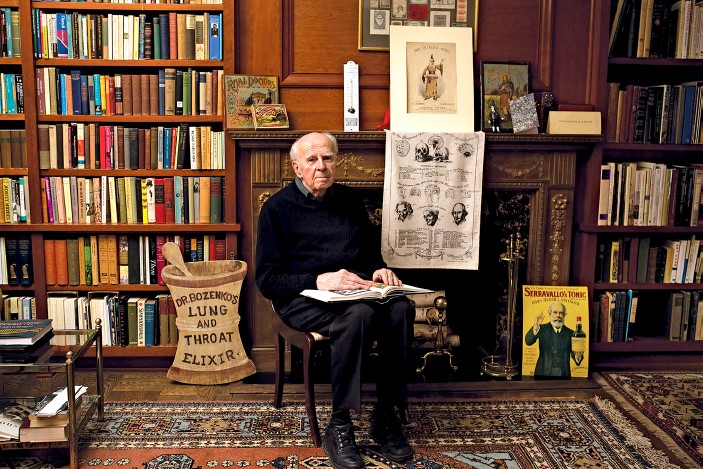
By Joseph Bishop, Princeton University, and the Librarys 2022 Audrey and William H. Helfand Fellow

Mr. Bishop completed his Fellowship residency in summer 2022 and will present his research by Zoom on November 8 at 4:00 (EST). To attend his talk, “Pharmaceutical Visions: How US Drug Companies and Ad Agencies Revamped Their Credibility by Marketing with Scientific Imagery,” register through the Academy’s Events page.

This spring I spent weeks immersed in the vast historical materials available at the New York Academy of Medicine (NYAM) Library. I had the honor of receiving the Audrey and William H. Helfand Fellowship to pursue a project that I believe would have interested Bill Helfand: an examination of the changes in medical advertising at the turn of the twentieth century. During his life, Helfand amassed an extensive collection of fascinating drug and medical memorabilia and visual art. Much of his collection and work illuminates the dynamic between drugmakers and the public during the late nineteenth and early twentieth century.  Significant portions of Helfand’s collection are available at NYAM, and I took advantage of the richly colored and wide-ranging collection of patent medicine trade cards. NYAM also has mountains of pharmaceutical, medical, and allied trade journals and magazines brimming with pharmaceutical advertisements.

At the turn of the twentieth century, the American public saw the rise in large corporate pharmaceutical companies, national and corporate advertising, and federal drug regulation. An important question is what prompted the transformations in medical visual culture that helped to portray drug companies as scientific and research-oriented. To answer that question, I compare late-nineteenth-century patent medicine trade cards with medical ads in LadiesHome Journal in the 1920s. This comparison reveals a transition from entertainment and fantasy to a preoccupation with scientific progress and medical authority. My research at NYAM has led me to conclude that drug companies and ad agencies emphasized scientific and medical imagery to revamp their medical credibility and professional image amid national drug regulation and the public’s anger with the industry’s past association with patent medicine.

American patent medicine companies faced scrutiny for producing nostrums with cryptic contents and questionable efficacy during the first decade of the twentieth century. They depended on local advertisement imagery that reflected the nineteenth-century public’s anxieties and aspirations. Popularizations of science were created for various reasons—from entertainment to informing citizens—but they all served to increase scientific and medical awareness within the American public. Philadelphia-based ad agency N. W. Ayer’s accounts show that patent medicine was their most lucrative commodity category, carrying 26 percent of their total revenue in 1878, and their second-most lucrative commodity in 1900, carrying 15 percent. By 1879 in the US, more than 400 religious weeklies each needed a steady flow of advertising revenue to stay in business. Newspapers generated patent medicine business, and medical advertisements sustained newspapers. Nostrum manufacturers developed new marketing techniques, created novel distribution systems, pioneered brand-name products, became an economic link between urban and rural centers, and expanded markets.

Americans living through the last two decades of the nineteenth century saw an explosion of advertising trade cards. Retailers gave away these pocket-sized cards, often stuffed in product packaging as a bonus and easily collectible by customers. Patent medicine companies used these cards extensively, as they proved to be a very successful sales medium. Many people collected cards into albums and created scrapbooks, which children sometimes received as birthday or Christmas gifts.

Image 1:“Ayer’s Ague Cure Is Warranted to Cure All Malarial Disorders”
Dr. J. C. Ayer & Co. (Lowell, MA)
Produced ca. 1875–1895
From the William H. Helfand Collection of
Pharmaceutical Trade Cards at The New York Academy of Medicine
Chromolithograph, 6.1 x 11.6 cm

Trade cards such as Ayer’s Ague Cure (Image 1) evoke an aura of connection with nature and adaptation to the surrounding environment. The image in the bottom right corner depicts an alligator and a couple of frogs discussing Ayer’s Cure as if they used it to protect themselves from malaria. The ad implies that Ayer’s product helps one adapt to one’s environment just as well as the alligators and frogs adapt to theirs.

Image 2: “Cas-car-ria Is Worth Its Weight in Gold”
Product: Cas-car-ria
Produced ca. 1875–1895
From the William H. Helfand Collection of
Pharmaceutical Trade Cards at The New York Academy of Medicine
Chromolithograph, 5.8 x 7.8 cm

On the other hand, Cas-car-ria’s trade card (Image 2) depicts a young girl holding a switch and a dog together, fending off miniature demons labeled with ailments. Cas-car-ria’s ad evokes notions of animal protection and self-protection, implying that when patients take Cas-car-ria, they harness the animal within, unleashing the strength required to fight off external demons.

The explosion of ads promoting proprietary medicines and their incessant hyperbole and mistruths eventually provoked strong public reactions that demanded transparency and regulation. There were always calls to rein in the quackery, but the inundation of promotion drove the regulation of patent medicine to become a public priority.

Image 3: “What is in your medicine cabinet?”
Ladies’ Home Journal, February 1924, p. 144
Manufacturer: E. R. Squibb and Sons
From Ohio State University

Examining Ladies’ Home Journal during the 1920s illustrates these changes in advertising.  At this point, advertisements offered a different portrayal of scientific medicine and appealed to a more docile public regarding medical professionals. A Squibb pharmaceutical ad depicts a well-organized medicine cabinet (Image 3). The caption asks, “What is in your medicine cabinet? Are they products your physician would approve?” This approval seeking from medical authority plays into the new values of placing faith in the trained judgment of scientific and medical authority. 

Image 4: “Zonite: the World War Antiseptic”
Ladies’ Home Journal, March 1924, p. 192
Manufacturer: Zonite
From Ohio State University

Ads for Zonite (Image 4) ran images of scientists wearing lab coats and examining test tubes, drawing a scientific aura into its products. Zonite also associated its product with scientific discoveries, like the Carrel-Dakin fluid (i.e., diluted bleach), a critical antiseptic used in World War I.

Image 5: “Why the familiar cake of Yeast Foam is now eaten as well as used in bread”
Ladies’ Home Journal, May 1921, p. 49
Manufacturer: Northwestern Yeast Co. (Chicago, IL)
From the University of Michigan

The ad for “Yeast Foam” (Image 5) also appeals to scientific and medical authority, depicting a man wearing a medical coat and peering into a microscope. In the foreground are two circular illustrations of microscopic specimens—one containing germs and the other germ-free. The ad portrays a professional man immersed in scientific work, suggesting that the product has been carefully vetted through scientific scrutiny for quality assurance.

Similarly, the Fleischmann advertisement (Image 6) depicts two men in lab coats working at a table equipped with flasks, a beaker, a microscope, and other scientific instruments. The caption below the image states, “Messages of startling importance from the laboratory of the scientist.” Text within the ad notes how Fleischmann’s Yeast cures various diseases. In the case of skin diseases, the ad relies on a general sense of medical authority: “Many physicians and hospitals are prescribing Fleischmann’s Yeast for impurities of the skin. It has yielded remarkable results.”

Image 6: “Ten or fifteen years of life”
Ladies’ Home Journal, September 1921, p. 45
Manufacturer: The Fleischmann Company (New York, NY)
From the University of Michigan

The values of corporate ad agencies following the patent medicine era are not only a reaction to muckraking journalism and reform movements. The use of scientific medical imagery conveying authority and professional judgment was also largely about revamping the medical credibility of US drug companies and corporate ad agencies; they benefited handsomely during the patent medicine era but later needed to diminish their connections to these fraudulent products. Ad agencies traced the American public’s anxieties and aspirations as they shifted from loose whimsy about panaceas in the late nineteenth century to a reverence for qualified scientific and medical experts and institutions at the beginning of the twentieth century. By tracing this transition in medical imagery, we can glean how drug companies and ad agencies shaped products to elevate their professional clout.

Library Research Fellowships for 2023

By Arlene Shaner, Historical Collections Librarian, Library Fellowship Coordinator

The NYAM Library has just awarded its 2023 residential research fellowships. The Audrey and William H. Helfand Fellowship in the History of Medicine and Public Health goes to Sean Purcell, Indiana University, for his project “Imaging Consumption: Seeing ‘The White Plague’ in American Medicine”; and the Paul Klemperer Fellowship in the History of Medicine to Anastasiia Zaplatina, Bielefeld University, for her project “The American Soviet Medical Society (1943–1947): Academic Exchanges between Allies and Their Cold War Legacy.” Congratulations to both recipients!

2023 Helfand Fellow Sean Purcell

Sean Purcell plans to dig deeply into the Library collections to find visual materials related to early twentieth-century tuberculosis research for an upcoming video art installation. He is especially interested in questions of how and why visual materials such as microscope slides, photographs, illustrations, and specimens were used in medical research, and who the subjects were.

2023 Klemperer Fellow Anastasiia Zaplatina

Anastasiia Zaplatina will be looking at archival and published materials related to the American-Soviet Medical Society, and at the Joseph Wortis Soviet Medicine and Psychiatry Papers as part of investigating the impact of this short period of collaboration between individuals in both countries, and specifically whether collaboration ultimately laid the groundwork for increased competition.

We invite all interested researchers regardless of academic affiliation to apply for one of the Library’s research fellowships. Our 2024 fellowship application process will open early in the summer of 2023, with an application deadline at the end of August and a decision in October, for a month’s residence at the Library during the following calendar year. Each fellowship provides $5,000 in support. At the conclusion of their time at the Library, each Fellow gives a presentation and writes a blog post on their work. For background on how these Fellowships arose, see The Faces Behind Our Fellowships; and for more information about the focuses of the awards, including a list of previous awardees and their projects, take a look at the Library Fellowships pages.

The Faces Behind Our Fellowships

By Arlene Shaner, Historical Collections Librarian

The Library has two residential research fellowships, the Paul Klemperer Fellowship in the History of Medicine and the Audrey and William H. Helfand Fellowship in the History of Medicine and Public Health. While there is plenty of information on our website about how to apply for our fellowships, there is no information there about the people for whom they are named, and it seems appropriate to share a little bit about them.

Paul Klemperer (1887–1964) spent much of his career at Mount Sinai Hospital, where he held the position of pathologist from 1927 until his retirement in 1955. Born outside of Vienna, Klemperer first enrolled at the University of Vienna, intending to become a lawyer. At the suggestion of his father, he took a class on psychoanalysis taught by family friend Sigmund Freud and began to study medicine instead. After receiving his medical degree in 1912, he spent two years studying pathological anatomy, and then served as a physician during World War I. In 1921, he emigrated to the United States, spending a year in Chicago before moving to New York, teaching briefly at the New York Post-Graduate Medical School before joining the staff at Mount Sinai. He also taught pathology at the College of Physicians and Surgeons of Columbia University for many years, and after retirement continued to teach the Albert Einstein College of Medicine.

Paul Klemperer, M.D. (1887–1964). NYAM Library Collections.

His students and colleagues were devoted to him. In 1962, the Academy presented him with the Academy Medal for Distinguished Contributions to Biomedical Science. In his remarks George Baehr, his colleague at Mount Sinai, noted that Klemperer’s skill as a pathologist combined with his skill as a teacher made him a much-loved figure in all the institutions to which he had a connection. Neuropathologist Stanley Aronson, in a 1989 reminiscence in the Mount Sinai Journal of Medicine, recalled him as “one who was shy yet effective, retiring yet generous, undemonstrative yet passionate, learned yet learning, always learning. For he was truly our teacher.”[1]

After he retired, Klemperer devoted significant time to the study of the history of medicine. He wrote the preface and introduction to the Academy’s publication of a translation of Giambattista Morgagni’s noted book on pathology, The Seats and causes of diseases investigated by anatomy, as well as translating five letters of Morgagni. He also wrote the introductions to several other volumes in the Academy’s history of medicine series. To honor his memory and his devotion to the history of medicine, some years after his death an anonymous group of donors endowed the fellowship that bears his name, first awarded in 1996.

William H. Helfand (1926–2018), a Philadelphia native, pursued a career as a pharmaceutical executive for Merck. His work dovetailed with his collecting interests in prints, posters, and such pharmaceutical ephemera as trade cards and almanacs, and he wrote extensively on their social history.[2] He and his wife, Audrey, endowed positions and fellowships at several institutions, including the Philadelphia Museum of Art, the Library Company of Philadelphia, and the Grolier Club. In 1998, the couple endowed the NYAM Library fellowship that bears their name, with the first fellowship awarded in the 1999–2000 academic year.

William H. Helfand (1926–2018). Image from the New York Times, October 5, 2018.

From the beginning, the Helfand fellowship supported research on the ways that visual materials enhance the study of the history of medicine, public health, and the medical humanities. Our own Library collections are far richer in these areas because Bill supplemented his endowment with gifts of materials from his own collections, Chief among these is the William H. Helfand Collection of Pharmaceutical Trade Cards, which is digitized and available here. In addition to trade cards, Bill gave the Library almanacs, broadsides, caricatures, prints, sheet music, and other medical ephemera. Our Helfand collection is one of many; others can be found at the Huntington Library, Yale University, Duke University, and the Library Company of Philadelphia.

If you are a scholar working on a history of medicine project, please consider our fellowships. Applications are being accepted until September 17, 2021, for a month’s residence at the Library. Successful applicants will be notified by October 22, and the next two fellows may work any time during the 2022 calendar year.

Lists of all the projects that have been supported through these endowments can be found on the fellowship pages for the Klemperer Fellowship and the Helfand Fellowship; application procedures are found there as well.

_________


[1] Baehr, George. “Citation and Presentation of the Academy Medal to Paul Klemperer, MD.” Bulletin of the New York Academy of Medicine 38, no. 4 (1962): 240; Aronson, S. M. “The legacy of Paul Klemperer.” The Mount Sinai Journal of Medicine, New York 56, no. 5 (1989): 347–350.

[2]William Helfand, a Collector Intrigued by Quackery, Dies at 92,The New York Times, October 5, 2018.

Apply for Our 2020 Fellowships

We’re pleased to announce that our two annual fellowships are open to applications!

nyam_rbr_106

The Drs. Barry and Bobbi Coller Rare Book Reading Room, where our fellowship recipients will conduct their research.

The Academy Library offers two annual research fellowships, the Paul Klemperer Fellowship in the History of Medicine and the Audrey and William H. Helfand Fellowship in the History of Medicine and Public Health, to support the advancement of scholarly research in the history of medicine and public health. Fellowship recipients spend a month in residence conducting research using the library’s collections.

Applications for our fellowships are being accepted now through late August for fellowships that may be used at any time during 2020.

Preference in the application process will be given to those whose research will take advantage of resources that are uniquely available at the Academy, individuals in the early stages of their careers, and, for the Helfand Fellowship, applications which include an emphasis on the use of visual materials held within the Academy’s collections and elsewhere. Applicants should provide information in their proposals about the collection items they plan to use, either by including a bibliography of resources they intend to consult or discussing those items in detail in the context of the application essay. Changes in the Library that are scheduled to take place beginning in the second half of 2019 will impact applicants whose projects rely heavily on 19th and 20th century serial literature or on monographs published during the second half of the 20th century. 

Applications are due by the end of the day on Friday, August 23, 2019. Letters of recommendation are due by the end of the day on Monday, August 26, 2019. Applicants will be notified of whether or not they have received a fellowship by Monday, October 4, 2019.

Prospective applicants are encouraged to contact Arlene Shaner, Historical Collections Librarian, at 212-822-7313 or history@nyam.org with questions or for assistance identifying useful materials in the library collections.

Click on their names to read blog posts about their projects from our most recent fellowship recipients, Matthew Davidson (Klemperer) and Tina Peabody (Helfand).

We look forward to hearing all about your projects!

The Enduring Impact of the X-Ray

Today we have part two of a guest post written by Dr. Daniel S. Goldberg, 2016 recipient of the Audrey and William H. Helfand Fellowship in the History of Medicine and Public Health. Part one can be read here.

X-ray exhibitions were hugely popular all over the country, and the greater NY area was no exception.  At a February 1896 demonstration run by Professor Arthur Wright, director of the Sloan Laboratory at Yale University, a newspaper reported that despite the auditorium being literally jam-packed, students were still crawling through windows 30 minutes into the lecture — and all this despite the fact that none of the audience, save those in the first few rows, could even hear Wright’s discussion.  The deans of multiple Yale schools (Divinity, Law, and Science), the head of the Yale Corporation, and the chief medical examiner were all in attendance.

bostonmedsurgjournal_feb201896_rontgenprocess_watermark

Perhaps the first published X-ray in the United States of a clinical condition. In “Rare Anomalies of the Phalanges Shown by the Röntgen Process,” Boston Medical and Surgical Journal 134(8), February 20, 1896: 198–99.

The pressing question is “why”? Why did X-rays exert such tremendous power across a wide spectrum of social domains? (X-rays were a constant topic of conversation in sermons and religious journals, in women’s journals, in influential satirical periodicals like Punch, and were the subject of a seemingly endless number of political and non-political cartoons, to name but a few).  Although historians of the X-ray have offered a number of plausible answers, I believe there is a key element left unexplored in the historiography: the intellectual frameworks, or ideas, relating to changing ideas of truth, doubt, and objectivity in U.S. society at the time.

Two of these frameworks are most useful in unpacking the stunning impact of the X-ray: the rise of mechanical objectivity, and what can be called “somaticism” within medicine and science.  Historians of science Lorraine Daston and Peter Galison explain that a new model of ‘objectivity’ begins to take hold during the middle decades of the 19th century.  Under this new model, the truth-value of scientific knowledge is a function of the investigator’s ability to remove or eliminate human, subjective influence from the knowledge-making process.  The fact that this is more or less impossible, and that X-rays can be manipulated in all sorts of ways was well-known to contemporaries and remained a source of anxiety for some time.  The important point is the ultimate goal: to let the mechanical processes of nature speak for themselves and reveal their truths.  Ideas of objectivity, as Daston and Galison point out, have for over four hundred years been connected to scientific images, which makes media like photography and X-rays especially significant.

By the end of the 19th century, ideas of mechanical objectivity begin to fundamentally reshape ideas of what is known and what is certain.  This is especially crucial in a century that features so much intense change, including but not limited to governments, family and labor structures, migration patterns, and, of course, industrialization and urbanization.  Late Victorians were beset with anxieties connected to their changing world, and they were especially concerned with artifice and deception — that the world was not what it seemed.  As such, intellectual frameworks that shaped the criteria for truth were hugely influential, and traveled well beyond narrow networks of scientists and medical men.

Somaticism integrates in important ways with constructs of mechanical objectivity.  Historians of medicine have documented the influence of somaticism (or, “empiricism,” as it is also sometimes termed) within medicine over the long 19th century.  The core of the framework is that truths about disease and the body are to be found in pathological anatomical objects.  The existence of these objects can then be clinically correlated with the illness complaints the patient has, or more likely had given that pathological objects are most likely to be located precisely during a postmortem — until the X-ray.  The truths of the sick body are to be found in the natural objects of disease, which makes seeing those objects so essential.  Laennec himself explained that the point of the stethoscope was not to listen; listening was merely a means to an end.  The point, as Jacalyn Duffin explains, was “to see with a better eye.”

Collectively, these frameworks go a significant length in explaining the enormous and enduring social impact of the X-ray.

X Rays for Consideration_watermarked

Article from the New York Record. May 1896.

For example, Morton’s clippings contain a May 1896 article from the New York Record entitled “X Rays for a Consideration: Light in a Human Kidney.”  The article details what may be the first private X-ray laboratory opened in New York City, founded by Mrs. M.F. Martin, and located at 110 East 26th Street.  The lab was intended solely for the use of physicians and hospitals.  One of its first patients was a doctor named George McLane, who traveled from North Dakota to have his kidney X-rayed for evidence of a possible stone.  A surgeon removed McLane’s kidney, and Morton placed it on a plate and subsequently irradiated it with X-rays.  The procedure “revealed the absence of any stone in the organ, demonstrating the entire reliability of doctors to prove the absence of stone in the kidney.”

The X-ray shines its light into the hitherto dark spaces inside the human body, revealing the truth of a disputed question: whether McLane suffered from a kidney stone or not.  The truth resides in the natural object itself, and the mechanism of the X-ray supposedly insulates the production of medical knowledge from the whims and artifices of the investigator (as compared to illustrations and drawings, for example).

Or, as Dr. McLane himself stated at the Post Graduate Hospital (the primary hospital at which Morton cared for inpatients):

“Dr. McLane spoke modestly at the Post Graduate Hospital about the risk he had taken in the name of science . . . ‘Hitherto a great many mistakes have been made owing to the inability of doctors to prove the absence of stone in the kidney . . .’  Now, by a very simple process, the truth can easily be determined.”

It is difficult to imagine how powerful it must have been, in 1896, to witness an X-ray operator remotely anatomize the living body.  Seeing inside the body had been a dream of physicians for centuries prior, and there is every reason to believe that its achievement has not eroded much of its social power.  Americans still perform significantly more medical imaging procedures than virtually any of our comparator societies, and what is most interesting is the evidence that this utilization is driven both by supply and demand.  That is, it is not merely that we have expensive X-ray and medical imaging machines — so we use them.  Across a wide variety of illness paradigms, illness sufferers and patients request medical imaging; they want it to be performed on their bodies.  The history of the X-ray helps us understand the enduring power of these tools, of what it means to delve into the penetralium.

The Early Days of the X-Ray

Today we have part one of a guest post written by Dr. Daniel S. Goldberg, 2016 recipient of the Audrey and William H. Helfand Fellowship in the History of Medicine and Public Health. Dr. Goldberg is trained as an attorney, a historian, and a bioethicist.  He is currently on the faculty at the Center for Bioethics and Humanities at the University of Colorado Anschutz Medical Campus.

After news of Wilhelm Röntgen’s discovery of X-rays was cabled across the Atlantic late in 1895, evidence suggests X-ray experimentation was taken up eagerly all over the U.S. almost immediately.  While scientists and physicians scrambled to build their own X-ray machines, newspapers in major cities throughout the country eagerly reported on their progress, with stories small and large appearing in nearly every significant daily from New York and Philadelphia to Chicago and St. Louis to San Francisco and Los Angeles.  Historians of the X-ray estimate that within only a year of Röntgen’s discovery, literally thousands of articles had been published on the X-ray in both lay and expert periodicals.  Even in the fertile print culture of 1896, this is a significant accounting.

Therein lies the methodological difficulty for the historian of the X-ray.  So often, the craft of history is a tedious search for small scraps of information that may not even exist.  Yet, as to X-rays, the problem is one of feast, not famine.  With so much print material appearing in so many different sources in so many different places all at the same time, sifting through the morass to articulate coherent and important narratives is difficult.

What makes this task far easier is a remarkable collection held at the New York Academy of Medicine Library.  The William J. Morton Collection is a small holding, consisting of only two boxes.  The second box is the true treasure, containing a single folder, approximately six inches thick.  Inside is an unbound series of pages consisting solely of newspaper clippings related primarily to early X-ray use in the U.S.  These are Morton’s clippings, and as far as is known, the order and arrangement of the pages is original to Morton himself.  The collection is astounding, for it represents something of an index or a cipher for the ferment of X-ray use in NYC in the first half of 1896.

Clippings

Newspaper clippings from the William J. Morton Collection, New York Academy of Medicine Library.

There is no question that New York City played an important role in early X-ray use, if for no other reason than the enormous shadow cast by the inventor, Thomas Edison.  There were, however, many other important figures involved in early X-ray use in NYC, including Nikola Tesla[1], Michael Pupin[2], and Morton.  Morton, the son of William T.G. Morton of anesthesia fame, was a prominent physician, a fellow of the New York Academy of Medicine, and a respected neurologist and electro-therapeutic practitioner.

Telegrams_watermark

A telegram dated January 2, 1896 from Dyer & Driscoll, attorneys for none other than Thomas Edison, indicated that Morton visited Edison’s workshop for the purpose of conducting experiments (almost certainly with X-rays) several days earlier.

Because Morton was unquestionably at the forefront of early X-ray experimentation in NYC, his curation is a reasonable index as to important events and moments in the early use of X-rays in NYC.  There are limitations to this approach, of course.  Morton was obviously interested in his own role in early X-ray experimentation, so there is something of a selection bias at work (although it should be noted that there are no shortage of clippings pertaining to Pupin’s important work).

The collection is full of interesting and significant stories in the early history of X-ray use.  For example, in March 1896, strongman Eugene Sandow, considered the father of modern bodybuilding, turned to Morton in an effort to locate the source of a frustrating pain he was experiencing in his foot.  Apparently Sandow had stepped on some broken glass, but even his personal physician could not specify the location of the glass in his foot.  The potential for the X-ray must have seemed obvious, and Sandow reached out specifically to Morton to see if he could be of help.  Morton was eager to oblige.  He turned the X-rays on Sandow’s foot and located the shard of glass disturbing Sandow’s equanimity.  A surgeon subsequently operated and removed the glass, and the story made national news.

How the photograph was made_watermark

The X-Ray of Eugene Sandow’s foot in process.

Interestingly, Sandow was apparently impressed enough with the powerful rays to send an unsolicited telegram to Edison, offering his services as a human subject for any X-ray experiments Edison wished to undertake.

SandowLetter_watermark

Letter to Thomas Edison from Eugene Sandow.

It is difficult to imagine how powerful it must have been, in 1896, to witness an X-ray operator remotely anatomize the living body.  Seeing inside the body had been a dream of physicians for centuries prior, and there is every reason to believe that its achievement has not eroded much of its social power.  Americans still perform significantly more medical imaging procedures than virtually any of our comparator societies, and what is most interesting is the evidence that this utilization is driven both by supply and demand.  That is, it is not merely that we have expensive X-ray and medical imaging machines; so we use them.  Across a wide variety of illness paradigms, illness sufferers and patients request medical imaging; they want it to be performed on their bodies.  The history of the X-ray helps us understand the enduring power of these tools.

Footnotes:
[1] Tesla was heavily involved in early X-ray experiments in his laboratory at 46 East Houston Street; much to Edison’s likely chagrin, given the frostiness of their relationship by the time. The New York newspapers constantly asked Edison about Tesla’s progress.
[2] Pupin, a Columbia University physicist, would in short order — in 1896, in fact —  go on to discover a way of substantially reducing the exposure time needed to produce an X-ray image from hours to minutes.  The basics of Pupin’s method are still used today.

Apply for our 2017 Research Fellowships

Does a one-month residence in The Drs. Barry and Bobbi Coller Rare Book Reading Room, immersed in resources on the history of medicine and public health, sound like a dream come true?

Rare book room

The Drs. Barry and Bobbi Coller Rare Book Reading Room

The Academy Library offers two annual research fellowships, the Paul Klemperer Fellowship in the History of Medicine and the Audrey and William H. Helfand Fellowship in the History of Medicine and Public Health, to support the advancement of scholarly research in the history of medicine and public health. Fellowship recipients spend a month in residence conducting research using the library’s collections.

Applications for our fellowships are being accepted now through late August for fellowships that may be used at any time during 2017.

Preference in the application process is given to early career scholars, although the fellowships are open to anyone who wishes to apply, regardless of academic status, discipline, or citizenship. While both fellowships are for researchers engaged in history of medicine projects, the Helfand Fellowship emphasizes the role of visual materials in understanding that history.

Applications are due by the end of the day on Friday, August 26, 2016. Letters of recommendation are due by the end of the day on Monday, August 29, 2016. Applicants will be notified of whether or not they have received a fellowship by Monday, October 3, 2016.

Prospective applicants are encouraged to contact Arlene Shaner, Historical Collections Librarian, at 212-822-7313 or history@nyam.org with questions or for assistance identifying useful materials in the library collections.

Spoiled by a Certain Englishman? The Copying of Andreas Vesalius in Thomas Geminus’ Compendiosa

Laura Robson, the author of today’s guest post, is our 2014–2015 Helfand Research Fellow. She completed her PhD in Classics at the University of Reading, UK.

“I wish the Epitome had not been spoiled so disgracefully by a certain Englishman (who I think lived with my brother for a time). He took what had been written with great care succinctly as a list in the Epitome and expanded it with excerpts taken from the books of the Fabrica… He utterly corrupted what had made it most praiseworthy and so roughly and absurdly copied what had been set forth with elegant drawing and engraving that he preserved no appearance of Oporinus’ majestic edition.”1

These were the concerns of Franciscus Vesalius, brother of the famous anatomist Andreas Vesalius, published in the preface to The China Root Epistle in 1546. Within only three years of the original publication of Andreas Vesalius’ De Humani Corporis Fabrica and its companion work, the Epitome, a number of medical authors had copied and reproduced the beautiful illustrations that had made Andreas Vesalius and his work so famous.2

Andreas Vesalius (1514-1564). De humani corporis fabrica libri septum. Basel: Johannes Oporinus, 1543. The most famous illustrations are the series of fourteen muscle men, progressively dissected. Some figures, such as this one, are flayed. Hanging the muscles and tendons from the body afforded greater detail, not only showing the parts, but how they fit together.

Andreas Vesalius (1514-1564). De humani corporis fabrica libri septum. Basel: Johannes Oporinus, 1543. Click to enlarge.

The Fabrica was one of the first anatomical treatises of the 16th century to present illustrations of the anatomised body in a naturalistic way. Vesalius promoted the dissection of the human body as the best way to learn about anatomy. By performing human dissections, he uncovered errors in the work of the ancient anatomist Galen, whose use of animals as dissection material to substitute for the lack of human cadavers had dominated the understanding of the body for centuries. Vesalius was caught in a conflict: how to show the anatomical errors in Galen’s treatises without going against such an important medical authority and potentially damaging his own medical career. He used the Fabrica to present his findings and to build on Galen’s important work.

The Fabrica contained 700 folio pages of Latin text and beautiful woodcut illustrations depicting the anatomical body in different poses. Readers were signposted to turn back and forth several times between image and text. This, as well as each image being accompanied by a letter key, encouraged a very active reading of Vesalius’ treatise.3 The work proved popular, with people taking a particular interest in the figures, although people copied, adapted, and reused both the images and text of Vesalius’ work in many different medical treatises. Due to the size and high cost of the Fabrica, cheaper copies were often more accessible than the original, even though the pirating enraged Vesalius and his close circle. Book piracy was common at this time. There was not the modern sense of intellectual property or copyright legislations. Licenses allowed particular printers to print works first, but the Venetian and imperial privileges obtained by the authors to try and protect their books from piracy did little to stop others from copying them.4

In fact, Franciscus Vesalius accused the wrong man of copying his brother’s work (suggesting, perhaps, that he had not seen a copy of the offending book). The only Englishman known to live with Vesalius was John Caius, when they lodged together in Padua during their studies. Caius went on to be physician to King Edward VI. There is no evidence that he pirated any version Vesalius’ work.5

The work Franciscus refers to is in fact Thomas Geminus’ Compendiosa.6 The first edition is predominantly made up of the Latin text of the Epitome and its illustrations, with the addition of many Fabrica figures. It is believed to be one of the first books to use copperplate illustrations.7 Geminus stated in his dedication that he followed Vesalius, but shortened his book to make it more useful to readers, in particular students.8 Reproducing the images of the Fabrica with the text of the Epitome meant that there was little interaction and connection between the two. However, the publication of the Compendiosa did bring Vesalius’ illustrations to a wider audience as the book was shorter and therefore cheaper than the original.

In order to make the work more accessible to those who could not read Latin, Geminus published an English edition of the Compendiosa in 1553. Nicholas Udall translated the short captions from the Fabrica figures into English. However, the main text of the Epitome was not translated. Instead the illustrations were placed after the text of Thomas Vicary’s The anatomie of mans body, first published in 1548.9 Geminus rearranged this text to follow the order of dissection for the parts of the body that decayed the fastest—the abdomen, the thorax, and the head. Although the images were not rearranged to fit this order, they connected more strongly to the text than in the Latin edition, as readers were signposted to particular figures discussing different parts of the body.

In his preface to the English Compendiosa, Nicholas Udall puts forward some interesting points about the uses of images and texts in medical manuals of his time. He said he did not know whether images or texts were more important when presenting anatomical information. He argued that information is set forth in writing for “high learning” and in pictures for the unlearned. He also explained that surgeons often performed duties like resetting bones by looking at figures alone.10 Surgeons had a low status in the medical profession at this time. They were not university educated like physicians and they were accused of having little knowledge about the science of medicine and healing. This suggests that readers who could not understand Latin, like surgeons, used anatomical figures and not written texts. I believe the English version of the Compendiosa was an attempt at encouraging these readers to read the text as well, by providing it in the vernacular language.

The coat of arms, left, and title page, right, of the Academy's copy of the 1559 English edition of Geminus’ Compendiosa.

The coat of arms, left, and title page, right, of the Academy’s copy of the 1559 English edition of Geminus’ Compendiosa. Click to enlarge.

In 1559 the English edition of Geminus’ Compendiosa was reissued.11 The annotated copy of this edition in the collection at the New York Academy of Medicine reveals hints as to how this anatomical text was used by readers at the time. The coloured and illuminated title page includes a portrait of Queen Elizabeth I. Slithers of gem stones have been attached to her necklace and the coat of arms opposite her on the adjacent page. The nude figures known as Adam and Eve are also coloured, and a reader inscribed the verse, “The Eyes of Them Both were opened, and They knew that They were naked: Genesis Chapter 3 Verse 7.”

Adam and Eve in the Academy's copy of the 1559 English edition of Geminus’  Compendiosa. Click to enlarge.

Adam and Eve in the Academy’s copy of the 1559 English edition of Geminus’ Compendiosa. Click to enlarge.

Two readers annotated this copy, both with different handwriting from the owner who in 1769 wrote his name—“G. Molesworth”—on the title page. One reader underlined key words and sections of the text, marking these with almost illegible notes in the margins. The other reader, though, focused on the illustrations. This second reader annotated the first three Vesalian musclemen images in the copy, adding the letters of the key, along with the Latin names for the body parts they represent.12 This English edition of the Compendiosa seldom uses the Latin names for parts of the body. So our reader did not get the information for his notes from this edition of the text. He must have consulted another text, such as the Latin edition of 1545, or even the original work of Vesalius’ Fabrica, in order to make his annotations.

Image of annotated muscleman figure  in the Academy's copy of the 1559 English edition of Geminus’  Compendiosa. Click to enlarge.

The annotated muscleman figure in the Academy’s copy of the 1559 English edition of Geminus’ Compendiosa. Click to enlarge.

This demonstrates the culture of active reading in the early modern period. This reader engaged with more than one treatise, perhaps even several works, when learning about the dissection of the body. He was familiar with the Latin language, and was therefore not one of the unlearned readers mentioned by Nicholas Udall in his preface to the work.

While Andreas and Franciscus Vesalius opposed the reproduction of the Fabrica and Epitome, the works that copied, adapted, and reused material from these texts allowed for the transmission of Vesalius’ knowledge of the body to a wider audience than the original works could reach. And this new audience interacted with the material, coming to a greater understanding of the dissected human body in the early modern period.

References

1. Andreas Vesalius (1546), Vesalius: The China Root Epistle, translated by Daniel H. Garrison (2015), p. 6.

2. Andreas Vesalius (1543a), De Humani Corporis Fabrica Libri Septem, Basel and Andreas Vesalius, (1543b), Andreae Vesalii Suorum de Humani Corporis Fabrica Librorum Epitome, Basel.

3. On active reading in Vesalius’ anatomical texts see, Nancy Siraisi (1994), “Vesalius and Human Diversity in De humani corporis fabrica”, in Journal of the Warburg and Courtauld Institutes, Vol. 57 p.64 and Sachiko Kusukawa (2012), Picturing the Book of Nature: Image, Text, and Argument in Sixteenth-Century Human Anatomy and Medical Botany, Chicago and London, p. 24.

4. On history of copyright and pirating see Christopher L. C. E. Witcombe (2004), Copyright in the Renaissance: Prints and Privilegio in Sixteenth-Century Venice and Rome, Leiden.

5. See Charles O’Malley (1955), “The Relations of John Caius With Andreas Vesalius and Some Incidental Remarks on the Guinta Galen and on Thomas Geminus,” in Journal of the History of Medicine and Allied Sciences Vol. 10.2 pp.147-172.

6. Thomas Geminus (1545), Compendiosa totius anatomie delineatio, aere exarata: Thomam Geminum, London.

7. Leroy Crummer (1926), “The Copper Plates of Raynalde and Geminus”, in Proceedings of the Royal Society of Medicine Vol 20.1 p. 53.

8. Thomas Geminus (1545), Compendiosa totius anatomie delineatio, aere exarata: Thomam Geminum, London, p. 1.

9. Thomas Vicary (1577 [1548]), A Profitable Treatise of the Anatomie of mans body: compyled by that excellent chirurgion, M. Thomas Vicary esquire, seriaunt chirurgion to king Henry the eyght, to king Edward the. vj. to Queene Mary, and to our most gracious Soueraigne Lady Queene Elizabeth, and also cheefe chirurgion of S. Bartholomewes Hospital. Which work is newly reuyued, corrected, and published by the chirurgions of the same hospital now beeing, London.

10. Thomas Geminus (1553), Compendiosa totius anatomie delineatio, aere exarata: Thomam Geminum, London, p.1.

11. Thomas Geminus (1559), Compendiosa totius anatomie delineatio, aere exarata: Thomam Geminum, London.

12. Ibid. p. Cii.

Apply for our 2016 Research Fellowships

Are you working on a history of medicine project that would be enhanced by spending a month mining our collections?

NYAM Library, Rare Book Room photos by Amy Hart © 2012We are now accepting applications for the Paul Klemperer Fellowship in the History of Medicine and the Audrey and William H. Helfand Fellowship in the History of Medicine and Public Health. Each fellow receives a stipend of $5,000 to support travel, lodging, and incidental expenses for a flexible period between January 1, 2016 and December 31, 2016. Fellows are expected to spend at least four weeks in New York City, working at The New York Academy of Medicine. Besides completing a research project, each fellow will be expected to make a public presentation at the Academy and submit a final report.

Both fellowships are designed to support researchers who can demonstrate how an opportunity to immerse themselves in our rich holdings will enhance their work. Preference in the application process is given to early career scholars, although the fellowships are open to anyone who wishes to apply, regardless of academic status, discipline, or citizenship. While both fellowships are for researchers engaged in history of medicine projects, the Helfand Fellowship emphasizes the role of visual materials in understanding that history.

The application deadline is Monday, August 17, 2015. Letters of recommendation must be received by Friday, August 21, 2015. Fellowship recipients will be notified by Thursday, October 1, 2015.

Perspective applicants are encouraged to contact Arlene Shaner, Reference Librarian for Historical Collections, at 212-822-7313 or history@nyam.org with questions or for  assistance identifying useful materials in the library collections.

2014–2015 Helfand and Klemperer Research Fellows

By Lisa O’Sullivan, Director, Center for the History of Medicine and Public Health

We are pleased to announce the 20142015 Helfand and Klemperer Research Fellows: Laura Robson (University of Reading) and Heidi Knoblauch (Yale University).

Rösslin, Eucharius. The byrth of mankynde, otherwyse named the womans booke. [London : Tho. Ray[nalde]], 1545.

The Audrey and William H. Helfand Fellowship in the History of Medicine and Public Health  focuses on the use of visual materials. Laura Robson will explore how medical works in the sixteenth century used images and texts from Andreas Vesalius’ anatomical treatise, the Fabrica. She will use Geminus’s Compendiosa (1545) and Raynalde’s translation of The byrth of mankynde (1545) to demonstrate the complex relationship between anatomical image and text and to unite the history of the book with the history of the representation of the body.

Louis A. Sayre Personal Casebook with multiple photographs, drawings, and ephemera of his patient, Aldoph Roussell ca. 1867

Louis A. Sayre Personal Casebook with multiple photographs, drawings, and ephemera of his patient, Aldoph Roussell ca. 1867

Our Paul Klemperer Fellow in the History of Medicine, Heidi Knoblauch, will use Lewis Sayre’s casebooks, the Photographic Review of Medicine and Surgery, and Bellevue Hospital Records. Looking especially at images and records from the Photographic Department at Bellevue Hospital (1868−1906), the first such department in a civil hospital in the United States, she will explore how 19th- and early 20th-century medical professionals in the U.S. used photographs of patients. What did physicians intend to do with photographs? What role did patients play in the collection of photographs? Her research will track how patients and physicians conceived the confidential nature of recording, collecting, and disseminating medical information (an ongoing question for medical archivists and historians).

Keep an eye out for guest posts from our fellows, who will also present their work at the end of their fellowships.