Naissance Macabre: Birth, Death, and Female Anatomy

Brandy Schillace, PhD, the author of today’s guest post, is the research associate and guest curator for the Dittrick Museum of Medical History. She will speak at our October 18th festival, Art, Anatomy, and the Body: Vesalius 500.

The dance of death: Death emerges from the ground and is greeted by a group of allegorical women, symbolizing the vices. Woodcut after Alfred Rethel, 1848. Credit: Wellcome Library, London

The dance of death: Death emerges from the ground and is greeted by a group of allegorical women, symbolizing the vices. Woodcut after Alfred Rethel, 1848. Credit: Wellcome Library, London. Click to enlarge.

The danse macabre, or dance of death, features whirling skeletons and other personifications of death stalking the living. These images appeared regularly in the medieval period, particularly after outbreaks of bubonic plague. One of the salient features was death and life pictured together, frequently in the form of a young and beautiful woman. The juxtaposition symbolized how fleeting life could be, and served as a warning against vice and vanity. While death and the maiden might remind viewers of their own mortality, another set of images became far more instructive to the preservation of life: death and the mother—the anatomy of the pregnant womb.

From Jacob Reuff’s The Expert Midwife. Image courtesy of the Dittrick Museum.

From Jacob Reuff’s The Expert Midwife. Image courtesy of the Dittrick Museum.

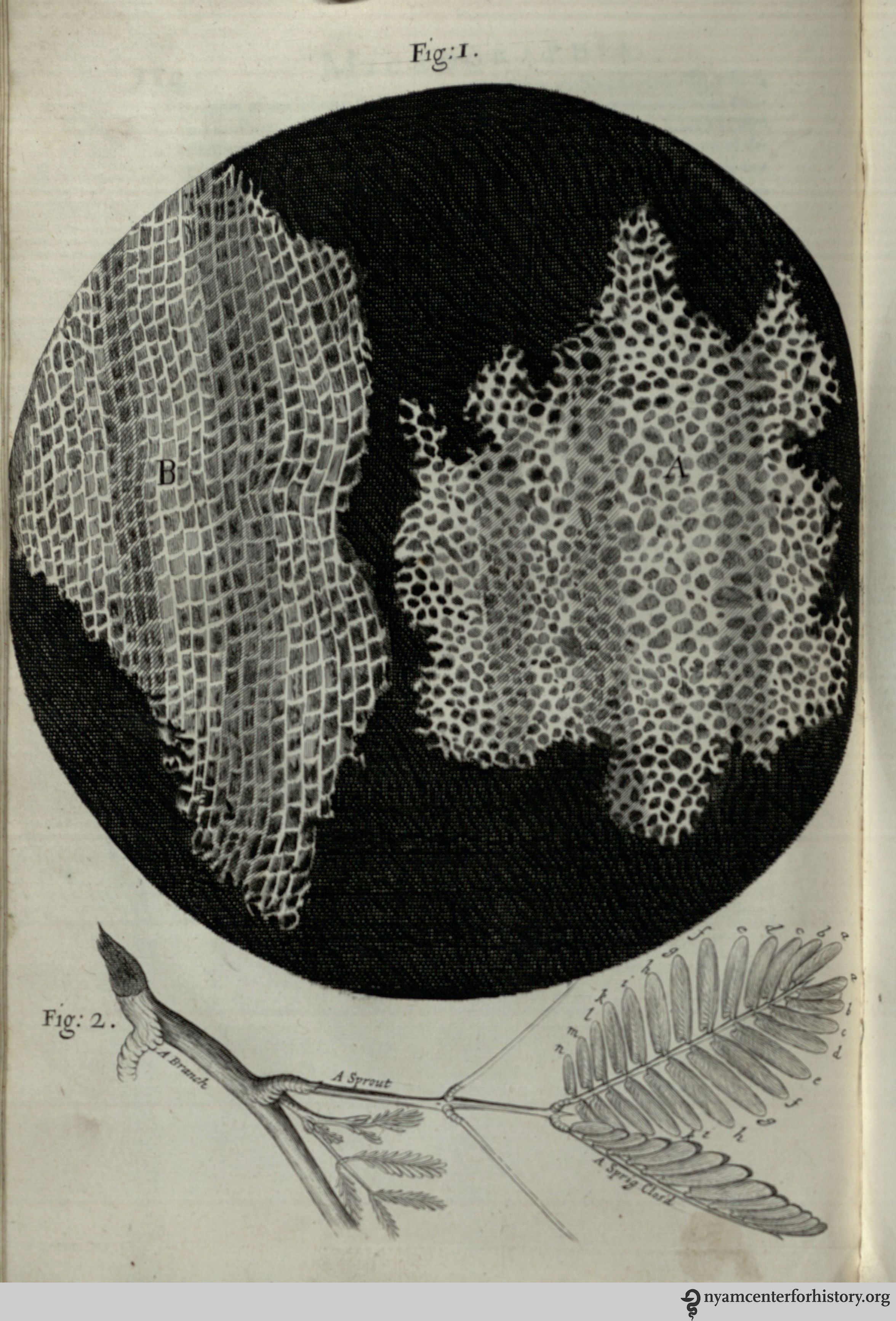
The 1500s saw the proliferation of full-figure anatomy. Jacob Reuff’s The Expert Midwife (and other texts like it) displayed women with their torsos peeled back, daintily displaying their inner organs. Plenty of scholarship has focused on the near-wanton and sexualized poses of these and of the “wax Venus” figures, some of whom appear to be in raptures despite being disemboweled. Male figures also appeared in full and sometimes opened—many of Vesalius’ plates in On the Fabric of the Human Body provide these interior views. The male gaze is often directed at the viewer or at the anatomy, while female figures tend to look askance (perhaps with modesty or shame at the revelation of their innards). By the 18th century, however, the whole had been replaced by sectioned and partial anatomies. No longer were the figures walking, dancing, or—in the case of women—curtseying. Instead, only the relevant bits appear in the pages of the atlas, which meant (in pregnant women) only the womb.

Easily the most famous works on pregnant anatomy in the 18th century, William Smellie’s A Sett of Anatomical Tables and William Hunter’s Gravid Uterus provide a portal for viewing key developments in the practice of 18th-century midwifery. In Tables, Smellie set out to demonstrate technique, but, as historian Lucy Inglis explained in a recent talk at the Dittrick Museum, Hunter was more interested in ensuring his fame by making scientific discoveries on the causes of maternal death in childbirth. In fact, the title Gravid Uterus suggests just how primary the womb had become; the women to whom they belonged are depicted headless, limbless, with bloodied cross-sections of stumped legs.

From Hunter’s Gravid Uterus. Image courtesy of the Dittrick Museum.

From Hunter’s Gravid Uterus. Image courtesy of the Dittrick Museum. Click to enlarge.

Neither anatomist provided entire forms—there was no expectation that they should. But Smellie’s models often included sheets of cloth to hide, but also to suggest, extremities. There is some debate about whether Hunter deliberately tried to achieve artistic or visceral impact,1 but unlike the birthing sheet, which hid the woman’s body from the midwife, the atlas rendered the female form more than denuded: It was naked of flesh, severed in places, the internal matter laid open for observation. At the same time, these female anatomies, like silent muses, were invaluable to the practice of midwifery, particularly as it pertained to difficult and dangerous cases. So what was gained—or lost—by these piecemeal renderings?

In February 2013, I worked with Lucy Inglis on a temporary gallery at the Dittrick that showcased both atlases, not for the sake of their authors, but to exhibit the work of the artist. Jan Van Rymsdyk—the artist behind the majority of figures in both atlases—had a “forensic eye.” He attended when Hunter obtained a new corpse and sketched as the dissections took place. Once, he watched a stillborn baby, more suited to the illustration, substituted within a dead woman’s womb. Lucy and I pondered the ramifications of this, the strange artificial quality of these posed cadavers. Enlightenment ideals required strict adherence to evidence, to the “real.” And yet, even here, anatomies were constructed by doctor and artist, a “dance” that renders plain the problems and process of birth at the moment of death.

In Dream Anatomy, historian Michael Sappol suggests that mastery over the dead body was akin to mastery over oneself, and even a kind of mastery over death.2 He notes, too, the attempts of early anatomy texts to shock the reader, and even the pleasure of shock; the sense that anatomists and anatomy artists wielded an erotic power in undressing the body.2 The detachment necessary to the task (and feared by a public concerned that dissection rendered doctors inhuman) cannot be universally applied to all, however. Van Rymsdyk suffered something akin to a breakdown from the hours spent hovering over dead women and their children with his palette of chalks—and Smellie turned his anatomical information into instruction for saving the lives of women and children. Even so, in the naissance macabre, artist and author reduce female anatomy to constituent parts: woman becomes womb, objectified as teaching tool…a mute muse, but a muse none the less.

References

1. McCulloch, N.A., D. Russell, S.W. McDonald. “William Hunter’s casts of the gravid uterus at the University of Glasgow.” Clinical Anatomy 14, no. 3 (2001): 210-217.

2. Sappol, M. (2006). Dream Anatomy. Washington, D.C.: Government Printing Office, 34.

Robert Hooke’s Micrographia (Item of the Month)

By Rebecca Pou, Archivist

The title page of Hooke's Micrographia.

The title page of Hooke’s Micrographia.

Robert Hooke was born on July 28 (O.S. July 18), 1635. To commemorate his birthday, we are featuring his book Micrographia as July’s item of the month.

Hooke published Micrographia in 1665 when he was 30 years old. At the time, Hooke was the curator of experiments for the Royal Society of London, which involved conducting several experiments a week and presenting them to the society. Hooke made many of the observations found in Micrographia through his activities for the society, and the Royal Society commissioned and printed the book.1

An extraordinary work, Micrographia details Hooke’s observations on objects as varied as the point of a needle, a louse, and the moon (he also utilized telescopes). The book includes 38 copperplate engravings of microscopic views based on Hooke’s drawings. Micrographia was not the first book of microscopic observations, but it was more successful and accessible than its predecessors. Who wouldn’t marvel at a close up shot of a flea?

Here is a selection of Micrographia’s plates (click to enlarge):

Fig. 1 shows a microscopic view of kettering-stone. In observation XV, Hooke notes, “We may here find a Stone by the help of a Microscope, to be made up of abundance of small Balls…and yet there being so many contacts, they make a firm hard mass…”

Fig. 1 shows a microscopic view of kettering-stone. In observation XV, Hooke notes, “We may here find a Stone by the help of a Microscope, to be made up of abundance of small Balls…and yet there being so many contacts, they make a firm hard mass…”

In his observation on cork, Hooke compared its structure to that of honeycomb and. He discovered plant cells, “which were indeed the first microscopical pores I ever saw, and perhaps that were ever seen…,” and coined the term “cell.”

In his observation on cork, Hooke compared its structure to that of honeycomb. He discovered plant cells, “which were indeed the first microscopical pores I ever saw, and perhaps that were ever seen…,” and coined the term “cell.”

For observation XXXIV, Hooke examined the eyes and head of grey drone-fly.

For observation XXXIV, Hooke examined the eyes and head of grey drone-fly.

Hooke seemed enamored with the white feather-winged moth, calling it a “pretty insect” and “a lovely object both to the naked Eye, and through a Microscope.”

Hooke seemed enamored with the white feather-winged moth, calling it a “pretty insect” and “a lovely object both to the naked Eye, and through a Microscope.”

The flea is one of several fold-out plates in the book. Again, Hooke has a scientist’s appreciation for the insect, commenting equally on its strength and beauty. He is particularly fascinated with the anatomy of its legs and joints, which “are so adapted, that he can…fold them short within another, and suddenly stretch, or spring them out to their whole length.”

The flea is one of several fold-out plates in the book. Again, Hooke has a scientist’s appreciation for the insect, commenting equally on its strength and beauty. He is particularly fascinated with the anatomy of its legs and joints, which “are so adapted, that he can…fold them short within another, and suddenly stretch, or spring them out to their whole length.”

In the last observations, Hooke turned his attention to celestial bodies. His study of the moon lead him to believe it might be covered in vegetation. He thought the hills seen in Fig. 2 “may be covered with so thin a vegetable Coat, as we may observe the Hills with us to be, such as the short Sheep pasture which covers the Hills of Salisbury Plains.”

In the last observations, Hooke turned his attention to celestial bodies. His study of the moon led him to surmise that the hills seen in Fig. 2 “may be covered with so thin a vegetable Coat, as we may observe the Hills with us to be, such as the short Sheep pasture which covers the Hills of Salisbury Plains.”

The National Library of Medicine’s Turning the Pages project has a selection of images from Micrographia available. It is well worth flipping through; you’ll find curator’s notes and you can even open the folded plates. If you are interested in looking at Micrographia in its entirety, contact us at history@nyam.org or 212-822-7313 to make an appointment.

Reference
1. Espinasee, Margaret. Robert Hooke. London: Heinemann, [1956].

Guest curator Riva Lehrer on Vesalius 500

Our “Art, Anatomy, and the Body: Vesalius 500” festival guest curator, artist and anatomist Riva Lehrer, describes some of her thinking about bodies, anatomy and art.

In 1543, when Andreas Vesalius published his De humani corporis fabrica (On the fabric of the human body) many contemporaries refused to accept his results. They contradicted canonical texts passed down over millennia: belief and expectation trumped direct experience and observation.

It’s easy to smile condescendingly at such pig-headedness. Yet we can scarcely look in the mirror without being caught in a fog of distortion. Every day we’re overloaded with information about how we should look and how our bodies should work. There are still plenty of ways in which our biases form medicine, and medicine, in turn, forms us.

"Circle Stories #4: Riva Lehrer" 1998  self portrait

“Circle Stories #4: Riva Lehrer” (1998).

I was born with visible disabilities. My body has always been seen as lacking, in need of correction, and medically unacceptable. My parents and doctors pushed me to have countless procedures to render it more “normal” as well as more systemically functional. These were two different streams of anxiety—how I worked and how I looked— yet they became inextricably woven together. My life in the hospital gave me a tremendously intimate view of medicine, as does the fact that I come from a family of doctors, nurses, and pharmacists. It gave me an acute awareness of how medical choices control and shape our bodies.

I first studied anatomy at the School of the Art Institute of Chicago and later at the University of Illinois at Chicago, as a visiting artist in the cadaver lab. I often think about what my first anatomy professor told me, many years ago. She remarked that when she was a child, people grew into their original faces. Whatever oddities they were born with formed what they looked like, year after year. Faces were hard-won and unique. But modern dentistry, nutrition, grooming—all the large and small interventions of medicine—made people look much more alike than they did sixty years ago.

In the 21st century, medicine is not just about the “correction” of significant impairments; personal perfectibility is as much the point of modern medicine as the curing of significant diseases. We view our bodies as lifetime fixer-upper projects.

Yet, it’s that very fluidity that opens profound questions about the identities our bodies express. Technologies such as radical cosmetic surgery, cyborgian interfaces, and gender reassignment procedures raise and complicate our expectations. Medicine offers new options if the inside of our bodies does not match the appearance of the outside. We live in a state of wild restlessness, trying to see and feel who we are. We see chimeras of possibility.

"At 54" Riva Lehrer 2012 self portrait

“At 54” by Riva Lehrer (2012).

My body was not normalized through all my surgeries; yet the original body I had would not have lived. It’s been changed so many times that I can’t even guess at what it would have been. My own mutability has given me a deep interest in the two-way relationship between one’s body and the course of a life.

I teach anatomy for artists at the School of the Art Institute and am a visiting artist in Medical Humanities at Northwestern University. My studio practice focuses on the intersection of the physical self and biography. I interview people in depth about the interweaving of their bodies and their stories. These interviews become narrative portraits, as I try to understand what can be known about a life in a single portrait image.

Join us as we explore the role of anatomy in identity formation through our celebration of the 500th anniversary of Vesalius’ birth. We’ve invited artists, performers, scholars, and historians to help us ask how our imaginations form our living flesh. Let’s all look in the mirror and ask, what are we really seeing, and what do we believe we see?

Some of the issues our speakers will explore include:

""Chase Joynt" by Riva Lehrer and Chase Joynt 2014

“”Chase Joynt” by Riva Lehrer and Chase Joynt (2014).

—How do we decide what is “lifesaving” and what is “elective” surgery when it comes to identity? Transgender performer Chase Joynt questions what it means to save a life, and how his dealings with the medical establishment led him to question such choices.

—How many of us were raised with the constant imprecation to stand up straight? Sander Gilman peers into the use of posture lessons in public schools to control the American body.

—Artist Steven Assael creates dramatic portraits of New Yorkers, from street performers to elderly eccentrics. His work shows us how identity travels from the inner self to the outer shell.  Assael is a long-time professor at New York’s School of Visual Arts, one of the last bastions of serious anatomical study in the U.S.

—Famed choreographer Heidi Latsky will discuss GIMP and how she creates dance for performers with a range of movements and morphologies. A performance and film excerpt bring us into the innovative strategies used by the GIMP collective.

—Many contemporary artists use anatomy in investigations of identity and formal exploration. Curator Ann Fox will present images from an international roster of artists. She will be joined by Taiwanese artist Sandie Yi, who will show work that deals with the intense difficulties of having a physically different body in China.

"Coloring Book" Riva Lehrer 2012

“Coloring Book” by Riva Lehrer (2012).

Graphic Medicine is a consortium of comics artists who explore medicine from the standpoint of doctor, nurse, patient and family member. The founders of Graphic Medicine, MK Czerwiec and Ian Williams, will discuss how the vulnerable body is rendered in comics form. Comics allow artists to move from the inside of the body to the outside in seamless transitions, to weave together objective perspectives and highly personal, subjective experiences.

Tracking the History of Cancer Drug Development

Lourdes Sosa, today’s guest blogger, is an associate professor in the department of management at the London School of Economics and Political Science.

Have new cancer drugs entered the market targeting ever-smaller portions of the total cancer patient population? If so, is this a symptom of a high-tech market phenomenon known to economists as submarket fragmentation?1 If we accurately answer these questions, we will better understand oncology drug discovery competition and thus will offer better strategic recommendations to enhance drug discovery efficiency.

My co-authors, Prof. Roberto Fernandez (MIT Work and Organization Studies), Prof. Andrew Lo (MIT Finance), and myself, Prof. Lourdes Sosa (LSE Department of Management), set about to answer these questions more than a year ago. As we began our research, our most important first step was to identify the anticancer drugs available in the US market since the birth of chemotherapy in the 1940s. A perfect data source became the Physicians’ Desk Reference (PDR®), an annual directory of approved drugs and full prescribing information that began publication in 1947.

Our next challenge came about immediately: where could we locate an accessible repository that held the entire collection to date? Although key local libraries offered us access to a large portion of the collection in print, we found in the New York Academy of Medicine Library full access to the entire collection. Furthermore, NYAM holds the collection in microfiche format, making it easy to browse from one year to another.

Starting a year ago, we began collecting data from the NYAM Library. We are now happy to report how our study is taking shape (we are also delighted to have an avenue to thank the support of Ms. Danielle Aloia and the team of expert librarians at NYAM).

The title page and an entry in from the 1949 Physician's Desk Reference.

The title page and an entry from the 1949 Physicians’ Desk Reference.

The figure below shows the oncology drugs available in the US market from 1947 until 2001 (data entry is still in progress). The process to identify these drugs started with the Product Category Index of the PDR®, where all cancer-related drugs can be found. We then read the full prescription information included in the product information section of the PDR® to extract the actual indications approved per drug. This latter step allowed us to make a precise decision on whether the drug was a treatment for cancer (as opposed to a treatment for a side effect or complication), and if so, to define for which cancer indications the drug was approved.

Courtesy of Roberto Fernandez, Andrew Lo, and Lourdes Sosa.

Courtesy of Roberto Fernandez, Andrew Lo, and Lourdes Sosa.

As can be seen in the figure, there is a big change in reporting in 1970. Starting that year the Product Category Index of the PDR® reported a category titled antineoplastics that made it straightforward to identify relevant drugs. In contrast, the categorization used in 1947–1969 has categories such as multiple myeloma and breast carcinoma listed separately. More importantly, during those earlier years a vast majority of drugs listed as cancer-related were in fact general-purpose drugs such as steroids, analgesics, and diuretics, which just happened to be novelties in the market.

As mentioned, we used the full prescription information to discern between the cancer-treating drugs that constitute the core of our study and those of either general application (e.g., steroids) or symptom-relief purpose (e.g., anemia treatments). The actual population of cancer-treating drugs for us to use is the black portion of the above figure shown with the legend “treating drugs.”

Our next step (after completing this exercise to year 2013) will be to calculate an index of coverage that proxies for the percentage of all cancer patients that each drug can treat. We will eagerly report on our progress as soon as we have preliminary results to share.

Reference

1. Sutton, J. 1998. Technology and Market Structure: Theory and Structure. MIT Press, Cambridge, MA.

Celebrate Andreas Vesalius’s 500th Birthday With Us on October 18

On October 18, our second-annual Festival for Medical History and the Arts, “Art, Anatomy, and the Body: Vesalius 500” will celebrate the 500th birthday of anatomist Andreas Vesalius.

Vesalius’ groundbreaking De humani corporis fabrica (The Fabric of the Human Body) of 1543 is a key Renaissance text, one that profoundly changed medical training, anatomical knowledge, and artistic representations of the body, an influence that has persisted over the centuries. Our Festival is one of a global series of celebrations of his legacy.

Our day-long event will explore the intertwined histories of art and anatomy, illustration and medicine, performance and the body, body snatching and dissection, identity and intersexuality, disability and representation, and contemporary visual arts and the body. Speakers, performers, and artists will be joined by anatomical cartoonists, 3D printing demonstrations, workshops, and more. Artist and anatomist Riva Lehrer will be our guest curator. Speakers and presenters will include Daniel Garrison, Steven Assael, Sander Gilman, Brandy Schillace, Lisa Rosner, Ann Fabian, Bill HayesMichael Sappol, Chase JoyntProof X, and  Kriota Willberg (look for a full list of speakers later this summer).

Follow our blog over the summer for guest posts from Festival participants and more on the wonderful Vesalius holdings in our collection.