Deafness as a Public Health Issue in the 1920s & 1930s (Part 1 of 2)

Today we have part one of a guest post written by Dr. Jaipreet Virdi-Dhesi, the 2016 Klemperer Fellow in the History of Medicine at the New York Academy of Medicine and a SSHRC Postdoctoral Fellow in the Department of History at Brock University in St. Catharines, Ontario. She is working on her first book, Hearing Happiness: Fakes, Fads, and Frauds in Deafness Cures, which examines the medical history of hearing loss and “quack cures” for deafness. Some of these cures are explored on her blog, From the Hands of Quacks. You can find her on twitter as @jaivirdi.

In 1935, physician Francis L. Rogers of Long Beach read a paper addressing the worrisome statistics of deafness. One study discovered nearly thirty-five thousand Americans were deaf. Another found that out of a million people tested for their hearing, 6% had significant hearing impairment. Yet another study reported three million people had some kind of hearing impairment. This “problem of deafness,” Rogers emphasized, “is primarily of public health and public welfare.” Not only were there too many people failing to adequately care for their hearing, but many cities, schools, and governments lacked the proper infrastructure to educate the public on the importance of hearing preservation. Indeed, as Rogers stressed: “Today the three great public health problems confronting the world are heart disease, cancer, and deafness.”[1]

Image 1

A window display in Detroit (Hearing News, June 1942)

The notion of deafness being statistically worrying as a public health issue actually dates to the late nineteenth century, especially to the work of otologist James Kerr Love of Glasgow. Love conducted several statistical studies of the ears of deaf schoolchildren, discovering that the majority of them were not completely deaf, but had some level of “residual” hearing. With proper medical treatment, the hearing could be intensified enough to warrant a “cure.” For other cases, children could be taught to make use of that residual hearing through invasive training using acoustic aids and other kinds of hearing technologies.

Love’s research concluded that many deafness cases could actually be relieved if the ears of children were examined early and frequently—that is, deafness could be prevented. His “prevention of deafness” concept was influential for the new generation of otologists in America, especially those who were members of the New York Academy of Medicine’s Section of Otology during the first three decades of the twentieth century.

To raise awareness on the necessity of proper medical examinations and frequent hearing tests, these otologists collaborated with social organizations such as the New York League for the Hard of Hearing, which was established in 1910. The League was a progressive group catering to the needs of hard of hearing or deafened persons who were raised in a hearing society rather than in a D/deaf community and communicated primarily with speech and lip-reading rather than sign language. Composed mostly of white, middle-class, and educated members who lost their hearing from illness, injury, or progressive deafness, the League strove to construct hearing impairment as a medical issue. They argued hearing impairment was not an issue of education or communication, but rather a handicap.

Image 2

An otologist examining a young patient’s ear (Hygeia, June 1923)

The collaboration between New York otologists and the League eventually created a national network of experts, social services, teachers, physicians, and volunteers who banded together to address the so-called “problem of deafness.” That is, the problem of how to best integrate the hard of hearing, the deafened, and to some extent, even the deaf-mutes, into society. One key achievement of the League was the establishment of hearing clinics to properly assess hearing impairment, especially in children, to ensure medical care could be provided before it was too late. This project was primarily spearheaded by Harold M. Hays (1880-1940), who was recruited as president of the League in 1913, becoming the first active otologist collaborating with the League. After the First World War, Hays set up a clinic for treating hearing loss in children at the Manhattan Eye, Ear, and Throat Hospital.

Image 3

Group hearing tests of schoolchildren, using an audiometer. Headphones are used first on the right ear, then the left. (Hygeia, February 1928)

Hays claimed that hearing impairment might be a handicap, but “the sad part of it is that 90 percent of all hearing troubles could be corrected if they were treated at the proper time.” With regular hearing tests, this was possible. Yet, as Hays argued, regular hearing tests were not considered on par with other hygienic measures under public health services:

We are saving the child’s eyes! We are saving the child’s teeth! Is it not worth while to save the child’s ears?”[2]

During the 1920s, Hays’ activism for regular hearing tests was so instrumental that in 1922, the League’s newsletter, The Chronicle, told its readers “we believe that the League would justify its existence if it did no other work than to prevent as much deafness as possible.”  To achieve this mandate, the League launched a large public campaign to raise awareness on the importance of medical care. Indeed, in one report for the League, Hays remarked that with the increased publicity, there were 10,000 calls to the League in 1918 alone inquiring about aural examinations. A steady increase in patients would follow: 17 clinic patients in 1924, 326 in 1926, and then 1,531 in 1934.

Another publicity campaign spearheaded by the League was the establishment of “Better Hearing Week” in 1926, a week-long awareness program (later renamed “National Hearing Week”). Held in October, the campaign included symposium discussions on the “Problems of the Hard of Hearing,” including topics on the relationship between the physician and his deafened patient, how the deafened could build their lives, and even on newest technological developments in hearing aids. October issues of The Bulletin (the renamed League newsletter) and the Hearing News, the newsletter of the American Society for the Hard of Hearing (ASHH) included reprints of letters from prominent leaders supporting the mandates of “Better Hearing Week,” including letters from President Roosevelt and New York Mayor LaGuardia.

Image 4

Advertisement for Western Electric Hearing Aid, the “Audiophone.” These before-and-after shots were powerful for demonstrating the effects of “normal” hearing, sending the message that outward signs of deafness, such as the “confused face,” could easily disappear once being fitted properly with a hearing aid. (Hearing News, December 1936)

The 1920s publicity campaigns were primarily focused on fostering ties between otologists and the League, in cooperation with hospitals and schools. In 1927, the League purchased audiometers and offered invitations to conduct hearing tests in schools across New York, so children with hearing impairment could be assessed accordingly. Two years later, the League worked with Bell Laboratories to further substantiate the conviction that deafness was a serious problem amongst schoolchildren and that something needed to be done.

At the same time otologists across America established joint ventures between organizations like the America Medical Association and the American Otological Society. They formed committees to write reports to the White House on the national importance of addressing the “prevention of deafness.” Wendell C. Phillips (1857-1934), another president of the League and the founder of ASHH, particularly emphasized the need to address the “psychologic conditions and mental reactions” of the deafened patient, for the tragedy of acquired deafness meant it is a “disability without outward signs, for the deafened person uses no crutch, no black goggles, no tapping staff.”[3] It was an invisible handicap that needed to be made visible if it was to be prevented, if not cured.

References

[1] The Federation News, August 1935.

[2] Harold M. Hays, “Do Your Ears Hear?” Hygeia (April 1925).

[3] Wendell C. Phillips, “Reminiscences of an Otologist,” Hygeia (October 1930).
Apply for our 2017 Research Fellowships

Does a one-month residence in The Drs. Barry and Bobbi Coller Rare Book Reading Room, immersed in resources on the history of medicine and public health, sound like a dream come true?

Rare book room

The Drs. Barry and Bobbi Coller Rare Book Reading Room

The Academy Library offers two annual research fellowships, the Paul Klemperer Fellowship in the History of Medicine and the Audrey and William H. Helfand Fellowship in the History of Medicine and Public Health, to support the advancement of scholarly research in the history of medicine and public health. Fellowship recipients spend a month in residence conducting research using the library’s collections.

Applications for our fellowships are being accepted now through late August for fellowships that may be used at any time during 2017.

Preference in the application process is given to early career scholars, although the fellowships are open to anyone who wishes to apply, regardless of academic status, discipline, or citizenship. While both fellowships are for researchers engaged in history of medicine projects, the Helfand Fellowship emphasizes the role of visual materials in understanding that history.

Applications are due by the end of the day on Friday, August 26, 2016. Letters of recommendation are due by the end of the day on Monday, August 29, 2016. Applicants will be notified of whether or not they have received a fellowship by Monday, October 3, 2016.

Prospective applicants are encouraged to contact Arlene Shaner, Historical Collections Librarian, at 212-822-7313 or history@nyam.org with questions or for assistance identifying useful materials in the library collections.

17th Century Recipes, Fit for a Gala

By Arlene Shaner, Historical Collections Librarian

The New York Academy of Medicine hosted its annual fund-raising gala at the Mandarin Oriental on June 14th.  Gala attendees had the opportunity to sample two treats based on recipes from one of our favorite manuscript receipt books.

The Academy Library has 37 manuscript receipt books, most of which contain a mix of culinary, medicinal and household recipes. Some of them have been featured already on our blog (see earlier posts on Mother Eve’s Pudding, and English Gingerbread). The Recipes Project also featured an interview with Anne Garner, our Curator of Rare Books and Manuscripts, about the print and manuscript historical recipe books in our collection.

One of our favorite manuscripts is A Collection of Choise Receipts from the late seventeenth century. Inspired by a recipe for Black Cherry Water in the manuscript, Pietro Collina and Matt Jozwiak created a signature cocktail, the “Choise Cherry Crush,” for gala guests. You can try your hand at mixing one up if you are so inclined.

Gala Cocktail Flyer-page-001

The adapted recipe for the Choise Cherry Crush, adapted from A Collection of Choise Receipts (1680)

The drink was inspired by this 1680 recipe:

choise-recipe-cherry-water copy

“Black Cherrie Water,” A Collection of Choise Receipts, 1680.

On their way out at the end of the evening, guests received bags with a pair of almond cookies also adapted from a recipe in Choise Receipts.

Postcard with cookies

The finished  give-away almond cookies, pictured with their recipe, adapted from A Collection of Choise Receipts (1680)

There are several recipes for cookies or little cakes made with almonds in the manuscript.  My favorite, “The Lady Lowthers Receipt for to make Bean Bread” a cookie that very much resembles a macaron in texture, takes its name from the slivered almonds that look like little beans that are mixed into the dough.

choise-bean-bread-0001 copy

“Lady Lowthers Receipt, for to make Bean Bread,” from A Collection of Choise Receipts, 1680.

The recipe for Almond Bisketts that we reproduced for the gala, however, seems to be missing a crucial ingredient: almonds!

Choise original almond biskett

The front of the recipe postcard produced for our give-away cookies for the gala.

Only when examining the full page of the manuscript, on which a very similar recipe for Almond Cakes appears directly above the one reproduced on the postcard, does it become clear that the “half a pound of fflower” referred to in this recipe would be made from ground almonds.  The adapted recipe printed on the back of our card makes that clear.

Choise adapted recipe

The adapted recipe, on the back of the postcard.

If you make a batch of these tasty cookies, let us know how they turn out!  Better yet, send us a picture and we’ll post it on Instagram.

Digitizing Medical Journals of State Societies

By Robin Naughton, Ph.D., Head of Digital

State medical journals digitized for the MHL collective project.

State medical journals digitized for the MHL consortium.

The New York Academy of Medicine Library is digitizing state society medical journals as part of a mass digitization project with the Medical Heritage Library (MHL), a digital curation consortium. The Academy Library is one of five collaborators on the project, along with the College of Physicians of Philadelphia; the Countway Library of Medicine at Harvard University; the Health Sciences and Human Services Library, University of Maryland; the Founding Campus; and the University of California at San Francisco.

Together, the MHL team is actively working to digitize 48 state society journals, more than 3,800 volumes that span much of the 20th century. Digitizing the state medical journals will provide open access to quality historical resources in medicine for researchers and the general public, letting them explore connections between medicine and society.

State medical journals digitized for the MHL collective project.

State medical journals digitized for the MHL collective project.

Evenly splitting the volumes among the MHL team makes the process of mass digitization more manageable and very collaborative. The Academy Library has already digitized almost 50% of the state medical journals assigned to it since Fall 2015. The journals are scanned by the Internet Archive (IA) and are publicly available as part of the Library’s and MHL’s collections on the IA site. Our digitized assets are open for anyone to access and use. Thus far, we have digitized journals representing 24 states and almost 238, 000 images.

The volumes are digitized in their entirety, showing the journals’ articles and  advertising. For example, in Alaska Medicine (vol. 29, 1987), as you read the article “Alaska State Hepatitis B Program – Past, Present and Future” by Elizabeth A. Tower, you can’t help but notice the advertisement for medical transcription. It is hard to resist the “Hello …. Museum of Primitive Civilizations and Hieroglyphs?”

Scan from Alaska Medicine, vol. 29, 1987.

Scan from Alaska Medicine, vol. 29, 1987.

State medical journals are valuable resources that should lead to many new and novel projects for researchers in the history of medicine. Look for more on the project as it progresses.

Explore our collection.

Get Crafty at the Museum Mile Festival on June 14

By Emily Miranker, Project Coordinator

When my office is perfumed by the smell of crayons and stocked with boxes of jumbo-sized sidewalk chalk, I know its Museum Mile Festival time. This year’s Museum Mile Festival takes place on Tuesday, June 14 from 6:00-9:00 pm, rain or shine.

Museum Mile (New York City’s Fifth Avenue from 82nd to 105th Street, which is technically three blocks longer than a mile) is one of the densest cultural stretches in the world.1 For the last 38 years, Fifth Avenue closes to traffic for a few hours on an early June evening. The eight major museums and their neighbors–that’s us!–throw open their doors and spill out onto the street in a block party.

Museum Mile at the New York Academy of Medicine. Courtesy of the Academy's Communications Office.

Museum Mile at the New York Academy of Medicine. Courtesy of the Academy’s Communications Office.

The first festival was held in 1979, the brainchild of the Museum Mile Association, to increase cultural audiences and garner support for the arts in time of great fiscal crisis in the city. The festival has since brought many New Yorkers and tourists to upper Fifth Avenue for the first time, and total attendance over the years has surpassed one million visitors.

Besides free admission to the museums along the mile, street performers, chalk drawing, live bands, balloons, and family-friendly activities abound. Dedicated to improving the health and well-being of people living in cities, the Academy has partners from the East Harlem Asthma Center of Excellence and Shape Up NYC joining us for the evening.

Getting physical with our community partners at Museum Mile. Courtesy of the Academy's Communications Office.

Getting physical with our community partners at Museum Mile. Courtesy of the Academy’s Communications Office.

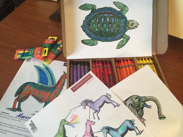
The Library has planned some special crafts for the festival. We have the perennial favorite: coloring pages based on images from our collections. Feel free to download your own pages any time from #ColorOurCollections online.

Coloring sheets fro the New York Academy of Medicine Library. Photo: Emily Miranker.

Coloring sheets from the New York Academy of Medicine Library. Photo: Emily Miranker.

Among the treasures of our collection are the anatomical flap books. These are detailed anatomical illustrations superimposed so that lifting the sheets reveals the anatomy and systems of the body as they would appear during dissection. We created a simple DIY version of a flapbook inspired by these remarkable figures from the 1559 edition of Geminus’ Compendiosa totius anatomiae delineatio, aere exarata. The sheets are quite delicate, so it’s rare to see intact versions like this 400 years after they were made. Make your own flapbook with us during the festival.

Male flap anatomy from The Academy's copy of the 1559 English edition of Geminus’ Compendiosa.

Male flap anatomy from The Academy’s copy of the 1559 English edition of Geminus’ Compendiosa.

Female flap anatomy from The Academy's copy of the 1559 English edition of Geminus’ Compendiosa.

Female flap anatomy from The Academy’s copy of the 1559 English edition of Geminus’ Compendiosa.

Make this flap anatomy craft with us at Museum Mile! Photo: Emily Miranker.

Make this flap anatomy craft with us at Museum Mile! Photo: Emily Miranker.

And there’s nothing like using your own body to create art—finger print art!2

Make fingerprint art with us at Museum Mile! Photo: Emily Miranker.

Make fingerprint art with us at Museum Mile! Photo and artwork: Emily Miranker.

We look forward to seeing you at Museum Mile!

References

1. “Museums on the Mile.” Internet Archive Wayback Machine (June 2011). Accessed June 3, 2016. https://web.archive.org/web/20120101013336/http://www.museummilefestival.org/museums/

2. “Fingerprint Fun.” Bookmaking with Kids (June 2010). Accessed June 6, 2016. http://www.bookmakingwithkids.com/?p=1826

The Foresight of Trans-vision: An Innovative Anatomy of the Eye

By Anne Garner, Curator of Rare Books and Manuscripts

Early European anatomical lift-the-flap books made use of technologies available during the 16th century: woodcut and engraving, combined with manual cutting and pasting.1 Flap anatomies like Geminus’ Compendiosa (1559) allowed readers to peel away the layers of the body to reveal different organs, but these flaps were made of paper and opaque, and didn’t allow the reader to view the strata of the body simultaneously.

Female flap anatomy from The Academy's copy of the 1559 English edition of Geminus’ Compendiosa.

Female flap anatomy from The Academy’s copy of the 1559 English edition of Geminus’ Compendiosa.

Fast forward four centuries, when an innovation in printing technology let readers take a deep dive through the layers of the body all at once.

Anatomical illustrators used transparencies to show the layers of the body as early as the 1920s. J.E. Cheesman published Baillière’s Synthetic Anatomy, a series of 14 booklets, in London from 1926 to 1936. The series used a set of glassine sheets to show what lay beneath the surface of the skin.

Forearm in Cheesman, Baillière's Synthetic anatomy, 1926-1936.

Forearm in Cheesman, Baillière’s Synthetic Anatomy, 1926-1936. Click to enlarge.

Hand in Cheesman, Baillière's Synthetic anatomy, 1926-1936.

Hand in Cheesman, Baillière’s Synthetic Anatomy, 1926-1936. Click to enlarge.

In 1942, Richard Lasker patented a new printing process for Milprint Inc., a Milwaukee-based company. He called this new method trans-vision. Trans-vision allowed for the printing of images on the inner surfaces of folded sheets of transparent acetate. These sheets could then be piled on top of one another so that they overlapped, enabling a multi-layered view with the top sheets depicting the most superficial layers of an object and the bottom sheets the deepest level.2

The patent application for Lasker’s trans-vision process used a cutaway illustration of a mattress, with different layers of acetate offering views of the mattress’ filling.3 Trans-vision’s medical applications proved significantly more useful: it made possible the representation of complex anatomical relationships to health professionals and public audiences alike.

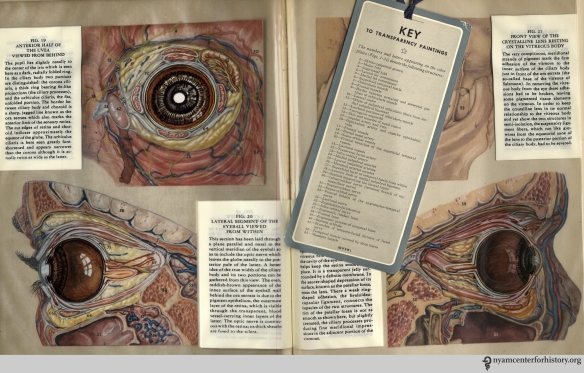
In 1943, Peter C. Kronfeld, an ophthalmology professor at the University of Illinois, published The Human Eye in Anatomical Transparencies. The book contains 34 color anatomical paintings printed using trans-vision.4 Each page offers a frontal and temporal view of the eye and its area, with transparent layers that can be peeled away by turning the pages. The paintings are printed on the inner side of the acetate to minimize damage from handling. The parts of the eye are numbered. Readers can use a bookmark key laid-in to identify the different parts by name.5

Figure I in Kronfeld, The Human Eye in Anatomical Transparencies, 1943. Click to enlarge.

Figure 1 in Kronfeld, The Human Eye in Anatomical Transparencies, 1943. Click to enlarge.

Kronfeld describes the project in The Human Eye’s preface:

It was at once obvious that the eye could advantageously be represented by this means, for it is a three-dimensional object in which great structural intricacy is combined with relatively small size. Ordinary drawings of its separate parts tend to isolate them too much from each other in the mind of the observer….The text has been so organized as to present not only a systematic account of ocular anatomy—taking up the various structures in a functionally logical order—but also a topographic treatment of the anatomy…which necessarily reveal the structures layer by layer in an order determined somewhat by the layers of dissection techniques.6

The paintings were made at twice the actual size by Gladys McHugh, an illustrator at the University of Chicago. The pioneering medical illustrator Max Brödel had been McHugh’s teacher and mentor at Johns Hopkins University, where she studied. It was Brödel who influenced McHugh to make her own dissections.

Figure II in Kronfeld, The Human Eye in Anatomical Transparencies, 1943. Click to enlarge.

Figure 2 in Kronfeld, The Human Eye in Anatomical Transparencies, 1943. Click to enlarge.

McHugh augmented her dissections of human eyes with specimens from pigs and monkeys. She describes her extensive dissection work in her introduction:

Over a course of time I obtained from baby autopsies ten good cases, making a total of twenty eyes and orbits. These I dissected layer by layer, making color notes and drawings from the fresh specimens. To develop a technique for separating the layers of the eyeball as intact semispheres, pigs’ eyes were employed. Also, to supplement my observation of the muscles and other structures not fully developed in the infant, monkey orbits were dissected.

As Professor Shelley Wall has argued, turning the pages in The Human Eye mimics the dissection process. As the layers on the recto side of the book are more deeply revealed with each page turn, the layers build on the opposite verso, allowing for the eye’s reconstruction.7 With The Human Eye, the union of format, technology, and material is harmoniously in sync.

Figues 3-6 in Kronfeld, The Human Eye in Anatomical Transparencies, 1943. Click to enlarge.

Figues 3-6 in Kronfeld, The Human Eye in Anatomical Transparencies, 1943. Click to enlarge.

Figure 14 in Kronfeld, The Human Eye in Anatomical Transparencies, 1943. Click to enlarge.

Figure 14 in Kronfeld, The Human Eye in Anatomical Transparencies, 1943. Click to enlarge.

Figures 19-22 in Kronfeld, The Human Eye in Anatomical Transparencies, 1943. Click to enlarge.

Figures 19-22 with bookmark key in Kronfeld, The Human Eye in Anatomical Transparencies, 1943. Click to enlarge.

By 1958, the book had enjoyed five editions.8 Students and educators embraced the text and its ingenious illustrations. In 1946, the trans-vision process was applied again to McHugh’s paintings in for The Human Ear in Anatomical Transparencies. Initially conceived as a wartime project useful to the aviation industry, the book’s value, as with The Human Eye, was in its power to demonstrate to both lay and specialized audiences the inner workings of organs not easily seen.

The finest examples of trans-vision printing occurred when the coffers of the pharmaceutical companies who published them were at their fullest. After The Human Eye and The Human Ear, medical illustrator Ernest Beck used trans-vision technology to produce more than 30 anatomical transparency projects published by Milprint for encyclopedias, pharmaceutical companies and other commercial concerns.9 A decade after Kronfeld, Ciba Pharmaceuticals published a 13-volume collection of anatomical illustrations using anatomical transparencies between 1953 and 1989. The illustrator of these, was a native New Yorker, fellow of the Academy, and former member of the Art Students League of New York. His name was Frank Netter.10

References

1. For more on this, see Andrea Carlino’s excellent Paper bodies: A catalogue of anatomical fugitive sheets, 1538-1687.

2. Wall, Shelley. “Mid-twentieth-century anatomical transparencies and the depiction of three-dimensional form.” Clinical Anatomy 23 (2010) 915-921. Accessed December 2015.

3. Wall, 917.

4. David Templeman, review of The Human Eye in Anatomical Transparencies by Peter C. Kronfeld. Optometry & Vision Science, 35 7 (1958). 388.

5. Wall, 919.

6. Kronfeld, Peter. The Human Eye In Anatomical Transparencies. Rochester, New York: Bausch & Lomb Press, 1943. iii.

7. Wall, 919.

8. David Templeman, review of The Human Eye in Anatomical Transparencies by Peter C. Kronfeld. Optometry & Vision Science, 35 7 (1958). 388-389.

9. Brierley, Meghan. Dialogue and Dissemination: The Social Practices of Medical Illustrators in the Pharmaceutical Context. Dissertation, University of Alberta. 2013. Accessed online May 31, 1916.

10. Frank Netter was the most prolific medical illustrator of the twentieth-century. During his more than sixty-years as a medical illustrator he produced more than 4,000 illustrations of the entire anatomic and pathologic character of the human body, system by system. His comprehensive and detailed illustrations, published in The Ciba Collection of Medical Illustrations, Clinical Symposia, and The Atlas of Human Anatomy educated generations of medical professionals and students all over the world.