Winsome Fetal Skeletons Bearing Scythes: Monro’s Traité d’ostéologie of 1759: Guest post by Morbid Anatomy

A note from the Center for the History of Medicine & Public Health: This is the last post in Morbid Anatomy‘s guest series leading up to our Festival of Medical History and the Arts. If you’ve enjoyed these posts as much as we have, don’t despair! Tomorrow’s event holds a full day of lectures and activities from Morbid Anatomy, Lawrence Weschler, and the Center. We hope you can make it! See the full schedule here.

Frontispiece
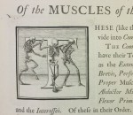
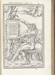
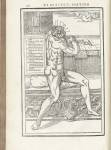
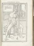
The NYAM rare book collection holds a gorgeous copy of the first French edition of Alexander Monro’s (1697–1767) celebrated Traité d’ostéologie (or “Anatomy of Bones”). Monro was trained in London, Paris, and Leiden before going on to become the first professor of anatomy at the newly established University of Edinburgh. It was under his leadership, and that of his successors, that the school went on to become a renowned center of medical learning.

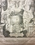
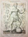
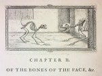
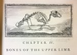
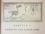
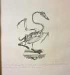
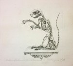
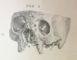
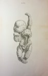
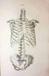
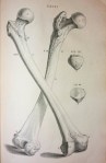
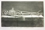
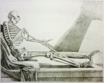
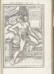
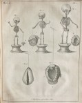
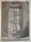
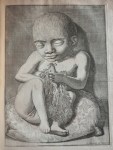
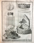
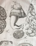
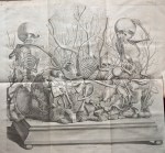
Monro originally published this book without images, thinking them unnecessary after William Cheselden’s lavishly heavily-illustrated Osteographia, or the anatomy of the bones of 1733 (more on that book at this recent post). The very fine copperplates you see here were added to the French edition by its translator, the anatomist Jean-Joseph Sue (1710–1792).

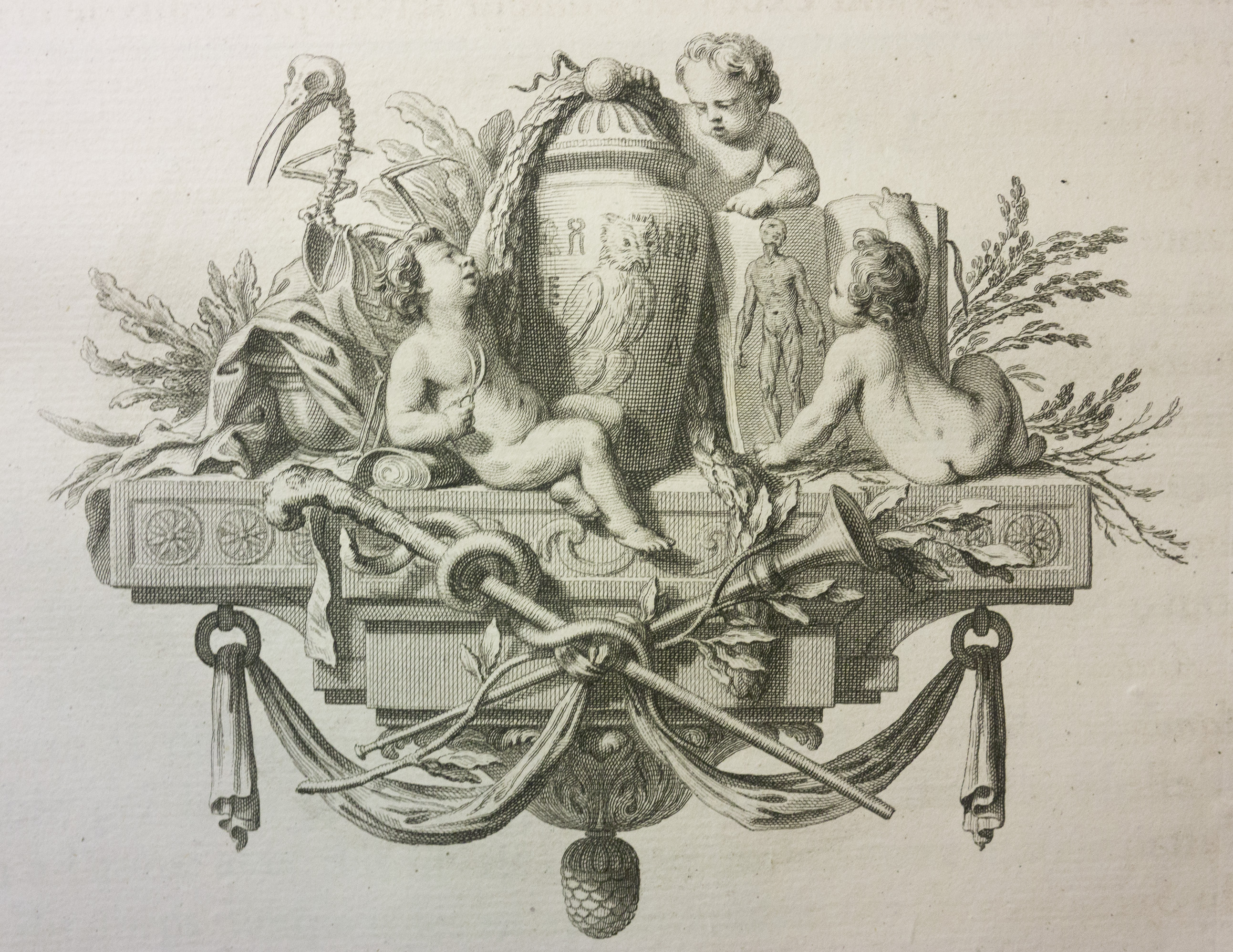
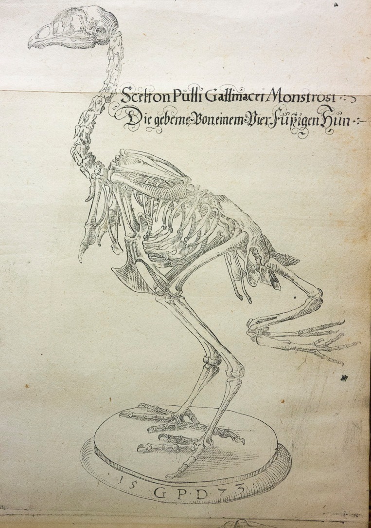
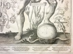
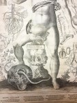
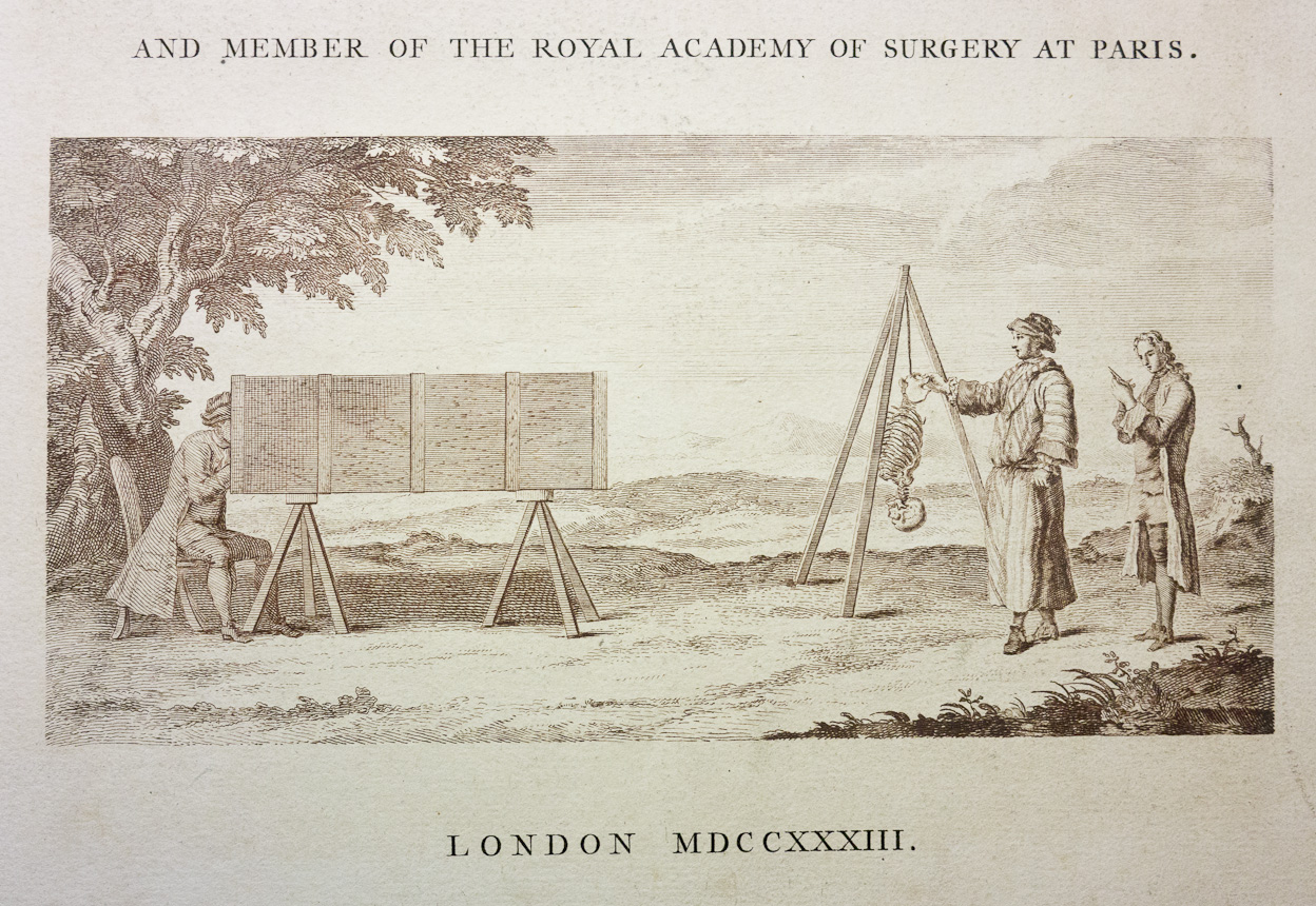
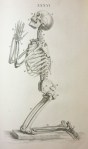
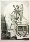
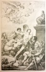
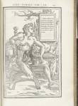
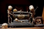
My favorite image in the book is a kind of memento mori–themed tableau morte of winsome, scythe-bearing fetal skeletons enigmatically arranged in a funereal landscape (images 1–3). I also love the frontispiece in which a group of plump putti proffer anatomical atlases and dissecting tools under the oversight of a skeletal bird (above).

This post was written by Joanna Ebenstein of the Morbid Anatomy blog, library and event series; click here to find out more. All images are my own, photographed at the New York Academy of Medicine.

A Renaissance Man at Work: Volcher Coiter’s “Externarum et internarum principalium humani corporis” of 1573: Guest post by Morbid Anatomy

Coiter2


Coiter10

One under-seen and fascinating book to be found in the NYAM rare book collection is Externarum et internarum principalium humani corporis partium tabulae published by Dutch Renaissance man Volcher Coiter (1534–1576) in 1573. Not only was Coiter renowned as an anatomist, surgeon, and physician accomplished in the fields of physiology, ornithology, and embryology; not only did he establish the study of comparative osteology and describe cerebrospinal meningitis before any of his peers; he was also an artist, and signed many of the finely drawn copper engravings in his books, including those you see here.


All images are my own, photographed at the New York Academy of Medicine, save the painted portrait of Coiter, which was sourced here. The caption, attributed to Dorothy M. Schullian, reads: “Coiter’s portrait (1575) in oils, attributed to Nicolas Neufchatel and representing him demonstrating the muscles of the arm, with the écorché he had constructed on his left and a shelf of medical classics behind him, is preserved in the Germanisches Nationalmuseum, at Nuremberg; there are later portraits at Weimar and Amsterdam.” (source for caption here)

Sources: Lessico Volcher CoiterWikipedia

This post was written by Joanna Ebenstein of the Morbid Anatomy blog, library and event series; click here to find out more.

Remmelin’s Dissectible Cosmic Anatomical Extravaganza: Guest post by Morbid Anatomy

Johan Remmelin (1583–1632) was town physician of Ulm and Plague physician of Augsberg. He was also the man behind both the concept and the original drawings (engraved by Lucas Kilian) for the ingenious moving-parts anatomical extravaganza Catoptrum microcosmicum, published in 1619, with numerous editions in many languages thereafter.  NYAM has both the 1619 and a 1639 edition in its rare book collection

title

This astounding book—in which flaps of paper can be drawn back to virtually dissect the human body—features a heady blend of the anatomical, the theological, and the metaphysical, beautifully expressing the worldview of Natural Philosophy, that precursor to science, which oversaw investigations into the human body in the early modern era. In this worldview, God and man, metaphor and the encountered world, were indivisible; the human being was the microcosm of all creation, so to understand the secrets of the human body would be to know the mind of God. Accordingly, as explained by Martin Kemp and Marina Wallace in their book Spectacular Bodies:

The purpose of anatomical images during the period from the Renaissance to the nineteenth century had as much to do with what we would call aesthetic and theological understanding as with the narrower intentions of medical illustration as now understood. . . .They were not simply instructional diagrams for the doctor technician, but statements about the nature of human beings as made by God in the context of the created world as a whole [as well as] the nature of life and death. . . .

It should not be surprising, then, that the dissectible humans herein are inextricably entwined with images of Jesus Christ (image 9,17); memento mori mottos (16) and imagery (images 12, 17); allusions to God and the angels (image 1); and even the head of the devil, serving as a kind of fig leaf covering the female sex organs in one instance (image 2). There are also numerous biblical references, including a serpent slithering through a human skull holding a branch from the tree of knowledge in its mouth (image 13), lest we forget that original sin introduced death and disease into our world in the first place; without it, we would still be luxuriating in Eden with no need for medicine, or, by extension, books such as this one. The book also contains the occasional inadvertent (?) eroticism, as the peeling back of obscuring layers brings you, in a sort of pre-modern striptease, to the unveiled sexual organs below (image 14 and 15).

If you page through all of the images below, you will get a sense of the carnivalesque exuberance and dynamism of this book; you can also virtually dissect them yourself by clicking here, or here, compliments of The Hardin Library of The University of Iowa, which was also a source for much of the factual content of this piece.

This post was written by Joanna Ebenstein of the Morbid Anatomy blog, library and event series; click here to find out more. All images are my own, photographed at the New York Academy of Medicine.

William Cowper’s Myotomia reformata: or an anatomical treatise on the muscles of the human body, Guest Post by Morbid Anatomy

Cowper01

William Cowper (c. 1666–1709) was a British surgeon and anatomist best known today for describing “Cowper’s Gland,” part of the genito-urinary system. He has also been described by at least one scholar as “the first of the surgeon-scientists of Great Britain… the first to bring the power of the experimental method to bear on practical surgical problems… [anticipating] the celebrated Hunterian school of surgery by more than half a century.” (In the last half of the 18th century, famous London surgeon John Hunter made his reputation in part by advocating for the scientific method in medicine.)

Cowper02

Cowper’s book Myotomia reformata: or an anatomical treatise on the muscles of the human body, first published in 1694 (NYAM’s copy is from 1724), is filled with grimly literal anatomical and dissection-themed initial capital letters, and charming, if somewhat rough, illustrations.

This post was written by Joanna Ebenstein of the Morbid Anatomy blog, library and event series; click here to find out more.

William Cheselden’s Memento Mori and Skeletons at Prayer: Guest Post by Morbid Anatomy

Cheselden 1

Cheselden 1

William Cheselden (1688–1752) is remembered today as one of the greatest English surgeons; he was surgeon to Queen Caroline, wife of King George II, to whom he dedicated the wonderful, epically scaled book Osteographia, or the anatomy of the bones  (1733), which is described by the NYAM Library as “one of the finest of English works containing anatomic illustrations.” The copperplate images were done by Cheselden and his engravers, Gerard van der Gucht and Mr. Shinevoet, with the use of the camera obscura—a pre-photographic drawing aid; this is delightfully alluded to in this wonderful image featured on the title page (images 1 & 2) .

Cheselden 2

Cheselden 2

The book is best known for its large scale and exquisite renderings of skeletons brandishing bones (image 3); leaning on skulls (image 4); with “the same proportions with the venus de Medicis” (image 5); “in the same proportions and attitude with the Belvidere Apollo” (image 6); or, most famously of all, “the side view of the skeleton of a very robust man” at prayer (image 7).

Less seen—yet equally delightful—are the wonderfully imaginative anatomically-themed initial capital letters (8-12); the playful chapter openers (13-19); the memento-mori themed end piece (30); and delicately exquisite animal skeletons (20-22) which fill the book. Despite—or perhaps because of—these luxurious touches, this large-scale atlas was a financial failure.

This post was written by Joanna Ebenstein of the Morbid Anatomy blog, library and event series; click here to find out more.

Jacques Gamelin’s Marauding Skeletons and Écorché Crucifixions: Guest Post by Morbid Anatomy

Gamelin 1

Gamelin 1

I was so excited to finally have the opportunity to pore over a book in the NYAM collection which I had long admired from afar: Jacques Gamelin‘s beautiful and lavish Nouveau recueil d’osteologie et de myologie (“New collection of osteology and mycology”) of 1779. This book, as explained to me by Arlene Shaner—acting curator and reference librarian for historical collections, at NYAM—was intended as a large scale, deluxe manual for artists interested in understanding human anatomy in order to create more convincing depictions of human figures.

The meat of the book, as it were, is a collection of extremely virtuosic anatomical renderings (12-15) showing skinned—or écorché—human figures in a variety of poses. But what is much more interesting—at least to me—is the assortment of animated skeletons which fill the opening pages; these fanciful figures are engaging in such activities as waking up in a cemetery to the trumpet of the resurrection (text reading: surgite mortui venite ad judicium, or “Rise up, come to the judgment of the dead”; image 6); brandishing anatomical drawings in what looks to be a dissection room littered with bones (3); and raping and pillaging the parties of fashionably bewigged lords and ladies (9, 10). I am also very drawn images playing on biblical themes, such as a calm Saint Bartholomew being flayed alive (an old staple of anatomical illustration; image 11), and a skinned and anatomized Christ on the cross (12) which evokes this more literal rendition, cast from a convicted murderer just a few decades after this book was published.

Interestingly, Gamelin is best remembered today not for this book, but as a painter and engraver of battle scenes, genre scenes, and portraits. He dedicated this book to his mentor, a certain Baron de Puymaurin (image 1 and 2), who had recognized Gamelin’s artistic abilities and funded his training when his father refused to do so. It is thought that Gamelin funded the book himself after inheriting a great deal of money upon his father’s death, which perhaps accounts for its delightful eccentricity.

This post was written by Joanna Ebenstein of the Morbid Anatomy blog, library and event series; click here to find out more.

17th Century Anatomical Striptease: Guest Post by Morbid Anatomy

Tab VI

Another series of most wonderful and enigmatic anatomical illustrations in the New York Academy of Medicine historical library collections are to be found in De humani corporis fabrica libri decem, tabulis XCIIX aeri incisis … exornati … Opus posthumum and Tabulae anatomicae (Venice, 1627) by Adriaan van de Spiegel (1578–1625), Giulio Cesare Casserio (1533–1616), and Daniel Bucretius (d. 1631). In this complex effort, van de Spiegel produced the text; Casserio commissioned the plates (for his own unrealized work); and the whole was published through the editorial offices of Bucretius, after both van de Spiegel and Casserio had died.

Tab XVI

These illustrations are described beautifully by Michael Sappol in the exhibition text for his National Library of Medicine exhibition  “Dream Anatomy” where he placed them under the header “Show-off Cadavers” and described them thusly:

The emergence of anatomical illustration in the period 1500-1750 coincided with a larger phenomenon, a new definition of personhood that was performed at court, in salons, coffeehouses, country estates, theaters, marketplaces, and at court. Inevitably anatomists took up, commented on, and played with, the contemporary obsession with self-fashioning and individuality—it was an era of manners, wit, foppishness, and coquetry. In the works of Giulio Casserio, John Browne and Pietro da Cortona, the illustrated anatomy book is a stage featuring posing, prancing cadavers. Animated with an exuberant vitality, the corpses perform an anatomical show for the reader’s gaze.

The images in this post are from the Tabulae anatomicae and show the anatomized body engaging in a sort of exuberant anatomical striptease. All images are drawn from the National Library of Medicine’s brilliant online exhibition Dream Anatomy; click on the exhibition name to see more!

Tab XV

Michael Sappol will be both speaking and screening films from the National Library of Medicine as part of the October 5th NYAM Festival of Medical History & the Arts; more on that here. Hope very much to see you there!

This post was written by Joanna Ebenstein of the Morbid Anatomy blog, library and event series; click here to find out more.

16th Century Anatomy and Pornography? De dissectione partium corporis humani libri tres, Charles Estienne, 1545; Guest Post by Morbid Anatomy

Estienne5caraglio


The first image above and those in the gallery below are illustrations from De dissectione partium corporis humani libri tres [Three Books on the Dissection of the Parts of the Human Body], published by Charles Estienne in Paris in 1545. Although this book came out 2 years after Andreas Vesalius’ landmark Fabrica, Charles Estienne’s Dissection was actually completed in 1539, so it is considered a work of pre-Vesalian anatomy.

Not only are these illustrations delightfully surreal, they also have a very surprising back story, creating an unlikely link between pornography and anatomy! This is revealed in the similarity of pose between the first image, a woodcut from Estienne’s work, and the second, a slightly earlier erotic engraving.

As explained on the Christie’s auction site:

The anatomical woodcuts in De Dissectione have attracted much critical attention due to their wide variation in imagistic quality, the oddly disturbing postures of the figures in Books 2 and 3, the obvious insertion in many blocks (again, in Books 2 and 3) of separately cut pieces for the dissected portions of the anatomy, and the uncertainty surrounding the sources of the images. The presence of inserts in main blocks would suggest that these blocks were originally intended for another purpose, and in fact a link has been established between the gynecological figures in Book 3, with their frankly erotic poses, and the series of prints entitled Gli amori degli dei [The Loves of the Gods], engraved by Gian Giacomo Caraglio after drawings by Perino del Vaga and Rosso Fiorentino.

A possible explanation of this interesting connection between pornography and anatomy is that the engraver of the female nude woodcuts did not have access to a model, and for the sake of expediency copied the general outlines of the female nudes from “The Loves of the Gods,” eliminating the male figures from the erotic illustrations. Another wood engraver, perhaps [Etienne de la] Rivière, would then have prepared the anatomical insert blocks showing the internal organs.

Still another explanation might have been that in an era in which there was little graphic erotica available the author and the publishers deliberately exploited the erotic undercurrents of this anatomical work as a way of expanding the market beyond medical students. Perhaps because of the erotic undertones the book sold unusually well for a dissection manual and anatomical textbook, causing the publishers to issue an edition in French only one year later, in 1546.

All images except the second are from De dissectione partium corporis humani libri tres, using the National Library of Medicine’s fantastic online resource Historical Anatomies on the Web; you can see all the images from Estienne’s book by clicking here.

The second image is by Jacopo Caraglio (engraver), after Rosso Fiorentino (artist): “Pluto and Proserpina,” 1527, from the series, The Loves of the Gods. It was exhibited in “‘An Earthly Paradise’: The Art of Living at the French Renaissance Court,” at the Herbert F. Johnson Museum of Art, Cornell University, January 16 – April 18, 2010, and is found on the Cornell University website.

This post was written by Joanna Ebenstein of the Morbid Anatomy blog, library and event series; click here to find out more.

Identification Anthropométrique, Alphonse Bertillon, 1893; Guest Post by Morbid Anatomy

Title pageAnother fascinating book from the NYAM’s holdings is French criminologist and anthropologist Alphonse Bertillon’s Identification anthropométrique; instructions signalétiques of 1893. Alphonse Bertillon (1853–1914) invented a criminal identification system using photography and physical measurements which allowed police to document and convict repeat criminals. His system was called signaletics or bertillonage and involved recording certain traits, such as measurements of the head and body and the shape of the ear, and converting them into a unique formula. The formula was paired with a photo of the individual and filed. The system was successful and widely used, but errors were possible, since different officers might obtain different measurements and bodies can change, and eventually fingerprints became the preferred method of identification. Bertillon also introduced the systematic use of photography to document crime scenes. The reductionism of such medicalized views of the body went on to inspire the works of the surrealists, as seen in works such as Salvador Dali’s “The Phenomenon of Ecstasy” (see last image) of 1933.

This post was written by Joanna Ebenstein of the Morbid Anatomy blog, library and event series; click here to find out more.

“Artist of Death” Frederik Ruysch at NYAM: Guest Post by Morbid Anatomy’s Joanna Ebenstein

Ruysch1

1720 frontispiece to Opera Omnia, 1721.

My very favorite figure operating at the intersections of art and medicine–and probably the most bizarre to the modern eye–is Dutch anatomist, artist, preparator, and early museologist Frederik Ruysch (1638-1731). A pioneer in the art of preserving the human body, he was famed for his uncannily life-like and imaginative human preparations (i.e. bits of bodies preserved for study) which he achieved through a combination of injections of colored wax and a secret alcohol-based preservation formula. He is best remembered today for his lavish memento mori-themed tableaux utilizing real human fetal skeletons and other bits of human remains (see images 15-18) which are beautifully explained by Steven Jay Gould in his book Finders, Keepers: Eight Collectors:

Ruysch made about a dozen tableaux, constructed of human fetal skeletons with backgrounds of other body parts, on allegorical themes of death and the transiency of life… Ruysch built the ‘geological’ landscapes of these tableaux from gallstones and kidneystones, and ‘botanical’ backgrounds from injected and hardened major veins and arteries for “trees,” and more ramified tissue of lungs and smaller vessels for ‘bushes’ and ‘grass.’

The fetal skeletons, several per tableau, were ornamented with symbols of death and short life–hands may hold mayflies (which live but a day in their adult state); skulls bemoan their fate by weeping into ‘handkerchiefs’ made of elegantly injected mesentery or brain meninges; ‘snakes’ and ‘worms,’ symbols of corruption made of intestine, wind around pelvis and rib cage.

Quotations and moral exhortations, emphasizing the brevity of life and the vanity of earthly riches, festooned the compositions. One fetal skeleton holding a string of pearls in its hand proclaims, ‘Why should I long for the things of this world?’ Another, playing a violin with a bow made of a dried artery, sings, ‘Ah fate, ah bitter fate.’

Rusych showcased his thousands of human preparations in his own cabinet of curiosities visited by medics and philosophers, as well as members of the aristocracy and royalty. Here, one could see not only his fantastic tableaux, but also his imaginative human specimens in glass jars, preserved organs, exotic birds, butterflies and plants. Ruysch published several lavishly illustrated guides to his cabinet; in image 1, above, you can see an allegorical view of his museum as depicted in a frontispiece to his Opera Omnia, 1721.

Very sadly, none of Ruysch’s astonishing tableaux are known to exist today, and are only known to us through book illustrations. One can get a sense of what the real thing probably looked like, however, in image 19, a contemporaneous 17th fetal skeleton tableau emblazoned with a memento-more-themed Virgil quote; this photo was featured in my recent exhibition The Secret Museum, on which you can find out more here. You can also still see visit many of Ruysch’s wet preparations in collections such as the St. Petersburg Kunstkammer (which has 916 of them), Museum Bleulandinum in Utrecht, and the Anatomisch Museum LUMC in Leiden.

All of these images, save the photo, are drawn from the exceptionally rich Ruysch holdings of the NYAM Historical collections. Hear more about Frederick Ruysch at the October 5th Festival of Medical History & the Arts, when Daniel Margoscy will speak on “The Anatomy of the Corpse: Ruysch, Descartes, and the Problem of Wax.”

This post was written by Joanna Ebenstein of the Morbid Anatomy blog, library and event series; click here to find out more.