Caring for a Collection of Seventeenth Century Ivory Manikins

By Scott W. Devine, Head of Preservation

The Gladys Brooks Book and Paper Conservation Laboratory recently completed the rehousing of a fascinating collection of seventeenth century ivory manikins (small sculptures which open to reveal details of human anatomy). As with most items that are treated in the conservation lab, recent consultation and study of the collection by a researcher provided the starting point for conservation assessment and a review of the current housing.

2_Number27_Closed_watermark

Each manikin includes delicately carved features and is often attached to a support of carved wood. Finely detailed pillows are a common feature on items in the collection. Webster Anatomical Manikin Collection #27.

1_Number27_Open_watermark

In most female manikins, the abdominal wall removes to reveal tiny painted organs and a small fetus connected by a linen cord. Webster Anatomical Manikin Collection #27.

History of Ivory Manikins

The renewed interest in human anatomy following the publication by Andreas Vesalius of De humani corporis fabrica in 1543 resulted in a growing demand for écorché drawings which depicted anatomical cross sections of the human body. In addition to drawings, sculptors in France, Italy and Germany began to specialize in detailed cross sections of specific organs which could be used for anatomical study. Out of this tradition of producing three-dimensional study models, either molded from wax or sculpted from wood or ivory, grew the art of carving ivory manikins:

Quite apart from the écorché figures, the ivory eyes, ears and skeletons, yet another product of the carver’s skill was produced in considerable numbers during the seventeenth and eighteenth centuries. This was a small manikin of a man or a woman measuring from 12 to 24 centimeters in length with the anterior thoracic and abdominal wall removable to reveal the viscera. By far the greater number of these lie supine on a stand or in a fitted case and are carved in ivory; some stand on a small pedestal. Although they do occur in pairs, male and female, it is more common for single female figures to be found and in almost every case the figure is represented in an advanced state of pregnancy; the foetus being attached to the uterus by a red cord or else loose within the cavity.[1]

The term manikin is preferred as it denotes a figure with articulated limbs, the moveable arms being essential for allowing the removal of the abdominal wall.

The New York Academy of Medicine Library holds seven manikins, including a rare male and female pair. The manikins do not contain physical markings to indicate artist or date of creation. We do know that one of the largest producers of ivory manikins was Stephan Zick (1639-1715) of Nürnberg and that the Zick workshop produced possibly more manikins than any other workshop in Germany.[2]

Significance and Use

Unlike the detailed écorché figures designed for study purposes, it is unlikely that the manikins were used for teaching or instruction. The lack of detail on the internal organs would limit their function in this capacity. Le Roy Crummer (1872-1934) describes a female patient who remembers learning about pregnancy in 1865 with the aid of an ivory manikin, although such instruction does not seem to be the intended use of the manikins.[3] It is possible that the manikins were considered objects of curiosity, collector’s items that perhaps represented a growing interest in women’s health and the physiology of pregnancy. It is also conceivable that the manikins were given as gifts to newly married couples as good luck tokens intended to signify a future of healthy childbirth. Regardless of the original purpose, as art form the manikins represent an intriguing merger of Baroque art and science.

Designing a New Enclosure

Maintaining complex three-dimensional moveable objects such as the manikins is similar to the work required to preserve rare books in good working condition. In both cases, proper storage and housing are critical for long term preservation.  Enclosures designed for the delicate manikins must account for many moving parts, including fragile ivory fingers and tiny internal organs. The previous temporary housing consisted of wrapping the manikins in acid-free tissue and tying labels to each manikin, stacking them in a Coroplast® polypropylene box.  While this solution protected the manikins during storage, it did not allow for easy viewing and required a complex unwrapping and re-wrapping procedure to access each manikin.

3_OriginalHousing_watermark

The previous temporary housing did not facilitate easy access and introduced the possibility of damaging the delicate manikins during the unwrapping process.

The new enclosure takes into consideration the needs of each manikin by creating a small custom designed tray with two types of polyethylene foam to make sure that each manikin fits securely inside each tray: dense Ethafoam® provides basic support and is lined with softer Volara® foam in areas where the foam directly touches the manikin. The trays are fitted with handles of linen tape that allow the tray to be removed from a larger housing without touching the manikin. The trays are designed to fit into pre-made archival boxes purchased from Gaylord Brothers. The pre-made boxes were retrofitted with Ethafoam® supports lined with Volara® foam. The addition of the Ethafoam® allows the boxes to be easily transported from the environmentally controlled stacks to the Rare Book Room, minimizing vibration and movement within the box.

4_BoxDesign_1_watermark

Yungjin Shin, Collections Care Assistant, designed the interior of the storage boxes, taking advantage of the box depth to fit as many trays in each box as possible. In this case, the manikin’s tortoise shell bed and pillow rest in a tray above the actual manikin, pictured in the next image. Webster Anatomical Manikin Collection #23.

5_BoxDesign_2_watermark

Chloe Williams, 2017 Pre-Program Intern, designed customized trays for each manikin, taking into consideration the contours of each object. Webster Anatomical Manikin Collection #23.

As an additional support, each tray includes a custom fitted pillow of Tyvek® filled with polyester batting that rests on top of each manikin. The pillows further minimize shifting within the box without introducing a rigid support that could damage the fragile ivory features of each manikin. Typical of most artifact housings, each box is labeled with a photograph of the contents so that there is no confusion about which manikin is inside.

6_BoxExterior_watermark

Boxes labeled with photographs allow for easy identification of contents without having to check inventory numbers or search for less obvious identification marks.

Gloves are used when the manikins need to be handled to reveal the intricate internal organs. In situations where the manikin needs to be removed from the tray, the placement of supports within each tray is intentional and designed to encourage the use of two hands when removing the manikin.

7_Gloves_watermark

The use of gloves when handling the manikins protects the item and allows for better control when handling the smooth ivory surface.

Working with this extraordinary collection has allowed the conservation staff to refine our skills in objects housing and to begin designing similar projects to preserve the rich collection of artifacts that complement the Academy Library’s rare book collection.

References:
[1] K.F. Russell. Ivory Anatomical Manikins. Medical History 1972; 16(2): 131-142.
[2] Eugene von Philippovich. Elfenbein. Munich: Klinkhardt und Biermann, 1981.
[3] Le Roy Crummer. Visceral Manikins in Carved Ivory.  American Journal of Obstetrics and Gynecology 1927; 13: 26-29.
Birds of New York

By Rebecca Pou, Archivist

The Academy sits directly across Fifth Avenue from one of the loveliest areas in Central Park: the Conservatory Garden. It’s a favorite lunchtime spot for Academy librarians, and lately we’ve been enjoying an array of avian visitors. We’re in the midst of fall migration and New York City is on the Atlantic Flyaway bird migration route, with hundreds of species of birds flying through.

Inspired by our feathered friends, I want to highlight a series of reports on New York state wildlife published in the 19th century that also features hundreds of species of birds, along with other animals, plants, minerals, fossils, and more. In 1836, the New York State Legislature approved a plan to conduct a geological and natural history survey of the state. This survey took place from 1836 through 1840 and resulted in an epic 22-volume set of reports on New York’s zoology, botany, mineralogy, geology, agriculture, and paleontology, published from 1842 through 1867.

Left: Plate 81, Great Blue Heron, Black-crowned Night Heron, & Great White Heron; Right: Plate 118, Ruddy Duck & Buffle-headed Duck.

The survey was part of a larger trend of state sponsored geological and natural history surveys, beginning with North Carolina’s geological survey in 1823. For the most part, these surveys focused on geological resources. As F. C. Newcombe points out in a 1913 Science article, the earliest surveys were comprehensive, writing “the prevailing idea in these early surveys in the various states seems to have been what we may designate by the word recognizance, including geology, physiography, botany, and zoology.” Over time, the scope of the surveys narrowed. Newcombe laments the decline in zoological and botanical efforts, though he singles out New York as “the only example known to the writer that from the first has continued its natural history studies.”[1]*

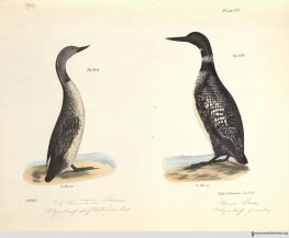
Left: Plate 137, Red-throated Loon & Great Loon; Right: Plate 40, Red-bellied Nuthatch & Red-throated Hummingbird.

Economic interests figured highly in New York’s survey, but it was also prompted by a desire for scientific knowledge. Curiously, the work begins with a far-reaching, 178 page introduction by Governor William H. Seward, detailing such varied topics as “a geographical and political description of the state;” “a notice of the theological profession;” “an account of the formation and establishment of the constitution of the United States;” and “notices of the application of the steam engine to navigation.” It is not until page 174 that Seward begins to discuss the survey, writing of geological studies that preceded it and the surveys of other states. Seward briefly describes some of the geological and mineralogical discoveries (including the disappointment of not finding coal) and makes little mention of other kinds of natural resources.

Left: Plate 50, Spotted Warbler, Blackburnian Warbler, & Black-throated Green Warbler; Right: Plate 79, American Ring Plover, Black-breasted Snipe, Killdeer.

Still, the survey did include zoology, botany, and even paleontology. The zoological study was led by the zoologist James E. De Kay and the reports were published in 5 parts, covering mammals, birds, amphibians and reptiles, fish, and mollusks and crustaceans.  All are beautifully illustrated, but the volume on birds is the largest, with 141 hand-colored lithograph plates. As De Kay explains in his preface, the illustrations are by the artist J. W. Hill and most were based on a living animal or mounted specimens. Initially, the illustrations were meant to be engravings and most of the mammals are engraved, but due to the expense and time involved, they switched to lithographs. There was some anxiety over this change, and De Kay writes “We hope that in the lithographies furnished by Mr. G. Endicott, the naturalist will not regret a departure from the original plan.” I can’t claim to be a naturalist, but I don’t think he should have worried; the plates are almost as striking as the birds we see in the park.

Left: Plate 18, Yellow-bellied Woodpecker & Crested Woodpecker; Right: Plate 4, Pigeon Hawk & Cooper’s Hawk.

Reference:
[1] Newcombe, F. C. “The Scope and Method of State Natural History Surveys.” Science, New Series, No. 956 (Apr. 25, 1913), pp. 615-622. Retrieved 9/21/2017.

* The work of the New York State Natural History Survey continues today in the New York State Museum; the Museum was established as part of the survey in 1836.

Wound Ballistics: The Science of Injury and the Mystery of Exploding Bullets

1018Johnkinder-FBToday’s guest post is written by John Kinder, Associate Professor of History and American Studies at Oklahoma State University. He is the author of Paying with Their Bodies: American War and the Problem of the Disabled Veteran (University of Chicago Press, 2015). On Tuesday, October 17, Kinder will give his talk, “A History of American War in Five Bodies.” To read more about this lecture and to register, go HERE.

On March 11, 1944, an American soldier in the 182d Infantry was digging a foxhole on the island of Bougainville when a Japanese bullet ricocheted and hit him in the ankle. The wound didn’t look that serious. There was almost no blood. Still, it was better to be safe than sorry. Medics bandaged the wound, loaded the soldier onto a litter, and started down the hill to the aid station. He was dead before they reached the bottom.

I recently discovered this story in a volume on wound ballistics published by the US Army Medical Department in the early 1960s. Wound ballistics is the study of the physiological trauma produced by modern projectile weapons. It achieved quasi-scientific status in the late nineteenth century, as military physicians and other self-proclaimed wound experts carried out experiments to measure and ultimately predict what happened when chemically-projected metal collided with living human tissue.

Early on, much of their research involved shooting ammunition into pine boards or the carcasses of  animals to estimate the casualty-causing potential of various armaments. Over time, however, wound ballisticians developed increasingly sophisticated techniques for mapping the body’s vulnerability to different weapons and fine-tuning the production of physiological trauma.

Dog

Microsecond X-ray of the femur of a dog after it has been shot by an 8/32-inch steel ball travelling at 4,000 feet per second. The bone has been shattered despite the fact that it was not actually hit by the steel ball. In order to understand the mechanisms of human injury, World War II-era scientists carried out ballistics experiments on a variety of “model” targets including living dogs, cats, pigs, and horses, as well as blocks of gelatin and tanks of water. 

In the process, they also managed to solve one of the most head-scratching mysteries in nineteenth-century military medicine. The mystery emerged in the mid-century, when growing numbers of observers began to notice a peculiar phenomenon: soldiers were dying from what initially appeared to be relatively minor “through-and-through” wounds. High-velocity bullets seemed to enter and exit the body with only minimal damage. Upon autopsy, however, surgeons discovered extensive internal trauma—pulped tissue, ruptured veins, shattered bones—far outside of the track of the bullet. How was this possible? As early as the 1840s, critics charged that the wounds must be the product of “exploding bullets,” which were subsequently banned by international treaty in 1868. In later years, physicians speculated that the internal explosions were caused by compressed air or heat, but nothing could be proven.

Cat

Microsecond X-ray of a thigh of a cat that has been shot by a 4/32-inch steel ball at an impact velocity of 3,000 feet per second. The dark area is the temporary cavity formed as the ball passes through the muscle tissues. X-rays like this one helped wound ballisticians explain the “explosive effect” that mystified nineteenth-century military physicians. 

By the 1940s, scientists were able to use X-rays and high-speed cameras to solve the mystery once and for all. They discovered that, around 200-400 microseconds after a high-speed bullet strikes a human body, a temporary cavity begins to form around the bullet path. This cavity, which expands and contracts in a fraction of a second, can be more than 20 times the volume of the permanent wound track, resulting in the explosive damage to nearby tissue and bone. And, thanks to the elasticity of human skin, the bullet’s entrance and exit wounds might be nearly closed over by the time the patient reaches medical attention. It was remarkable discovery—not least because it affirmed wound ballisticians’ belief that, when it came to understanding injury, the human eye was no match for a scientist and a machine.

To this day, practitioners of wound ballistics like to justify their work in humanitarian terms. The goal of their research, they often say, is to help military surgeons and body armor manufacturers cut down on unnecessary deaths. All of this is true—to a certain extent. From the very start, however, the field of wound ballistics has played a more ominous role in military history. If wound ballistics is the science of injury, it is also the science of injuring others. Understanding the body’s vulnerabilities has allowed warring nations to develop deadlier antipersonnel weapons: armaments designed to pulverize, poison, burn, shred, emulsify, and eviscerate the bodies of one’s enemies.

No doubt, some readers might be wondering about the soldier at Bougainville, the one who died after a light wound to the ankle. Was he too a victim of the “exploding bullet” phenomenon? As it turns out, his death can be chalked up to a more quotidian threat: human error. Today, we can only speculate about the medics’ actions: perhaps they were in a hurry, or perhaps they were exhausted after a brutal day of fighting, or perhaps—and this is my guess—they were so used to seeing war’s butchery that this soldier’s injury appeared inconsequential by comparison. Whatever the reason, they failed to apply a tourniquet to the wounded man’s leg.

Shortly after the litter party started down the hill, the soldier’s ankle began to hemorrhage. As blood drained from his body, he said that he felt cold. Within minutes, he was dead.

References:
1. International Committee of the Red Cross. Wound Ballistics: an Introduction for Health, Legal, Forensic, Military and Law Enforcement Professionals (film). 2008.
2. Kinder, John. Paying with Their Bodies: American War and the Problem of the Disabled Veteran. Chicago: University of Chicago Press, 2015.
3. Saint Petersburg Declaration of 1868 (full title: Declaration Renouncing the Use, in Time of War, of Explosive Projectiles Under 400 Grammes Weight”). November 29-December 11, 1868.
4. United States Army Medical Department. Wound Ballistics. Washington DC: Office of the Surgeon General, Department of the Army, 1962.

Images:
Dog X-ray: Newton Harvey, J. Howard McMillan, Elmer G. Butler, and William O. Puckett, “Mechanism of Wounding,” in United States Army Medical Department, Wound Ballistics (Washington DC: Office of the Surgeon General, Department of the Army, 1962), 204.
Cat X-ray: Ibid, 176.

Open Access to Your State Medical Society Journals

By Robin Naughton, Head of Digital

In 2015, The New York Academy of Medicine Library embarked on a mass digitization project with the Medical Heritage Library (MHL), a digital curation consortium.  Over the course of two years, the Academy Library along with MHL collaborators digitized state society medical journals from 48 states, the District of Columbia and Puerto Rico.  The Academy Library contributed state medical journals from 37 states, which accounted for 716 volumes of the digitized content now available.   Today, you can find, 97 titles, 3,816 volumes and almost 3 million pages of digitized journals on the Internet Archive.

Digitizing the medical journals of state societies has been an amazing experience for the Library and it is a significant contribution to preserving our cultural heritage and making it accessible to anyone with an internet connection.  Researchers and the general public now have access to a major resource on medical history that includes journals from the 19th and the 20th centuries that would not otherwise be available to the public.  “One of the great values of having the state medical journals online is the willingness to provide full-text digital content for materials that would normally be available only with limited content because they are still in copyright,” says Arlene Shaner, Historical Collections Librarian.

Dr. Daniel Goldberg, Associate Professor at University of Colorado, Denver and 2016 Academy Library Helfand Fellow, agrees:

“As an intellectual historian, medical journals in general are really important for my work because they can reveal much about significant ideas and concepts circulating in medical discourse.  I am working on several projects where the specific local and state histories are crucial to the story I am trying to tell, so having full access to digitized state medical journals will be enormously helpful.  I continue to be so grateful for the important work of the MHL and its partners!”

A quick exploration of the journals can be the catalyst for a deeper research project across many disciplines.  For example, what style and design trends can be identified from the covers of the Illinois Medical Journal?

SMJournals
Illinois Medical Journal through the years.

We invite you to explore the journals, use them, and share with us how they’ve impacted your work: https://archive.org/details/nyamlibrary

Charles Terry Butler and the “War before the War”

By Paul Theerman, Associate Director

The centenary of the United States entry into World War I was this past April. But wars—even those having such sharp cease-fires as this one did, on November 11, 1918—rarely have well-defined beginnings and endings. Even before the official American entry, Americans served in France from the outbreak of the war in 1914. Expats in Paris formed the American Ambulance (the term then meant field hospital), which spun off the American Field Service, charged with transporting wounded soldiers from the front line and providing immediate care. In direct combat, the famed Lafayette Escadrille was founded in 1916, made up of volunteer American air fighters under French command, who battled the Germans up until actual American military deployment two years later. And in the realm of battlefield medicine and surgery, Americans served as volunteers in France from 1914 up to 1917. One of the most noted was Dr. Joseph A. Blake (1864–1937) who, at the outbreak of war, resigned from his prominent surgical positions at Presbyterian Hospital and Columbia College of Physicians and Surgeons, and went to France. There he successively headed up three volunteer hospitals in Neuilly, Ris-Orangis, and Paris, up until his induction to the American military medical corps in August 1917 where he continued his work.

christmas_watermark

“Merry Christmas to J.A.B” [Joseph A. Blake, chief surgeon and hospital director], December 1916. Image: Charles Terry Butler papers, New York Academy of Medicine Library.

Blake had an outstanding reputation, so much so that he readily attracted both funds and workers. One such surgeon was Charles Terry Butler (1889–1980) whose memoir, A Civilian in Uniform (1975), and personal papers are held in the Academy Library. Butler was born in Yonkers, New York, to a prominent family. He was the son of lawyer William Allen Butler, Jr., whose father, William Allen Butler, Sr., both lawyer and author, was himself the son of Benjamin Franklin Butler, U.S. attorney general in the Andrew Jackson and Martin Van Buren administrations. Charles Butler led a life among the New York elite. As one example, he remembers that his family hosted William Howard Taft to dinner during his presidency.[1] Butler went to Princeton University, where he graduated in 1912, and then to medical school at Columbia University College of Physicians and Surgeons. After his graduation in 1916, he was due to take up an internship at Presbyterian Hospital that July. He postponed it to January in order to serve under Blake, then at the Anglo-French volunteer hospital in Ris-Orangis, France, some 25 miles southeast of Paris. As Butler put it:

My two year internship would be put off six months, but here was the opportunity to learn the treatment of serious war wounds under a great surgeon, perhaps my only chance to have such training, and if the United States were forced into the war, I would be much more useful to the Army.[2]

Blake promised Butler scant remuneration, 400 francs travel expenses each way, and 100 francs a month salary, relying on his “contribution” to aid the cause.[3]

Butler_watermark

Charles Terry Butler identity card for Ris-Orangis hospital, June 1916. Image: Charles Terry Butler papers, New York Academy of Medicine Library.

Butler left for Liverpool on May 27, and—after a long period of negotiating his credentials to enter France, as authorities were concerned about German infiltrators—he arrived at the Ris-Orangis hospital on June 10. A converted college, long empty before its refitting, the hospital was organized by two English patrons and operated by private donations and support from the French military. The hospital held about 200 beds, with a surgical theater and supporting radiology and bacteriological facilities, as well as, of course, kitchens and laundries.

with patient_watermark

Charles Terry Butler dressing a wound with the aid of two nurses, 1916. Image: Charles Terry Butler papers, New York Academy of Medicine Library.

ward_watermark

A recovery ward, 1916. The flags of Britain and France are mounted at the window, as this hospital was a joint effort: operated within the French military hospital system, sponsored by private British philanthropy, and staffed by American surgeons. Image: Charles Terry Butler papers, New York Academy of Medicine Library.

Butler’s letters home trace his awakening to war and medicine. Within a week, he wrote to his uncle Clare:

The hospital has about 200 beds, and on my arrival I was put in charge of two wards with over 90 beds and some 80-odd patients. It was some contract to start with, and for two or three days I hardly knew whether I was coming or going. I did about forty dressings a morning with three nurses to help me, and two getting their patients ready for dressing ahead of me and bandaging up when I was through. It took over three hours of hard, steady work.[4]

After a month, to his mother:

Last Sunday, 65 new blessés arrive—the majority of them frightfully wounded. They come by ambulance from a distributing railroad station some 6–7 kilometers away. Arriving in bunches of four or eight, they are sent immediately to their beds. Most of the orderlies had been given leave that day, so we doctors had to turn to and help carry them to the wards. (It isn’t particularly easy carrying a large man on a heavy stretcher with his trappings up three flights of stairs.) There they are undressed; their clothes put in a bag, tagged, and sent to be sterilized and cleaned; and then bathed. . . . The next thing is food. Many have not had anything for 24 hours or more while en route from the front or the last hospital. Then the surgeon comes along. Dressings, casts, splints, etc. are removed so as to see the condition and nature of the injury. It would be impossible to describe the state of some of the wounds—many not having been dressed for several days, some even for 10 or 14 days. A hasty and rather superficial cleansing must suffice for the time being, until the patient comes back from the X-ray room. … All the wounds are terribly infected, and a large percentage have foreign bodies (balls, pieces of shell, clothing, stones, dirt, etc., etc.) lodged…. [Surgery followed, aided by X-ray and fluoroscopy.] The recoveries are wonderful. Men whom no one would expect to live, ordinarily, in a civil hospital, hang by a hair for days and come around O.K.[5]

Butler noted that the average length of stay at the hospital was almost 50 days.

lg group_watermark

The staff of the Ris-Orangis Hospital, 1916. Dr. Joseph A. Blake, director, is the central figure (second row, seated); Charles Terry Butler is the third man to his left. Image: Charles Terry Butler papers, New York Academy of Medicine Library.

Ris-Orangis was considered one of the most successful hospitals in the war. [One of the founders, Harold J. Reckitt, wrote a detailed history of the hospital, V.R. 76: A French Military Hospital (1921)]. Butler spent most of his time dressing wounds, with little occasion for actual surgery. He returned to New York in January 1917 to take up his internship at Presbyterian. But upon the American entry into the war in April 1917, he was commissioned a first lieutenant with the United States Medical Corps, serving into 1919—the topic of a future blogpost. Butler’s experience at Ris-Orangis was crucial to his surgical accomplishments in this second phase of war service. After the war, he entered private practice, but by 1923 ill health—apparently resulting from wartime conditions—led Butler to retire. Moving to the Ojai Valley of Ventura County, California, he became a prominent civic and cultural leader up to his death in 1980.

References:
[1] Butler, Charles Terry. A Civilian in Uniform. Butler, 1975, p. 28.
[2] A Civilian in Uniform, p. 49.
[3] Blake to Butler, 29 April 1916, A Civilian in Uniform, p. 49.
[4] Butler to “Uncle Clare” [Clarence Lyman Collins (1848–1922)], 17 June 1916, A Civilian in Uniform, p. 57.
[5] Butler to “mother” [Louise Terry Collins (1855–1922)], 7 July 1916, A Civilian in Uniform, p. 62–64.

Images:
Charles Terry Butler, “Ris-Orangis, France, 1916,” photographic album. Charles Terry Butler papers. New York Academy of Medicine Library.