Caring for a Collection of Seventeenth Century Ivory Manikins

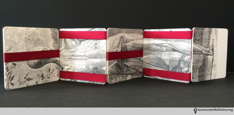
By Scott W. Devine, Head of Preservation

The Gladys Brooks Book and Paper Conservation Laboratory recently completed the rehousing of a fascinating collection of seventeenth century ivory manikins (small sculptures which open to reveal details of human anatomy). As with most items that are treated in the conservation lab, recent consultation and study of the collection by a researcher provided the starting point for conservation assessment and a review of the current housing.

2_Number27_Closed_watermark

Each manikin includes delicately carved features and is often attached to a support of carved wood. Finely detailed pillows are a common feature on items in the collection. Webster Anatomical Manikin Collection #27.

1_Number27_Open_watermark

In most female manikins, the abdominal wall removes to reveal tiny painted organs and a small fetus connected by a linen cord. Webster Anatomical Manikin Collection #27.

History of Ivory Manikins

The renewed interest in human anatomy following the publication by Andreas Vesalius of De humani corporis fabrica in 1543 resulted in a growing demand for écorché drawings which depicted anatomical cross sections of the human body. In addition to drawings, sculptors in France, Italy and Germany began to specialize in detailed cross sections of specific organs which could be used for anatomical study. Out of this tradition of producing three-dimensional study models, either molded from wax or sculpted from wood or ivory, grew the art of carving ivory manikins:

Quite apart from the écorché figures, the ivory eyes, ears and skeletons, yet another product of the carver’s skill was produced in considerable numbers during the seventeenth and eighteenth centuries. This was a small manikin of a man or a woman measuring from 12 to 24 centimeters in length with the anterior thoracic and abdominal wall removable to reveal the viscera. By far the greater number of these lie supine on a stand or in a fitted case and are carved in ivory; some stand on a small pedestal. Although they do occur in pairs, male and female, it is more common for single female figures to be found and in almost every case the figure is represented in an advanced state of pregnancy; the foetus being attached to the uterus by a red cord or else loose within the cavity.[1]

The term manikin is preferred as it denotes a figure with articulated limbs, the moveable arms being essential for allowing the removal of the abdominal wall.

The New York Academy of Medicine Library holds seven manikins, including a rare male and female pair. The manikins do not contain physical markings to indicate artist or date of creation. We do know that one of the largest producers of ivory manikins was Stephan Zick (1639-1715) of Nürnberg and that the Zick workshop produced possibly more manikins than any other workshop in Germany.[2]

Significance and Use

Unlike the detailed écorché figures designed for study purposes, it is unlikely that the manikins were used for teaching or instruction. The lack of detail on the internal organs would limit their function in this capacity. Le Roy Crummer (1872-1934) describes a female patient who remembers learning about pregnancy in 1865 with the aid of an ivory manikin, although such instruction does not seem to be the intended use of the manikins.[3] It is possible that the manikins were considered objects of curiosity, collector’s items that perhaps represented a growing interest in women’s health and the physiology of pregnancy. It is also conceivable that the manikins were given as gifts to newly married couples as good luck tokens intended to signify a future of healthy childbirth. Regardless of the original purpose, as art form the manikins represent an intriguing merger of Baroque art and science.

Designing a New Enclosure

Maintaining complex three-dimensional moveable objects such as the manikins is similar to the work required to preserve rare books in good working condition. In both cases, proper storage and housing are critical for long term preservation.  Enclosures designed for the delicate manikins must account for many moving parts, including fragile ivory fingers and tiny internal organs. The previous temporary housing consisted of wrapping the manikins in acid-free tissue and tying labels to each manikin, stacking them in a Coroplast® polypropylene box.  While this solution protected the manikins during storage, it did not allow for easy viewing and required a complex unwrapping and re-wrapping procedure to access each manikin.

3_OriginalHousing_watermark

The previous temporary housing did not facilitate easy access and introduced the possibility of damaging the delicate manikins during the unwrapping process.

The new enclosure takes into consideration the needs of each manikin by creating a small custom designed tray with two types of polyethylene foam to make sure that each manikin fits securely inside each tray: dense Ethafoam® provides basic support and is lined with softer Volara® foam in areas where the foam directly touches the manikin. The trays are fitted with handles of linen tape that allow the tray to be removed from a larger housing without touching the manikin. The trays are designed to fit into pre-made archival boxes purchased from Gaylord Brothers. The pre-made boxes were retrofitted with Ethafoam® supports lined with Volara® foam. The addition of the Ethafoam® allows the boxes to be easily transported from the environmentally controlled stacks to the Rare Book Room, minimizing vibration and movement within the box.

4_BoxDesign_1_watermark

Yungjin Shin, Collections Care Assistant, designed the interior of the storage boxes, taking advantage of the box depth to fit as many trays in each box as possible. In this case, the manikin’s tortoise shell bed and pillow rest in a tray above the actual manikin, pictured in the next image. Webster Anatomical Manikin Collection #23.

5_BoxDesign_2_watermark

Chloe Williams, 2017 Pre-Program Intern, designed customized trays for each manikin, taking into consideration the contours of each object. Webster Anatomical Manikin Collection #23.

As an additional support, each tray includes a custom fitted pillow of Tyvek® filled with polyester batting that rests on top of each manikin. The pillows further minimize shifting within the box without introducing a rigid support that could damage the fragile ivory features of each manikin. Typical of most artifact housings, each box is labeled with a photograph of the contents so that there is no confusion about which manikin is inside.

6_BoxExterior_watermark

Boxes labeled with photographs allow for easy identification of contents without having to check inventory numbers or search for less obvious identification marks.

Gloves are used when the manikins need to be handled to reveal the intricate internal organs. In situations where the manikin needs to be removed from the tray, the placement of supports within each tray is intentional and designed to encourage the use of two hands when removing the manikin.

7_Gloves_watermark

The use of gloves when handling the manikins protects the item and allows for better control when handling the smooth ivory surface.

Working with this extraordinary collection has allowed the conservation staff to refine our skills in objects housing and to begin designing similar projects to preserve the rich collection of artifacts that complement the Academy Library’s rare book collection.

References:
[1] K.F. Russell. Ivory Anatomical Manikins. Medical History 1972; 16(2): 131-142.
[2] Eugene von Philippovich. Elfenbein. Munich: Klinkhardt und Biermann, 1981.
[3] Le Roy Crummer. Visceral Manikins in Carved Ivory.  American Journal of Obstetrics and Gynecology 1927; 13: 26-29.

Preservation Week: Health Pamphlet Rehousing Project Moves Forward with Support from the National Endowment for the Humanities

By Yungjin Shin, Collections Care Assistant

To celebrate Preservation Week, sponsored by the ALA’s Association of Library Collections and Technical Services, we would like to highlight our work with our Health Pamphlet Collection.

One of the major preservation projects at the Gladys Brooks Book and Paper Conservation Laboratory is the Health Pamphlet Rehousing Project, which is funded in part by a grant from the National Endowment for the Humanities (NEH). The Health Pamphlet Collection includes approximately 50,000 health-related pamphlets dating from the 19th to the early 20th century.

The current process involves multiple steps: pulling the pamphlet boxes from the stacks — cleaning the pamphlets and assessing them for future treatment — transferring the pamphlets to envelopes with custom fitted supports — updating the bibliographic information in the online catalog — building custom designed storage boxes — labeling the envelopes and boxes— rearranging as needed —and re-shelving to the new location.

Here is a behind-the-scenes video that shows the overall process, start to finish.


The project is currently scheduled to be completed in January 2018.

Preservation week

What’s Your Job? Interview with Book Conservator Christina Amato

By Christina Amato, Book and Paper Conservator, with Emily Moyer, Collections Care Assistant

The New York Academy of Medicine Library is well known for its world-class collections and serves patrons from all over the world. We strive to make our collections as visible and accessible as possible, and a lot of work goes on behind the scenes towards this end. The Gladys Brooks Book and Paper Conservation Laboratory fulfills one component of this equation, attending to the physical well-being of collections materials.

Christina Amato, cleaning old glue from the spine of a book.

Christina Amato, cleaning old glue from the spine of a book.

The Gladys Brooks Book and Paper Conservation Laboratory was created in 1982, and occupies a bright, well-equipped space overlooking Central Park. Currently, one full-time conservator and two part-time collections care assistants work to preserve the collection of over 550,000 volumes. Christina Amato has worked as a book conservator here for approximately three years. Recently, she sat down with Collections Care Assistant Emily Moyer to discuss her work.

EM: How did you get into the field of conservation?

CA: People come to the field from a variety of backgrounds, in part because conservation crosses many disciplines, including science, art history, and studio art. I come from an art background, having received my BA in studio art from Bard College. It was really through an interest in materials (specifically paper, leather, and vellum) that I became involved in bookbinding. I received a diploma in bookbinding from the North Bennet Street School in Boston, which led me to many wonderful internships in book conservation.

There are actually many different possible paths to a career in conservation. The American Institute for Conservation of Historic and Artistic Works (AIC) has compiled a guide to education and training in conservation.

E.M.: Why is conservation important?

Many books that come up to the conservation lab are too fragile to be handled by readers; our work makes these materials accessible. This is important not just for readers that come and use our collection in person, but also for preparation for digitization projects and exhibitions. Much of our work focuses on preventative care, which ensures that materials remain in good condition for current and future use. This can include rehousing (creating enclosures for materials), regulating and monitoring environmental conditions, and disaster planning and response.

E.M.: What are you working on now?

Currently I am working on a copy of The byrth of mankynde, otherwise named The womans booke, by Eucharius Rösslin, London, 1545.  The book arrived in the lab broken in half:

The byrth of mankynde, otherwise named The womans booke, before treatment.

The byrth of mankynde, otherwise named The womans booke, before treatment.

It had been subjected to several unsuccessful attempts to repair the damage over the years. Several layers of Japanese paper had been glued over the spine, and only a few small fragments of the original spine piece were left.

The byrth of mankynde, otherwise named The womans booke, before treatment.

The byrth of mankynde, otherwise named The womans booke, before treatment.

After disbanding, and mending paper tears and fragile edges throughout the text block, I carefully documented the original sewing pattern, so that I could resew it in the same way.

Next, I dyed leather to match the original binding, which I will use to recreate the spine. After it is complete, the book will receive a new, custom enclosure, and will be ready to be used again.

Leather dying in progress.

Leather dying in progress.

E.M.: What are some interesting things you’ve worked on at the Academy?

A favorite book that I worked on was a copy of The Physiology of Digestion by William Beaumont, published in Vermont in 1847. Very shortly after I finished working on it, I listened to an archived episode of the Radiolab program called Guts. Our historical collections librarian, Arlene Shaner, spoke of a small, purple, cloth-bound book about digestion; it only very slowly dawned on me that it was the very same book that was on my bench.

Before and after, The Physiology of Digestion, Beaumont, William. Vermont, 1847.

Before and after, William Beaumont’s The Physiology of Digestion, Vermont, 1847. Click to enlarge.

Another favorite was a small volume entitled Geburtshulfliche Taschen-Phantome by Koichi Shibata, published in Munchen in 1892. We were so charmed by this little obstetrics text that we recreated the movable paper baby for a public workshop.

Koichi Shibata, Geburtshulfliche Taschen-Phantome, after treatment (left). The moveable paper baby (right).

Koichi Shibata, Geburtshulfliche Taschen-Phantome, after treatment (left). The moveable paper baby (right).

E.M.: Why do you like working in conservation?

Working in conservation can be very satisfying; the outcome of our work is unusually tangible. Working to preserve materials for (and from!) future readers can be creatively challenging, and requires flexible thinking. I like that it is a profession that combines working with your hands with an intellectual component. And of course, it is very rewarding to take a book that is too fragile to be handled and render it usable again.

Happy Preservation Week!

By Emily Moyer, Collections Care Assistant, Gladys Brooks Book & Paper Conservation Laboratory

PreservationWeek2015_logoSponsored by the American Library Association’s Association of Library Collections and Technical Services (ALCTS), Preservation Week aims to raise awareness of the importance of preservation and education in providing collections for future generations.

Every week is preservation week in the Gladys Brooks Book & Paper Conservation Laboratory at The New York Academy of Medicine. Preservation efforts include cleaning, stabilization, and rehousing; monitoring environmental conditions; education on the care and handling of materials; item-level treatments; and disaster preparedness. We work together to try to prevent future deterioration of materials and mitigate risks to the collection.

This behind-the-scenes video shows a day in the conservation lab here at the Academy: creating slings for our 60,000+ health pamphlet collection, shrink wrapping brittle periodicals and books, mounting facsimile images for an exhibition, refoldering and dry cleaning pamphlets, mending a manuscript cookbook, and rebacking a 19th-century medical student notebook.

Happy Preservation Week!


The Bookplates of Medical Men (Item of the Month)

By Erin Albritton, Head of Conservation, Gladys Brooks Book & Paper Conservation Laboratory

Book owners have forever endeavored to find ways to identify volumes as their own. With the invention of the printing press, however, books became more plentiful and an owner’s need to identify his or her own copy even more pressing. The earliest examples of printed bookplates (also known as ex-libris) come from Germany and date to the 15th century, just as printing began to take off in Europe. Over the following centuries, the use of bookplates became widespread throughout Europe and eventually followed colonists to America.1

In the summer of 2014, conservators in the Gladys Brooks Book & Paper Conservation Laboratory began treating three scrapbooks containing 184 bookplates.2 Frank Place, Jr., NYAM’s reference librarian from 1905 to 1945, collected them and donated the books to the library sometime in the mid-20th century. In compiling his collection, Mr. Place mounted the bookplates onto recycled paper pamphlet covers (measuring approximately 5.25” x 8”) and stored them alphabetically in two-ring binders,3 which were actively damaging the plates’ fragile paper supports and making it difficult to use the collection.

Binder showing ring mechanism and resulting damage to paper supports.

Binder showing ring mechanism and resulting damage to paper supports.

Example of pamphlet cover Mr. Place reused as a support for his bookplates.

Example of pamphlet cover Mr. Place reused as a support for his bookplates.

To remedy these issues and minimize the risk of future damage, conservators modified the binders while retaining as much of the original binding structure as possible, replacing the ring mechanisms with fixed posts and hinging the paper supports onto stubs.

Top: Volume 3, before treatment, in its original ring binder. Bottom: Volume 1, after treatment, in a modified post binding with hinged plates.

Top: Volume 3, before treatment, in its original ring binder. Bottom: Volume 1, after treatment, in a modified post binding with hinged plates.

Before and after binder modification.

Before and after binder modification.

Because the original binder spine pieces were too big for the modified structures, conservators encapsulated the pieces in Mylar and affixed them to protective four-flap enclosures.

Mylar-spine wrappers with encapsulated original spine pieces.

Mylar-spine wrappers with encapsulated original spine pieces.

Mr. Place’s charming collection in NYAM’s library offers a window into what some scholars have referred to as the “golden age of bookplate enthusiasm,”4 which spanned from the end of the 19th century through the first decades of the 20th century. During this time, societies for the collection and exchange of ex-libris sprang up across Europe and the United States. Collectors prized plates for their aesthetic value as miniature pieces of art and often acquired them, not for use in identifying their books, but for the sole purpose of organizing, exhibiting, and exchanging them with others. Many collectors limited their acquisitions to bookplates representing a particular theme and, here, it is no surprise that Mr. Place specialized in the plates of medical practitioners and institutions. Correspondence (included in the scrapbooks) between Mr. Place and other collectors (specifically H. M. Barlow, secretary at the Royal College of Physicians, and Dr. Henry de Forest, a prominent New York physician5) indicates that he was not only interested in growing NYAM’s collection but, in the spirit of the times, was also an active contributor to the collections of others.

While small by comparison to other collections, Mr. Place’s scrapbooks offer some wonderful examples of the broad ranging sizes and styles of bookplates—from modest ornamental name labels:

Ornamental name labels of (left) Samuel Smith Purple, MD and (right) Robert Latou Dickinson, MD.

Ornamental name labels of (left) Samuel Smith Purple, MD and (right) Robert Latou Dickinson, MD.

to elaborately illustrated panels, depicting anything from coats of arms to the owner’s occupation and hobbies. Not surprisingly, in the case of medical bookplates, images such as Hippocrates, microscopes, and the caduceus, along with skeletons, skulls, and beakers tend to figure prominently.

This slideshow requires JavaScript.

NewYorkAcademyOfMedicine_watermarkIt is a pity we do not know whether, in addition to collecting, Mr. Place ever commissioned his own miniature work of art to grace the inside covers of his personal volumes. Thanks to a bookplate, however, we are forever reminded of his contribution of this delightful little collection to NYAM’s library.

Treatment of the third and final scrapbook is in process and the entire collection will be available for use in early 2015. In the meantime, a list of all 184 bookplates can be obtained by contacting history@nyam.org or calling 212-822-7313.


Notes

1. The oldest known American bookplate dates to 1679 and takes the form of a simple label indicating the owner’s name. Curtin, R. G. (1910). “The Book-Plates of Physicians, with Remarks on the Physician’s Leisure-Hour ‘Hobbies’.” Philadelphia: J.B. Lippincott Co. (Reprinted from International Clinics, Vol. II, 20th Series), p. 239.

2. Thanks to Saira Haqqi, 2014 Gladys Brooks Book Conservation Intern, for her work conserving volume 2 of this collection.

3. In an address to the Historical Section of the Philadelphia College of Physicians on November 22, 1907, physician and bookplate collector Roland G. Curtin advises collectors to hinge their plates onto paper cards (measuring 7.5” x 9.5”) and to keep the cards loose, thus enabling the inspection of the backside of plates as well as the display of single plates without endangering the entire collection. Ibid. 253. It seems likely that Mr. Place read Dr. Curtin’s address (a reprint, cited above, was accessioned into NYAM’s collection in 1910) and was endeavoring to follow this advice when arranging his own collection.

4. Pincott, A. “American Bookplates.” Rev. of American Bookplates by W.E. Butler. Print Quarterly, Vol. 18, No. 3 (Sept. 2001), p. 351. http://www.jstor.org/stable/41826267.

5. See blog post by Arlene Shaner, Reference Librarian for NYAM’s Historical Collections, discussing a bookplate and correspondence from Dr. Henry de Forest: https://nyamcenterforhistory.org/2013/03/06/biblioclasts-bibliosnitches-beware/.

Registration Open for Vesalius 500 Workshops

Registration is now open for our hands-on art and anatomy workshops, presented as part of our Vesalius 500 celebrations on October 18, 2014. Create your own articulated anatomical figure or “exquisite corpse” at the Gladys Brooks Book & Paper Conservation Laboratory; learn Renaissance drawing techniques with medical illustrator Marie Dauenheimer; or explore the anatomy and art of the hand with physical anthropologist Sam Dunlap.

Spaces are strictly limited so register soon. Registration at one of the workshops includes free entry to the Festival. You can register for the Festival (without workshop attendance) here.

From the Cradle to the Grave: Session One: The Cradle

Moveable baby and female pelvis from one of NYAM’s 19th century obstetrics texts, Dr. K. Shibata's Geburtschülfliche Taschen-Phantom, or the Obstetrical Pocket-Phantom.

Moveable baby and female pelvis from one of NYAM’s 19th century obstetrics texts, Dr. K. Shibata’s Geburtschülfliche Taschen-Phantom (Obstetrical Pocket-Phantom).

Working with NYAM’s conservation team, celebrate Vesalius’s life with a hands-on workshop producing your own articulated anatomical figures in the Gladys Brooks Book & Paper Conservation Laboratory.

Time: 11am-1pm
Cost: $55
Includes: All materials, and free entry to the Festival.
Maximum participants: 12
Register here

During the morning’s Cradle workshop, we will construct paper facsimiles of a moveable baby and female pelvis from one of NYAM’s 19th century obstetrics texts, Geburtschülfliche Taschen-Phantom (or the Obstetrical Pocket-Phantom). The book was written by Dr. K. Shibata, a Japanese author studying in Germany, and was published first in German before being translated into English and Japanese.

Participants will have time to make at least one paper baby and pelvis, which can be produced as paper dolls or magnets.

From the Cradle to the Grave: Session Two: The Grave

An exquisite corpse made by staff of the Gladys Brooks Book & Paper Conservation Laboratory.

An exquisite corpse made by staff of the Gladys Brooks Book & Paper Conservation Laboratory.

Working with our conservation team, celebrate Vesalius’s life with a hands-on workshop producing your own “exquisite corpse” in the Gladys Brooks Book & Paper Conservation Laboratory.

Time: 2:30pm-4:30pm
Cost: $55
Includes: All materials, and free entry to the Festival.
Maximum participants: 12
Register here

During the afternoon’s Grave workshop, we focus on producing a Vesalian-themed exquisite (or rotating) corpse. Loosely based on the surrealist parlor game in which a picture was collectively created by assembling unrelated images, this workshop will employ a special, rotating binding structure and mix-matched facsimile images from NYAM’s rare book collections to allow students to create their own unique, moveable pieces of art.

Renaissance Illustration Techniques Workshop with Marie Dauenheimer, Medical Illustrator

Students at medical illustrator Marie Dauenheimer's workshop at last fall's Festival.

Students at medical illustrator Marie Dauenheimer’s workshop at last fall’s Festival.

Time: 10am-1pm
Cost: $85
Includes: All materials, and free entry to the Festival.
Maximum participants: 15
Register here

Artists and anatomists passionate about unlocking the mysteries of the human body drove anatomical investigation during the Renaissance. Anatomical illustrations of startling power vividly described and represented the inner workings of the human form. Leonardo da Vinci’s notebooks were among the most magnificent, merging scientific investigation and beautifully observed drawing.

Students will have the opportunity to learn and apply the techniques used by Renaissance artists to illustrate anatomical specimens. Using dip and technical pens, various inks and prepared paper students will investigate, discover, and draw osteology, models, and dissected specimens from various views creating an anatomical plate.

Understanding the Hand, physical anthropology workshop with Sam Dunlap, Ph.D.

Dr Sam Dunlop leading a workshop at last year's Festival.

Dr Sam Dunlop leading a workshop at last year’s Festival.

Time: 2:30pm-5:30pm
Cost: $75
Includes: All materials, and free entry to the Festival.
Maximum participants: 15
Register here

The hand as an expression of the mind and personality is second only to the face in the Renaissance tradition of dissection and illustration that continues to inform both art and science. Basic anatomical dissection, illustration, and knowledge continue to be fundamental in many fields from evolutionary biology to surgery, medical training, and forensic science. This workshop will offer participants the opportunity to explore the human hand and its anatomy, which will be demonstrated with at least three dissections.  Opossum (Didelphis virginiana) and orangutan (Pongo pygmaeus) forelimbs will be available along with other comparative skeletal material. We will discuss hand evolution, embryology, and anatomy, and the artistic importance of the hand since its appearance in the upper palaeolithic cave art. We will also analyze the hand illustrations of da Vinci, Vesalius, Rembrandt, and artists up to and including the abstract expressionists.