Table Reading

Our October 2014 “Art, Anatomy, and the Body: Vesalius 500″ festival guest curator, artist and anatomist Riva Lehrer, describer her first experience with cadavers and how that shaped her thinking about bodies, anatomy, and art.

"In the Yellow Woods," by Riva Lehrer. Click to enlarge.

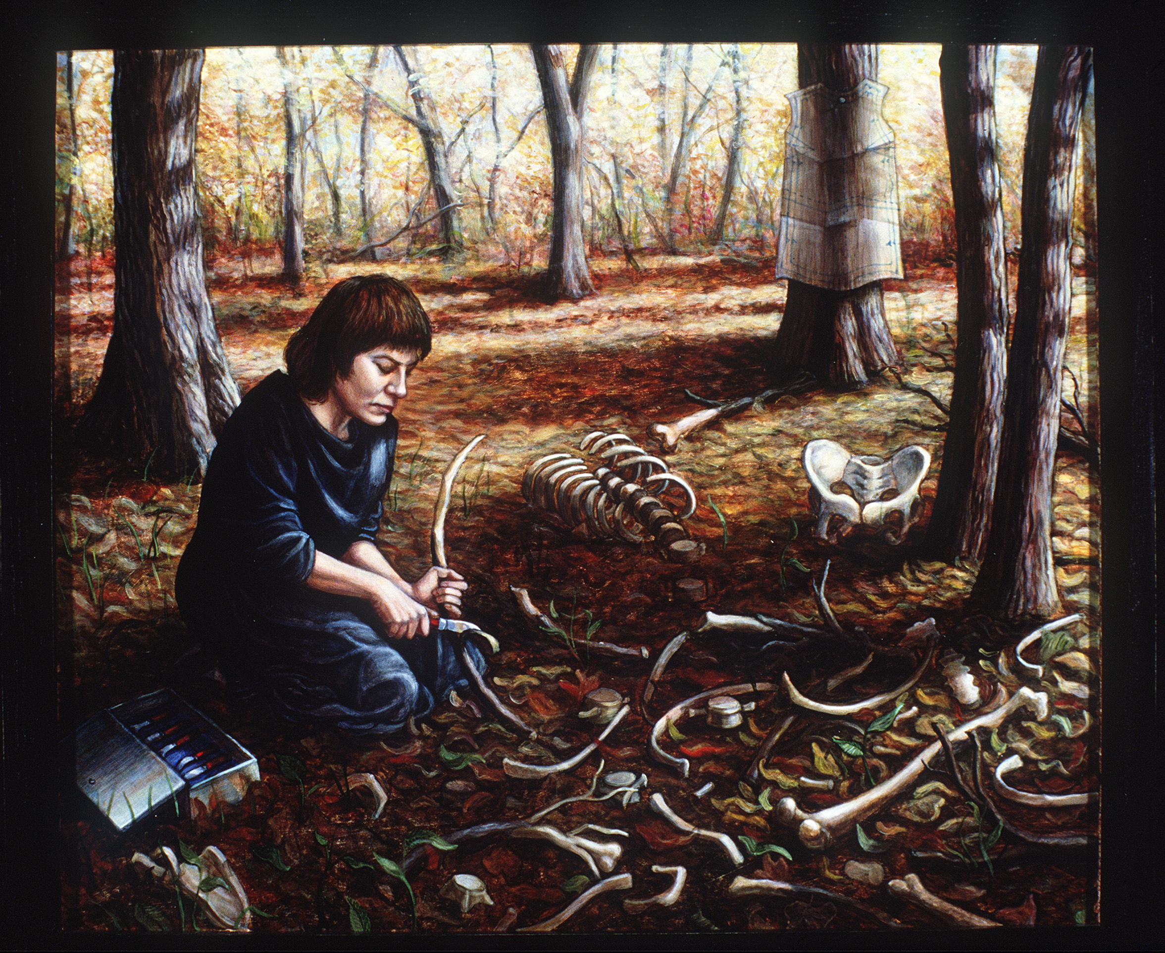
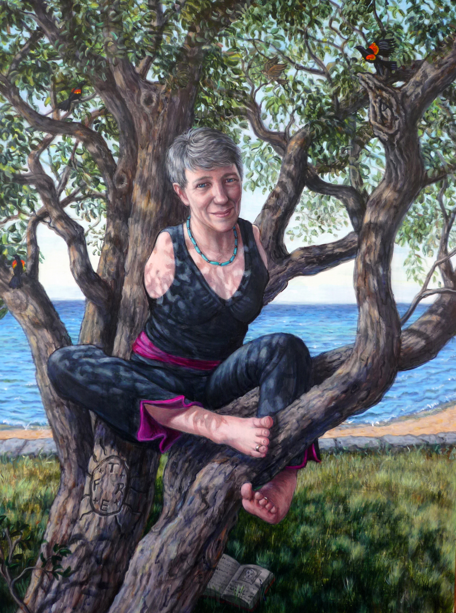
“In the Yellow Woods,” by Riva Lehrer. Click to enlarge.

The first time I ever saw a cadaver was a day in early September of 2006. The light was perfect—a glowing blue and gold herald of the coming Jewish New Year. I walked into lab behind Dr. Norm Lieska, head of Gross Anatomy at University of Illinois at Chicago, and a group of M1 students, all gangly in their brand-new starched white coats and spotless scrubs. The laboratory was a sort of extended corridor, comprised of a series of interlocking rooms, lit by high, industrial windows like those in an old factory. Burnished shafts of sunlight slanted across the rows of steel tables, skimming across the unzipped body bags. Each contained a cadaver that had been preserved and prepped for student exploration. For the main, though, they were pristine; head and hands demurely wrapped, all original parts on board.

I’d been warned that I might be nauseated or disgusted by the bodies. I braced myself to be sickened by the miasma of chemicals in the air. I did not expect to be overwhelmed by the sheer generosity represented in that room. Twenty-five people had decided that we needed to understand the human body in the most direct and unmediated way. They’d signed donation papers that gave us the right to read the history of their own flesh. I felt the impact of that gift even from my first steps into lab.

The dark vinyl of the body bags appeared as if gilded. This was the last moment they would all appear the same. We would pull down the zippers, and reveal the wild variations within. I am not in any way a religious person, but I thought: if I felt this kind of awe in synagogue, I’d be a very different kind of Jew. I was at the lab as part of my position as visiting artist in Medical Humanities at the Medical School of the University of Illinois at Chicago. Each cadaver in Gross Lab was assigned to a team of about 10 students; at the start of the semester I’d been assigned to one such team. These students worked on the same person for the entire year. Scalpels peeled away each archeological layer, skin down to the deepest core. It was a bizarre form of intimate knowledge—both closer and more abstract than their inhabitants had had in life. I began to focus on comparing the bodies from table to table, and to show the members of my team that each cadaver had its idiosyncrasies. None of them were ringers for the photographs in their Color Atlas of Human Anatomy.

"Theresia Degener," by Riva Lehrer. International Human Rights lawyer Theresia Degener is one of the drafters of the United Nations Convention on the Rights of Persons with Disabilities. As a member of the German generation of children whose mothers were given thalidomide, Degener  accomplishes all she wants to do through a range of inventive strategies.  Click to enlarge.

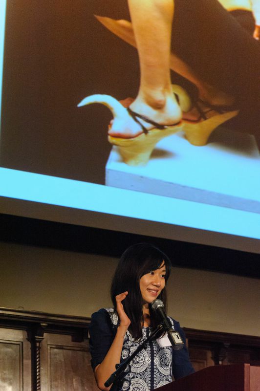
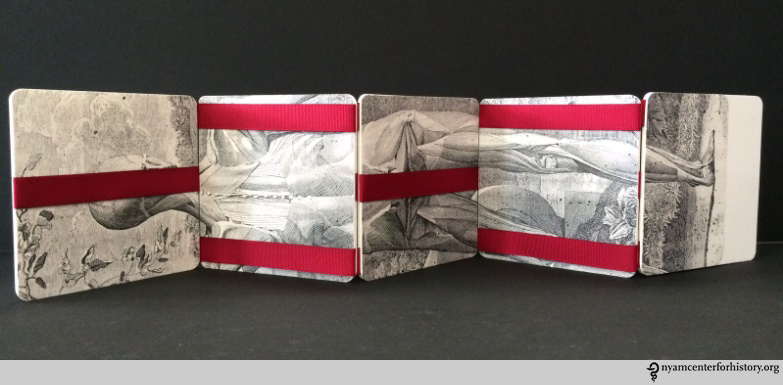
“Theresia Degener,” by Riva Lehrer. International Human Rights lawyer Theresia Degener is one of the drafters of the United Nations Convention on the Rights of Persons with Disabilities. As a member of the German generation of children whose mothers were given thalidomide, Degener accomplishes all she wants to do through a range of inventive strategies. Click to enlarge.

In all the time I was there, though, I never saw a body anything like mine. I was much too intimidated to ask why. Perhaps a body that was too different from those dissection pictures could not function as a primer? (Oddly enough, when I visited a different cadaver lab last year, a bare scoliotic spine was on a table in the back of the room, picked clean of the body in which it had dwelt).

I was the visiting artist in Medical Humanities at the Medical School of the University of Illinois at Chicago for four years, during which I taught figure drawing and portraiture for med students. I’ve gone on to teach those classes at Northwestern University School of Medicine, and as the professor of anatomy at the School of the Art Institute of Chicago (I’m on leave now to pursue other projects). Each year of teaching and study has only increased my sense of wonder at what a living body can do. All bodies (human and animal) are so densely woven with function, yet can accommodate such dysfunction.

I’ve asked my students at both medical schools whether I’m the only disabled person they interact with outside of clinical rotations. The answer is yes. I wonder if my professional presence changes what they think when they begin clinicals, though I also wonder if they begin silent diagnoses when I walk into the classroom. My SAIC students do often seem startled on their first day. (Though maybe that’s just an effect of the tables full of bones. Hard to tell with the young and ironic.) They may not have medical knowledge, but they are trained observers, and mine is the body at the center of the room, at least until our model climbs onto the platform in his/her birthday suit.

Riva Lehrer with students at SAIC in 2012.

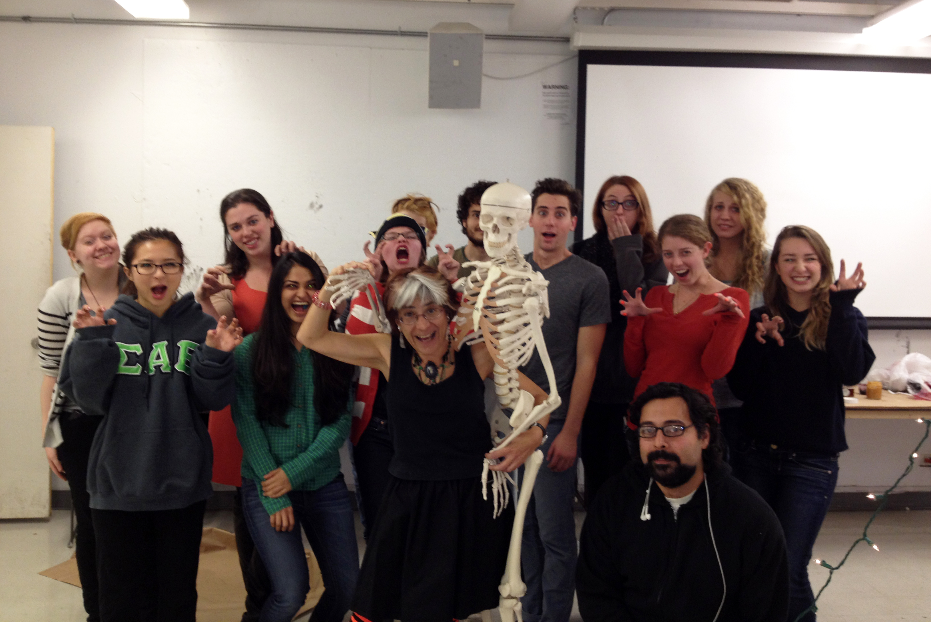
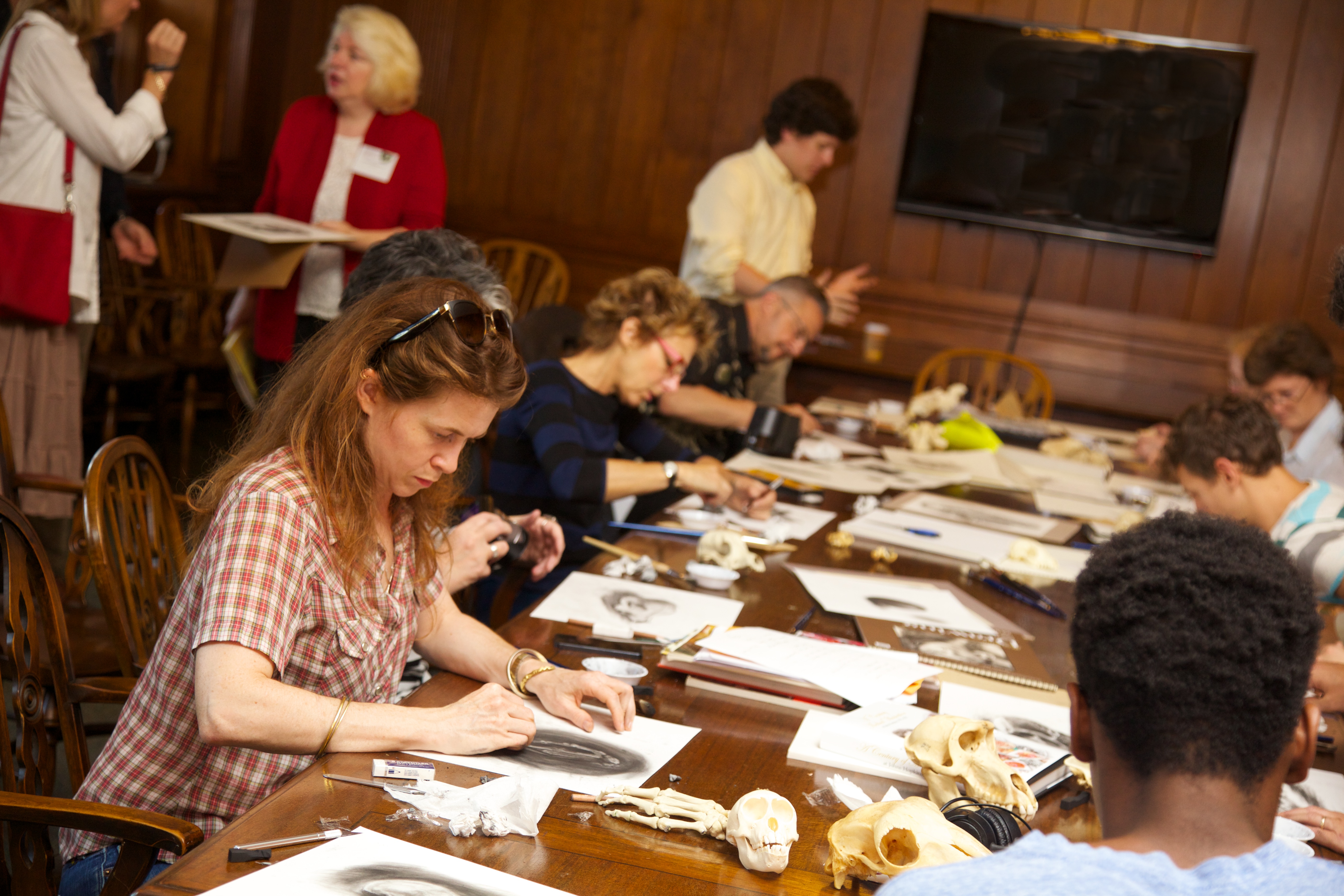
Riva Lehrer with students at the School of the Art Institute of Chicago in 2012.

For years, I was the elephant in the room. Eventually, I stopped pretending I wasn’t there and began to use myself as an exemplar. This doesn’t come easy—sometimes, my attempts at coping through humor sound like outtakes from Young Frankenstein—but it does produce a willingness on the part of students to ask uncomfortable questions. As the cadavers prove year after year, normal is a matter of degree. Our bodies let us live so many ways. Healing is creativity made manifest.

I’m writing this just before another New Year. I hope that 2015 brings you joy of your own mysteries, and that you will follow those secret trails through your own glowing, shadowed, and gilded rooms.

Brains, Brawn, & Beauty: Andreas Vesalius and the Art of Anatomy

By Rebecca Pou, Archivist, and Johanna Goldberg, Information Services Librarian

For our October 18 festival, Art, Anatomy, and the Body: Vesalius 500, we exhibited items from the library’s collections showing the history of anatomical illustration. You can still visit the New York Academy of Medicine to view the exhibit in person on the ground floor. If you can’t make it, we offer a digital version below.

The exhibit on display at the new York Academy of Medicine.

The exhibit on display at the New York Academy of Medicine.

In 1543, Andreas Vesalius was a 28-year-old professor of surgery and anatomy at the University of Padua, one of Europe’s best known medical schools. That year, he published his most famous work, De humani corporis fabrica, translated as On the Fabric of the Human Body. Vesalius dedicated the work to Charles V; he subsequently received the appointment of physician to the imperial family.

Working from three images from the Fabrica—a skeleton, a figure of muscles, and an illustration of the brain—this exhibit shows the many ways Vesalius’ work built on past anatomists, and exerted its influence well into the future.

Images from great works in our collection, from Magnus Hundt’s 1501 Antropologium to Dominici Santorini’s 1775 Anatomici summi septemdecim tabulae, show the evolution of artistic style and scientific understanding. Some show examples of “borrowing” Vesalius’ images and placing them in new contexts.

Click an image to view the gallery.

Reflections on “Art, Anatomy and the Body: Vesalius 500”

Our “Art, Anatomy, and the Body: Vesalius 500″ festival guest curator, artist and anatomist Riva Lehrer, reflects on the event.

Riva Lehrer, left, with Lisa O'Sullivan, director of the Center for the History of Medicine and Public Health. Photo by Charles Manley.

Riva Lehrer, left, with Lisa O’Sullivan, director of the Center for the History of Medicine and Public Health. Photo by Charles Manley.

My approach as co-curator of “Art, Anatomy and the Body: Vesalius 500” was to ask how we use anatomy today to understand what it means to be human. Throughout history, we’ve used metaphor to organize our concepts of the body. We’ve imagined it as a vessel full of roiling humors, as an elaborate clock, as a regulated factory, as a robot and a computer, to name just a few. Even anatomical study is affected by metaphor and symbolism, and often guides what we see.

As science creates new perspectives on human (and non-human) anatomy, society responds by re-imagining new possibilities. When we internalize these visions we live differently in our bodies. “Art, Anatomy and the Body: Vesalius 500” brought together artists, writers, and scholars to discuss about how we see ourselves now and how we construct ourselves as public bodies.

As emcee, I had the pleasure of introducing Ann Fox, Sandie Yi, Dan Garrison, Sander Gilman, Nuha Nazy, Dima Elissa, Bill Hayes, Steven Assael and Alice Dreger, who were among the more than 25 festival participants.

Chun-Shan “Sandie” Yi. Photo by Charles Manley

Chun-Shan “Sandie” Yi. Photo by Charles Manley

Curator Ann Fox and artist Chun-Shan “Sandie” Yi started the day with an overview of contemporary artists who explore identity through medical and anatomical imagery, including artists who tackle our continuing discomfort with HIV/AIDS, a disease that from the very first raised issues of identity and ostracism. Sander Gilman furthered our thoughts about unacceptable bodies by discussing how teaching posture in schools has been used to control and regulate bodies, and make them socially predictable.

Dan Garrison brought us back to the core of the festival via the origin of Vesalius’ Fabrica. He posited some very intriguing ideas around who Vesalius may have been; he may have had a variant body (possibly dwarfism), and this may have contributed to how he created his great work.

Dan Garrison. Photo by Charles Manley.

Dan Garrison. Photo by Charles Manley.

Identity is always a struggle between the specifics of individuality and alliance with group affiliation. This echoes the direction of modern medicine; treatments are becoming increasingly targeted to individual bodies, whether through genetics, prosthetics, or adapted drug regimens. We glimpsed this future during the ProofX presentation, with Nuha Nazy and Dima Elissa. ProofX uses 3D printing to produce extremely precise implants, surgical models, and adaptive devices for a wide range of conditions. It’s anatomical interface at an unprecedented level.

ProofX's 3D-printing demonstration.

ProofX’s 3D-printing demonstration. Photo by Charles Manley.

Bill Hayes engages medical history in order to understand his own biography. His research blends with memoir and hands-on experience, as witnessed by The Anatomist, the remarkable story of Henry Gray, author of Gray’s Anatomy. Hayes steps across the boundary between modern identity and historical precedent, here discussing the history of exercise, in order to show us how we arrived at our present state.

Anatomy and poetics also wove together in the work of Steven Assael. His paintings and drawings are highly (and gracefully) accurate, yet manage to be astute and nuanced examinations of his subjects’ personalities. He transfixed the audience by the lushness of his technique and the drama of his compositions.

Alice Dreger. Photo by Charles Manley.

Alice Dreger. Photo by Charles Manley.

Our excellent final event was a talk by bioethicist Alice Dreger. She traced the origins of contemporary medical photography as well as taking a fresh look at traditional anatomical illustration. Dreger has thought deeply about how we signal our identity through bodily choice. She raised questions about what caused doctors to lose touch with the vulnerability of people in medical settings, and to describe variant bodies in dehumanizing ways. She also pointed out that doctors often can’t admit the taboo pleasure of viewing physical anomalies, and how that covert pleasure affects their relationships with patients.

The human body has its secret, unspoken existence and its public presentation, meant to be decoded by other human beings. Anatomy would seem to be an objective bridge between the two, yet can be just as complicated and interpretive as any form of art. Our festival let us perceive the dialogue between poetics and science, and between inner and outer realities of the body.

For more images of the festival, visit our Facebook page.
Read a summary of the festival by presenter Kriota Willberg: Part 1 and Part 2
Click here for a blog from Hyperallergic.
Read “Seeing is Believing: New York Academy of Medicine’s Vesalius 500th Year Celebrations,” an article by presenter Brandy Schillace, PhD.

Vesalius 500: Art, Anatomy, and the Body

By Paul Theerman, Associate Director, The Center for the History of Medicine and Public Health

Join us this Saturday, October 18, for our second annual Festival of Medical History and the Arts, Art, Anatomy, and the Body: Vesalius 500. Register here.

Vesalius500STD_05_30_14This year, we celebrate the 500th birthday of Andreas Vesalius, the path-breaking anatomist whose 1543 book, De humani corporis fabrica (The Fabric of the Human Body), opened up new worlds in the understanding and representation of the human body. The festival’s presentations will focus on the cultural understanding of the body throughout history. We will have rare books on display, including one of our copies of Vesalius’ Fabrica; the Drs. Barry and Bobbi Coller Rare Book Reading Room will be open for visitors; and we will offer four hands-on workshops, still open for registration (festival entrance is included in the price of the workshops).

For more information, including the full schedule and participant biographies, see Vesalius 500.

To whet your appetite, look at our earlier blog posts by those joining us at the festival:

And don’t forget The Fabrica of Andreas Vesalius or our Vesalius 500 Workshops, presented by Sam Dunlap, Marie Dauenheimer, and the staff of our Gladys Brooks Book & Paper Conservation Laboratory.

Many others will present, as well:

  • Eva Åhrén on specimens in medical museums
  • Steven Assael on observing bodies
  • Alice Dreger on medical photography
  • Dima Elissa and Nuha Nazy on 3-D printing and anatomy
  • Ann Fox and Chun-Shan “Sandie” Yi on bodies in contemporary art
  • Daniel Garrison on translating Vesalius’ masterpiece
  • Heidi Latsky with Tiffany Geigel and Robert Simpson on The GIMP Project
  • Michael Sappol on making bodies transparent

See you on Saturday!

The Talented Dr. Knox

Lisa Rosner, PhD, author of today’s guest blog, will present “The True and Horrid Story of the Burke and Hare Anatomy Murders” at our October 18th festival, Art, Anatomy, and the Body: Vesalius 500.

Engraving of Dr. Robert Knox. From our online collection The Resurrectionists.

Engraving of Dr. Robert Knox. From our online collection The Resurrectionists.

Dr. Robert Knox, the anatomist whose cadaver purchases kept William Burke and William Hare in the murder business, has always been an enigma. Born in Edinburgh, Scotland, he served in the army and studied in Paris before returning home to set up a private anatomical school. He taught hundreds of students, lecturing twice a day in addition to holding separate dissection classes. He was curator of the surgical museum, wrote articles on human and comparative anatomy for scientific societies, and was in the process of seeing several books on anatomy through publication. His supporters claimed he knew nothing about the murders; his detractors argued that he simply turned his blind eye—for he had lost an eye to smallpox as a child.

Plate II in Knox's Man: His Structure and Physiology, shown flat and with lifted parts. Click to enlarge.

Plate II in Knox’s Man: His Structure and Physiology, shown flat and with lifted parts. Click to enlarge.

What we can see, using the extensive collection of Robert Knox materials in the New York Academy of Medicine Library, is just how talented an anatomist Robert Knox was. His edition of Hippolyte Cloquet’s A System of Anatomy is more than just a translation: it is instead a critical analysis of contemporary anatomical knowledge, enriched by examples from Knox’s own research and teaching. The same is true of his edition of Friedrich Tiedemann’s The Plates of the Human Arteries, prepared with two of his students, Thomas Wharton Jones and Edward Mitchell. The catalogue he prepared for the anatomical and pathological museum of the Royal College of Surgeons of Edinburgh is filled with his detailed insights: on anomalies of the biceps flexor cubiti, on the precise position relative of a fatal brain tumor, and on popliteal aneurism. Knox discussed the implications of these, and many more of his anatomical and surgical observations, in several series of articles for the London Medical Gazette. We can follow his teaching methods in The Edinburgh Dissector, the handbook he wrote for the use of his dissecting classes. “Nobody could ever say that he gave a dry lecture, or one that was not specially instructive,” reported his former student, Henry Lonsdale. Even in the midst of the detailed description that makes up most of the Edinburgh Dissector, Knox’s love of his subject shines through, as in his description of the bones of the foot, which “when well formed yields in beauty and perfection to no part in the human body.”

Could such a passionate observer of all subjects anatomical really have missed the fact that sixteen of his own “subjects” had been murdered? Contemporaries from Home Secretary Sir Robert Peel (founder of the London Metropolitan Police) to the Edinburgh evening papers refused to believe it and called for wider investigation. On the advice of legal counsel, Knox refused to answer any questions—just as he had refused to ask any, his professional rivals muttered darkly, when presented with Burke’s and Hare’s murder victims. There was no real case against him, and there are no records of any deliberations by the prosecuting attorneys. We will probably never know what Knox knew or when he knew it.

"Execution of the notorious William Burke the murderer, who supplied Dr. Knox with subjects." Engraved print in The Resurrectionists collection. Click to enlarge.

“Execution of the notorious William Burke the murderer, who supplied Dr. Knox with subjects.” Engraved print in The Resurrectionists collection. Click to enlarge.

The anatomical career of the talented Dr. Knox survived the Burke and Hare scandal, but it did not long survive the change in medical teaching and practices that followed it. He had a second career as a public teacher and lecturer: his books A Manual of Artistic Anatomy and Great Artists and Great Anatomists: A Biographical and Philosophical Study sold very well. But he never achieved the academic position he had striven for, and his research agenda, like his sixteen most famous subjects, died at the hands of Burke and Hare.

For more on Robert Knox and the Burke and Hare murders, visit our online collection, The Resurrectionists.

On Presenting Resisterectomy

Chase Joynt, co-author of today’s guest blog with Dr. M. K. Bryson, will present Resisterectomy at our October 18th festival, Art, Anatomy, and the Body: Vesalius 500.

Chase Joynt, left, and Dr. M. K. Bryson, right.

Chase Joynt, left, and Dr. M. K. Bryson, right.

Chase Joynt:

I announced my desire to find a collaborator for my then-still-hypothetical project Resisterectomy at every available opportunity. Lulls in dinner party conversation were filled with the always laughter-stopping question: “Does anyone know someone who has had a mastectomy and a hysterectomy who might be willing to talk about their experiences?” Anecdotes shared about eccentric distant relatives who happened to be both cancer survivors and watercolor painters were followed up with: “Do you think that person might be interested in working on a project with me?” And friends unfamiliar with the artistic process of starting a project from a place of utter-not-knowing (and/or perhaps at best “a hunch”) continued to entertain my quest suspiciously, albeit with sympathy. The most frequent reactions to my casual inquiries were blank stares and occasional bursts of conversational sarcasm directed at the seemingly impossible identifactory requirements of the project’s specificity. One day however, after lobbing the question into a blue-couch-filled Toronto living room, I was met with an animated, sarcasm-free answer: “You need to talk to my friend Mary Bryson.” Within hours of sending Mary the initial “Hello, how are you, might you be interested in chatting about these things?” e-mail, I was met with some necessary and critical resistance.

Dr. M. K. Bryson:

When I first heard from Chase (“I am looking for a woman who has had both a mastectomy and a hysterectomy.”), I was simultaneously deeply skeptical and intensely interested in his project. And even though I really did not, and do not, “feel like a woman” I assumed that because of Chase’s up-front trans* alliances, the complexities of our potential dialogue would find plausible vocabularies if not any shared experiences. I don’t in any case expect shared experiences no matter how self-evident they may appear to be. And besides, I had by then had at least a year’s worth of experiences interviewing participants in the Cancer’s Margins research project—Canada’s first ever nationally funded research project focused on LGBT experiences of breast and gynecologic cancer.

I knew several things by then about bodies, cancer, and the impact of mastectomy and hysterectomy. For one thing, my research interviews confirmed what I learned from my own cancer experiences—that for people with histories that overlap in minor or major ways with trans* health, the simple “fact” of the double-duty these surgeries take up—that mastectomy and hysterectomy are both cancer surgeries and also surgeries related to trans* health—means that these surgeries are already much more culturally complex than is typically within healthcare providers’ understanding and training. I knew that gender is very definitely implicated in how cancer patients experience cancer-related treatments and surgeries generally, and very specifically, that cancer patients’ histories of gender will shape what is meaningful about mastectomy and hysterectomy in ways that reveal the impact of trans* culture in the larger world of gender. I have always been very fond of exploring both/and relationships that organize how people located in precarious communities experience our lives and therefore, how organizations and institutions that create systems of care need to think about caring for marginalized people.

CJ:

It has been two years since our first meeting, and Resisterectomy continues to tour galleries, festivals, and schools internationally. In May 2014, we were invited to present the work as a part of the Sexuality Studies Summer School at the University of Manchester. Unbeknownst to the organizers, the occasion marked the first time since its creation that we were able to talk about the work together in public. As a result of living and working on opposite coasts, we rely on Skype and DropBox for our project-related intimacies, and I often tour and speak alone. Presenting a collaborative work alone is a complicated and precarious endeavor. How can I speak to the specificities of the project without problematically narrating (and therefore truncating) the experiences of someone else? And yet simultaneously, how can I protect that person by speaking to the assumptions so easily made about their experiences on account of their physical absence from these encounters? After our presentation in Manchester, Mary approached me at the reception with a smile, “I didn’t know you talked about the fact that this project was hard!” I smiled, “If there is one thing that every person in every room has thus far agreed upon, it is that talking about this project is hard,” I said.

JOYNT_RESIST_POSTER

Resisterectomy poster.

MKB:

I have been thinking for a while about the academic work that I am doing concerning cancer, gender, and marginalization under the general umbrella of “An Archive of the Talking Dead.” There is something absolutely unique in my experience of talking about cancer research and cancer experiences compared to talking about any other difficult, painful, or harrowing experience. North America is in many ways a culture obsessed with cancer and with mortality—and specifically, with avoiding cancer despite the fact that almost everything we do, like aging or driving a car, is something over which we have almost no control, and which increases our risk of cancer. In Resisterectomy, there is for most people who view the multimedia installation, a story of a trans* person (Chase) and a story of a cancer patient (Mary), both of whom have had a mastectomy and a hysterectomy. But that’s not how I see it at all. I am a trans* person for whom, having a mastectomy did double-duty as breast cancer surgery. However, when Chase and I are in the same space – either because our photographs are hanging on the wall, or our faces broadcast on a screen where the Resisterectomy video feed is playing, then the inevitable assumptions about Who-is-What overwrite what can be made visible in those spaces, and the play with what might be possible is cloaked by conventions. And so there we are.

What is a residual for me, every time I hear about one of Chase’s adventures installing Resisterectomy, or talking about the art with folks, is that he and I have enacted a mode of caring for each other’s responses. Resisterectomy then acts as a kind of Live Case History where a very diverse group of people gets to think, again, about things—about stories—that might benefit from a hell of a lot more energy and creativity. Chase and I took a huge risk in just saying, “Hello. Let’s compare notes. And actually, let’s mix up these stories we think we already know how to tell.” Let’s take great care in the curation of difficult stories—from the Archive of the Talking Dead… Any doctors or nurses in the house? Pay special attention. How could you talk to your patients as if you might be very surprised to learn who they are, and how their life stories are impacted by the changes health inevitably brings? And most of all, learn to enjoy how hard it needs to be. Learn to love what you don’t yet know about me.

Revisiting the Fabrica Frontispiece

Jeffrey M. Levine, MD, AGSF, author of today’s guest post, will present “Revisiting the Frontispiece: Vesalius’s Jewish Friend and the Impact of the Inquisition” with Michael Nevins, MD, at our October 18th festival, Art, Anatomy, and the Body: Vesalius 500.

Between the first edition in 1543 and the second edition in 1555, the frontispiece of Andreas Vesalius’ classic masterpiece, De humani corporis fabrica, was recut with many subtle variations in both style and content. I am thrilled to be presenting “Revisiting the Frontispiece: Vesalius’s Jewish Friend and the Impact of the Inquisition” at Art, Anatomy, and the Body: Vesalius 500 with my colleague and mentor, Dr. Michael Nevins. Together we will examine and compare the frontispieces and offer theories as to why differences appear. We propose, for example, that some changes to the second edition were in reaction to the Inquisition, which was revived by Pope Paul III.1

Today’s guest post introduces selected features of the frontispiece of the Fabrica’s first edition. This intricate and multilayered composition features the master Vesalius dissecting a young female corpse, her abdomen flayed open. They are surrounded by a multitude of spectators crowded into a three-tiered wooden scaffold built into a semicircular amphitheater of Corinthian columns. At top-center is the decorative escutcheon that bears the name of the book and the author. Above is the family coat-of-arms of Andreas Vesalius flanked by two putti, the chubby male children who were often a feature of Renaissance art, and two gargoyles. Below is the face of Jupiter, the Roman king of gods.

The frontispiece to the 1543 Fabrica in our collection.

The frontispiece to the 1543 Fabrica in our collection. Click to enlarge.

The frieze sitting atop the columns contains symbols including a bucranium, or ox skull with garlands hanging from its horns, which was the symbol of the University at Padua,2 and a winged lion representing the evangelist St. Mark, the symbol of neighboring Venice.3 The columns are flanked by two men, one naked with tense muscles and a worried look, the other relaxed and smartly dressed.

To the right of the skeleton bearing a risus sardonicas, a man in a truncated conical hat recoils as if in terror, squinting and raising his left hand in a defensive gesture. In his 1964 biography of Vesalius, Charles O’Malley identifies this figure as Vesalius’s Jewish friend, Lazaro de Fregeis, who assisted with the Hebrew nomenclature in the Fabrica.4 The only woman other than the corpse appears as a mysterious figure peeking between the columns. There are two Franciscan Monks among the spectators, neither exhibiting much interest in the dissection. Below right is a pickpocket caught in the act. On the opposite side, a leashed monkey screams in protest, and under the table two men fight over the dissecting tools.

There is much more to learn about the frontispiece of the first edition of the Fabrica, and even more when compared to the second edition. To find out more about the changes to the second edition frontispiece, and how they may have contained coded messages reflecting tensions of 16th-century Italian society, particularly in the context of the situation of European Jewry, come to our presentation at the New York Academy of Medicine’s Vesalius 500 celebration on October 18.

References

1. Historical overview of the Inquisition. 2001. Available at: http://galileo.rice.edu/lib/student_work/trial96/loftis/overview.html. Accessed September 23, 2014.

2. Padova Terme Euganee. University of Padua – Palazzo Bo. Available at: http://www.turismopadova.it/en/university-padua-palazzo-bo. Accessed September 23, 2014.

3. Imboden D. Winged Lion of St. Mark. Durant Cheryl Imboden’s Venice Visit. Available at: http://europeforvisitors.com/venice/articles/winged_lion_of_st_mark.htm. Accessed September 23, 2014.

4. O’Malley C. Andreas Vesalius of Brussels, 1514-1564. Berkeley: Univ. of California Press; 1964.

Introducing Graphic Medicine

Ian Williams and MK Czerwiec, authors of today’s guest post, co-run the website GraphicMedicine.org. They will present “Graphic Medicine and the Multiplanar Body” at our October 18th festival, Art, Anatomy, and the Body: Vesalius 500.

The 2010 Comics & Medicine gathering before Senate House.

The 2010 Comics & Medicine gathering before Senate House.

In the summer of 2010 a group of scholars, health care professionals, and comics artists gathered in Senate House, London. This brutal-looking art deco building, said to have inspired George Orwell’s “Ministry of Truth,” represented Gotham City Courts in the films Batman Begins and The Dark Knight. Those gathered, however, were not particularly interested in superheroes. They focused on graphic memoirs of illness, a modern phenomenon born of the counterculture in the 60s and 70s that has gathered momentum over the last 20 years.

Among the 75 delegates from around the world were the authors of this blog entry. The lead organizer of the conference was Ian Williams, a doctor and comics artist, creator of The Bad Doctor (2014, Myriad Editions). MK Czerwiec (pronounced sir-wick), aka Comic Nurse, has been making comics about her work in HIV/AIDS and hospice care since the late 1990s as a way of processing these caregiving experiences. We have now worked together for four years, talking and writing about the interplay between the comics and health care. We make comics, collaboratively and separately, and will give a talk on October 18th at “Art, Anatomy, and the Body: Vesalius 500” about Graphic Medicine, the field we helped pioneer.

The Bad Doctor. Cover by Ian Williams.

The Bad Doctor. Cover by Ian Williams.

MK Czerwiec teaching at Northwestern Feinberg Medical School. Still from BBC story by Katie Watson.

MK Czerwiec teaching at Northwestern Feinberg Medical School. Still from BBC story by Katie Watson.


Often when we describe Graphic Medicine, people say that comics must make an excellent educational medium for patients, especially those with poor literary skills and marginalized groups such as drug addicts, teenage mothers, or the mentally ill. While comics have certainly been used to reach these audiences, the idea behind this response is freighted with assumptions about comics, their target demographics, and the literacy skills or aesthetic proclivities of the social groups so named.

Stack of medically-themed graphic novels. Photo by Ian Williams.

Stack of medically-themed graphic novels. Photo by Ian Williams.

We regard comics as a sophisticated, rich, and adaptable system through which to explore the complex issues of health care. Our primary interest has been the use of graphic illness narratives to provide new knowledge about the illness experience and commentary on the pervading cultural conceptions of disease and health care. We are also interested in the psychological process of making comics. We have also been teaching using comics—both making them and reading them—in medical schools in the US and UK.

Binky Brown Meets The Holy Virgin Mary cover by Justin Green

Binky Brown Meets The Holy Virgin Mary cover by Justin Green

In 1972 Justin Green became the first comics artist to unburden his psychological troubles onto the page, creating Binky Brown Meets the Holy Virgin Mary. This inspired subsequent generations of artists to articulate their corporeal experiences in words and pictures, a process that Elisabeth El Refaie refers to as “pictorial embodiment.”1 More than 40 years later, the myriad comics titles that appear each year include stories of disease or trauma, known as “graphic pathographies,”2 in which the authors give highly subjective accounts of their own illnesses or caregiving experiences. The production of these works involves the repeated drawing of the author’s or subject’s body over a prolonged period, which may have interesting effects on how the artist perceives the body. The relentless decision-making process forces the artist to examine fears, suffering, anger, disgust, disappointment, and grief and distill the whole into a succinct series of sequential panels through which to transfer the narrative to the reader.

2014 Comics & Medicine poster. Art by Lydia Gregg.

2014 Comics & Medicine poster. Art by Lydia Gregg.

Since the London gathering, we have held international conferences in Chicago, Toronto, Brighton, and Baltimore. The movement is growing and what was initially viewed by some as a novelty interest is gaining respect in academia. As the nature of literacy changes, moving from the textual towards the image, comics is once again in ascendance, gaining new readers who might have previously dismissed the medium.


References

1. El Refaie, E. (2012). Autobiographical comics: Life writing in pictures. Jackson: University Press of Mississippi.

2. Green, M. J., & Myers, K. R. (2010). Graphic medicine: Use of comics in medical education and patient care. BMJ, 340, c863.

Registration Open for Vesalius 500 Workshops

Registration is now open for our hands-on art and anatomy workshops, presented as part of our Vesalius 500 celebrations on October 18, 2014. Create your own articulated anatomical figure or “exquisite corpse” at the Gladys Brooks Book & Paper Conservation Laboratory; learn Renaissance drawing techniques with medical illustrator Marie Dauenheimer; or explore the anatomy and art of the hand with physical anthropologist Sam Dunlap.

Spaces are strictly limited so register soon. Registration at one of the workshops includes free entry to the Festival. You can register for the Festival (without workshop attendance) here.

From the Cradle to the Grave: Session One: The Cradle

Moveable baby and female pelvis from one of NYAM’s 19th century obstetrics texts, Dr. K. Shibata's Geburtschülfliche Taschen-Phantom, or the Obstetrical Pocket-Phantom.

Moveable baby and female pelvis from one of NYAM’s 19th century obstetrics texts, Dr. K. Shibata’s Geburtschülfliche Taschen-Phantom (Obstetrical Pocket-Phantom).

Working with NYAM’s conservation team, celebrate Vesalius’s life with a hands-on workshop producing your own articulated anatomical figures in the Gladys Brooks Book & Paper Conservation Laboratory.

Time: 11am-1pm
Cost: $55
Includes: All materials, and free entry to the Festival.
Maximum participants: 12
Register here

During the morning’s Cradle workshop, we will construct paper facsimiles of a moveable baby and female pelvis from one of NYAM’s 19th century obstetrics texts, Geburtschülfliche Taschen-Phantom (or the Obstetrical Pocket-Phantom). The book was written by Dr. K. Shibata, a Japanese author studying in Germany, and was published first in German before being translated into English and Japanese.

Participants will have time to make at least one paper baby and pelvis, which can be produced as paper dolls or magnets.

From the Cradle to the Grave: Session Two: The Grave

An exquisite corpse made by staff of the Gladys Brooks Book & Paper Conservation Laboratory.

An exquisite corpse made by staff of the Gladys Brooks Book & Paper Conservation Laboratory.

Working with our conservation team, celebrate Vesalius’s life with a hands-on workshop producing your own “exquisite corpse” in the Gladys Brooks Book & Paper Conservation Laboratory.

Time: 2:30pm-4:30pm
Cost: $55
Includes: All materials, and free entry to the Festival.
Maximum participants: 12
Register here

During the afternoon’s Grave workshop, we focus on producing a Vesalian-themed exquisite (or rotating) corpse. Loosely based on the surrealist parlor game in which a picture was collectively created by assembling unrelated images, this workshop will employ a special, rotating binding structure and mix-matched facsimile images from NYAM’s rare book collections to allow students to create their own unique, moveable pieces of art.

Renaissance Illustration Techniques Workshop with Marie Dauenheimer, Medical Illustrator

Students at medical illustrator Marie Dauenheimer's workshop at last fall's Festival.

Students at medical illustrator Marie Dauenheimer’s workshop at last fall’s Festival.

Time: 10am-1pm
Cost: $85
Includes: All materials, and free entry to the Festival.
Maximum participants: 15
Register here

Artists and anatomists passionate about unlocking the mysteries of the human body drove anatomical investigation during the Renaissance. Anatomical illustrations of startling power vividly described and represented the inner workings of the human form. Leonardo da Vinci’s notebooks were among the most magnificent, merging scientific investigation and beautifully observed drawing.

Students will have the opportunity to learn and apply the techniques used by Renaissance artists to illustrate anatomical specimens. Using dip and technical pens, various inks and prepared paper students will investigate, discover, and draw osteology, models, and dissected specimens from various views creating an anatomical plate.

Understanding the Hand, physical anthropology workshop with Sam Dunlap, Ph.D.

Dr Sam Dunlop leading a workshop at last year's Festival.

Dr Sam Dunlop leading a workshop at last year’s Festival.

Time: 2:30pm-5:30pm
Cost: $75
Includes: All materials, and free entry to the Festival.
Maximum participants: 15
Register here

The hand as an expression of the mind and personality is second only to the face in the Renaissance tradition of dissection and illustration that continues to inform both art and science. Basic anatomical dissection, illustration, and knowledge continue to be fundamental in many fields from evolutionary biology to surgery, medical training, and forensic science. This workshop will offer participants the opportunity to explore the human hand and its anatomy, which will be demonstrated with at least three dissections.  Opossum (Didelphis virginiana) and orangutan (Pongo pygmaeus) forelimbs will be available along with other comparative skeletal material. We will discuss hand evolution, embryology, and anatomy, and the artistic importance of the hand since its appearance in the upper palaeolithic cave art. We will also analyze the hand illustrations of da Vinci, Vesalius, Rembrandt, and artists up to and including the abstract expressionists.

Postures of Childhood: A Conversation

This blog post presents a discussion between Riva Lehrer, artist and anatomist and our “Art, Anatomy, and the Body: Vesalius 500” festival guest curator, and Sander Gilman, distinguished professor of the liberal arts and sciences and professor of psychiatry at Emory University. Dr. Gilman will present “STAND UP STRAIGHT: Toward a History of the Science of Posture” at our October 18th festival. Register here.

Riva Lehrer:

Sander, when reading a scholar’s work, I often wonder whether it relates to personal experiences that set them on the path of intellectual obsession. For me, your work is so empathetic on the subject of difference it’s as if you’ve lived in the bodies of those you’ve written about. It’s fascinating to find out where that path started for you.

Riva Lehrer as a young child. Photo courtesy of Riva Lehrer.

Riva Lehrer as a young child. Photo courtesy of Riva Lehrer.

It seems that both of us were confronted with the problem of difference beginning in elementary school. Mine began right away. From kindergarten through eighth grade, it didn’t matter whether I was in math class, or English, or social studies; I knew at some point an aide would show up at the classroom door and call my name. All us kids knew this; twice an hour, someone would get pulled out of class and sent to the big open room on the third floor. There, we’d get down on one of the vinyl mats on the floor and start following orders.

These were our daily physical therapy sessions. Almost every student at Randall J. Condon School for Handicapped Children went through this same routine. Most of us had some variety of orthopedic impairment. Condon punctuated our academics with treatments for these perceived aberrations. My brothers were not disabled. They went to regular schools, where their growing bodies were exercised in gym class. This may have been wretched on its own terms but was at least somewhat communal, being arranged around games and team sports. Here, in PT, it was isolating.

Sander Gilman:

Why is gym always the horror! When you are in third grade gym is a horror in most cases any way—except for the two guys you always get chose first for ALL the teams — but when you wear high boots with VERY long laces that had to be cross tied all the way to the top and those boots had metal braces in them, even going into the locker room was a horror. Last one in (on purpose) and last one dressed. And then gym itself—jump, climb, run. But you run like a duck, the gym teacher shouted: STAND UP STRAIGHT!

RL:

A class at the Condon School. Photo courtesy of Riva Lehrer.

A class at the Condon School, with Riva Lehrer kneeling, second from left. Photo courtesy of Riva Lehrer. Click to enlarge

Well, we never played any games together. Whenever I showed up, there’d be 8 or 10 kids already in the PT suite, mired in separate islands of exercise mats, or on high tables that put them at arm’s level with the physical therapists. We half-ignored each other, though if someone let out a loud enough squawk that faux-privacy would end. As a rule, we were an obedient lot; splaying like starfishes on huge medicine balls, lifting our knees, doing wobbly push-ups, clutching squishy objects to build up our hand strength. In the 1960s, most disabled children weren’t even given basic academic instruction; Condon was unusual in its goals to give us some kind of mainstream education. But it was clear that in the battle between teaching disabled children how to read and pushing our bodies towards normalcy, the toss would always land us back on a vinyl mat.

In that room, every weakness and failure of our bodies was brought to our attention, and then set upon by the therapists. I was told I walked as if I had a broken leg, dragging it a half-step behind me.

SG:

In truth, a duck was not wrong. I waddled without my shoes and indeed with them. Standing was hard, running was difficult and the worst part of it was being always the one who was different. I could never quite stand up the way the gym teacher or others wanted me to. Now I know that all third graders KNOW that they are too different, too visible, too comical, but somehow I knew I really was odd. STAND UP STRAIGHT! Still haunts me.

RL:

Kids at Condon used to be called abnormal. Condon was a refuge of sortsat least we weren’t called the brutal names people used outside of school. The PT suite was the only place when I ever saw some of my friends out of their wheelchairs. If a kid could stand at all, the PTs made us watch ourselves walk. (it turned our most of my friends were taller than me; my assumption that I was one of the taller kids in class was an illusion). The room was divided by long metal poles that formed a narrow corridor ending in a tall mirror. I’d start at the far end, clutching the steel poles and trying to get my legs to regulate themselves as instructed. My reflection swayed and bounced as if I were on a ship in my own personal storm.

Riva Lehrer teaching at the School of the Art Institute of Chicago, circa 2008. Photo courtesy of Riva Lehrer.

Riva Lehrer teaching at the School of the Art Institute of Chicago, circa 2008. Photo courtesy of Riva Lehrer.

Until I stopped growing (ending up at 4′ 9″), and my spine was less curved, my limp was most the obvious sign of my disability. Thing was, my limp didn’t hurt. I didn’t even feel it. I only saw it in that tall mirror, where I watched myself list and sway, buffeted by those invisible shipboard winds. I seldom thought about the way I walked at all, but my doctors did, and operated. Nothing made much difference. A year after surgery, my limp always came back, tenacious as malaria.

I am not one who thinks that impairments should not be treated, or that bodies should not be given the chance to experience individual interpretations of health. But health cannot take its bearings from the polestar of normal. Bodies should be supported and encouraged according to specific, idiosyncratic parameters. What was missing from those well-meaning treatments at Condon was any pleasure in the body itself. These were the bodies we’d had at birth. According to our parents, teachers, and doctors, we’d come ashore in broken vessels.

For us, posture regulation, gait repair, and physical therapy rested on a bedrock of shame. We were not given the option of simply loving our bodies as-is, and exercising those bodies out of delight and wonder for what our bodies could do.

SG:

The thing is that that sense of being odd never leaves you as, perhaps, we never stop being third graders when we look deep into our souls. When I started my new project on the history and meaning of posture, the title seemed obvious: STAND UP STRAIGHT! We all write autobiographies, even those who avoid writing autobiographies. That is true of artists as well as scholars.

RL:

Our early lives taught us both that crooked is a posture that tilts your head and gives you a most unexpected view of the world.