Guest author Kim van Alkemade has a doctorate in English from the University of Wisconsin-Milwaukee and is a professor at Shippensburg University in Pennsylvania. Orphan #8 is her first novel.
“They weren’t treatments,” I interrupted, surprising both of us with my vehemence. “It was an experiment. I was experimented on, not treated.”1
The premise of my historical novel Orphan #8 is this: in 1919, four-year-old Rachel Rabinowitz is placed in a Jewish orphanage in New York where the fictional Dr. Mildred Solomon is conducting X-ray research using the children as her subjects. Years later, Rachel, who has become a nurse, is given the opportunity for a reckoning with her past when old Dr. Solomon becomes her patient. While the novel is fiction, medical research on children in orphanages was a common practice, and a child like Rachel Rabinowitz would not have been unique at the time. Not only were children “used as subjects in a number of experiments involving X-rays”2 but a “preponderance of the children subjects were poor, institutionalized, mentally ill, physically disabled, or chronically ill.”3

A dormitory in the Hebrew Infant Asylum. From Annual Report 1914 Hebrew Infant Asylum of New York.
The inspiration for the novel arose from research I was doing about Jewish orphanages for a family history project. In the archives of the American Jewish Historical Society, I read that Dr. Elsie Fox, a graduate of Cornell Medical School, X-rayed a group of eight children at the Home for Hebrew Infants in New York City, resulting in persistent alopecia. Upon the transfer of these children to the Hebrew Orphan Asylum in October 1919, the Board of Trustees discussed what to do in the “matter of the children received with bald heads.” On November 9, 1919, they entered into their meeting minutes a letter from the Home for Hebrew Infants “assuming responsibility… for the condition of these children.” The letter refers to an enclosure of data about the eight children, as well as a letter from Dr. Fox detailing her X-ray treatments. Unfortunately, the enclosures were not entered into the minutes. On May 16, 1920, the matter was put to rest when the Trustees “ordered that children afflicted with alopecia should have wigs made, and be boarded out, if possible.”4

Detail of the Meeting Minutes of the Board of Trustees of the Hebrew Orphan Asylum. Courtesy of the American Jewish Historical Society.
Dr. Solomon blinked, confused. She stared at me as if trying to focus on print too small to read. “You were one of my subjects?”
I nodded, imagining for a moment that she recognized me: her brave, good girl. She lifted her hand to my face, bent my head back to expose the underside of my chin. Her thumbnail circled the scars there, tracing the dimes of shiny skin. Then she placed her fingers against my drawn eyebrows and wiped away the pencil. Finally, she reached up to my hairline and pushed along the brow. My wig shifted. She pulled her hand back in surprise. It wasn’t tenderness I saw in her face, not even regret. Fear, maybe? No, not even that.
“So the alopecia was never resolved? I was curious about that, always meant to follow up. What number were you?”
I adjusted my wig. “Number eight.”5
Though I invented the character of Dr. Mildred Solomon before I discovered more about Dr. Elsie Fox, it turned out the real person was similar to my fictional character. Elsie Fox was born in Vienna, Austria, in 1885. When she graduated from Cornell with her medical degree in 1911, she was one of 8 women in a class of 53 graduates. She became a fellow of the New York Academy of Medicine in 1916, and was a member of the Bronx Roentgen Ray Society.6 A published medical researcher, she went on to become the Director of the Harvey School for the Training of Analytical and X-ray Technicians in Manhattan and was a Roentgenologist at City Hospital. She was 58 when she died in June 1943.
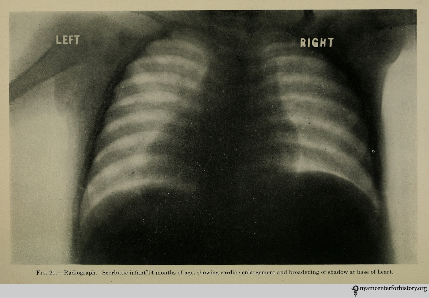
From Hess, Alfred F., M. D. Scurvy, past and present. Philadelphia, J.B. Lippincott Company, 1920.
In my novel, I paired the fictional Dr. Solomon with a character closely based on a real orphanage pediatrician of the time. Dr. Alfred F. Hess was attending physician to the Hebrew Infant Asylum and a renowned researcher into childhood nutritional diseases. He was the innovator of an infant isolation ward at the orphanage in which babies were kept in separate glassed-in rooms to avoid the spread of disease. Hess is well-known for a quote in which he extolled the advantages of conducting research on “institutional children” who provided the advantage of belonging to “the same stratum of society,” being “reared within the same walls,” and having the “same daily routine, including similar food and an equal amount of outdoor life.” He concluded: “These are some of the conditions which are insisted on in considering the course of experimental infection among laboratory animals, but which can rarely be controlled in a study in man.”7

Glassed-in babies, from Annual Report 1914 Hebrew Infant Asylum of New York.
Dr. Hess’s approach to the study of scurvy, which involved inducing the condition in children and then experimenting with various cures, was controversial even in his lifetime. In 1921, Hess was criticized “for using ‘orphans as guinea pigs’ in studies of the dietary factors in rickets and scurvy” by “withholding orange juice from institutionalized infants until they developed the characteristic small hemorrhages associated with the disease.”8

From Hess, Alfred F., M. D. Scurvy, past and present. Philadelphia, J.B. Lippincott Company, 1920.
“My name is Rachel, I’ve told you that. But you don’t care, do you? Even now, I’m just a number to you. All the children at the Infant Home were nothing more than numbers to you.” I thought of the tattoo on Mr. Mendelsohn’s frail arm. “Just numbers, like in the concentration camps.”
She gripped the sheets. “How can you say such a thing? You were in an orphanage, not some concentration camp. They took care of you, fed you, clothed you. Jewish charities support the best orphanages, the best hospitals. Even this Home is as good as it gets for old people like me. You have no right to even mention the camps.”
Of course the orphanage wasn’t a death camp, I knew that, but I wasn’t backing down. “You came into a place where we were powerless, you gave us numbers, subjected us to experiments in the name of science. How is that different?”9
When I would tell people about the medical experimentation on children depicted in my novel, they would often say it sounded like something the Nazis would do. As first I was impatient with the comparison: these experiments were conducted well before the rise Hitler in Germany, and the doctors conducting the research, many of them Jewish themselves, intended to advance medicine for the benefit of all children. Yet, as I thought about it from the point of view of one of the child subjects, I wondered if that distinction would matter.
It is easy for contemporary readers to conflate all medical experimentation on children with the atrocities of the Holocaust, but even after “the world was outraged at the murders carried out in the name of science by Nazi physicians during World War II,”10 some American doctors continued to use orphans, prisoners, and other disenfranchised populations in medical research without their consent. In my novel Orphan #8, I bring this aspect of medical history to general readers through the use of narrative and story. Medical students and physicians may also find that fiction provides an opportunity to explore these complex issues with empathy and imagination and to engage a wider community in the discussion of medical ethics.
References
1. van Alkemade, Kim. Orphan #8 (New York: William Morrow, 2015), 232.
2. Lederer, Susan E. and Michael A. Grodin. “Historical Overview: Pediatric Experimentation.” In Grodin, Michael A. and Leonard H. Glantz. Children as Research Subjects: Science, Ethics, and Law (New York: Oxford University Press, 1994), 10.
3. Lederer and Grodin, 19-20.
4. Executive Committee Minutes 1909-1930. Hebrew Orphan Asylum Collection, Archives of the American Jewish Historical Society, Center for Jewish History, 15 West 16th Street, New York, NY.
5. van Alkemade, 173.
6. The Bulletin of the New York Academy of Medicine. September 19 (1943): 676.
7. Lederer, Susan E. “Orphans as Guinea Pigs: American Children and Medical Experimenters, 1890-1930.” In Roger Cooter, ed. In The Name of the Child: Health and Welfare, 1880-1940 (New York: Routledge, 1992), 115.
8. Lederer and Grodin, 13.
9. van Alkemade, 282.
10. Lederer and Grodin, 16.