
Heidi Knoblauch, the author of today’s guest post, is our 2014–2015 Klemperer Research Fellow. She is a Ph.D. candidate in the History of Science and Medicine Department at Yale University.
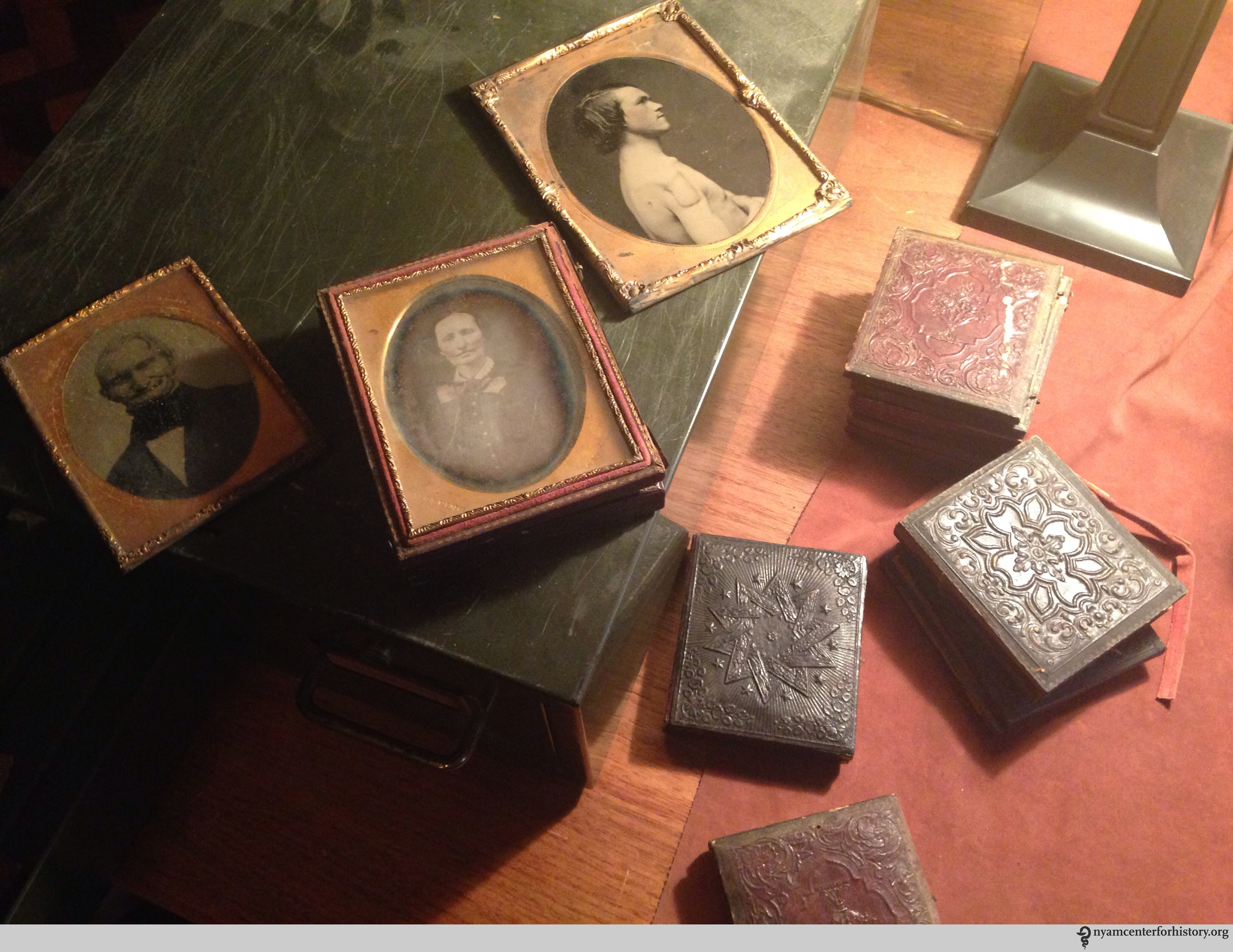
Tucked away in the New York Academy of Medicine’s special collections is a small green metal box, simply labeled “daguerreotypes.” The box contains twelve photographs and one painting. A few are images of doctors, but most are of patients.

The small green metal box, simply labeled “daguerreotypes.” Click to enlarge. Photo by Heidi Knoblauch.
You would not necessarily know these photographs were of patients unless you looked closely for a misshapen nose, outline of an excision, or nondescript facial scars. The subjects’ posing more closely resembles 19th-century photographic portraits circulated between family members than the poses we currently associate with a clinical image. These poses are accentuated by the fact that most of the photographs are housed in hinged frames with gold matting.
These photographs straddle the line between the medical and the personal that was becoming more defined during the 19th century. They blend intentional subjectivity with a new technology used to make what contemporary physicians described as a “more perfect record.”
During the 19th century, medical men collected photographs of patients and pasted them into personal scrapbooks, case records, and put them on display. These personal collections of notable cases represent not only the use of photographic technologies in consultation, but also the continuation of an engrained practice of collecting that began long before the advent of the daguerreotype. Like all archives and collections, they highlight the inclusion of things meant to be remembered and exclusion of things meant to be forgotten.
Another view of the special collection. Click to enlarge. Photo by Heidi Knoblauch.
Tracking the social practices associated with amassing medical collections is crucial for understanding this small box of photographs, almost all of which lack identifying information. These photographs have the potential to help us sketch out the formation of communities of collecting and exchange during the middle of the 19th century and to think about how doctors interpreted their relationships with their patients.
The famed surgeon Valentine Mott was one of many physicians who collected surgical and pathological specimens—including the images in the small green box. His museum, which was located at the University Medical College, was composed mainly of pathological specimens from surgical operations, collected in part from his students, who submitted dissections through an annual competition. Like many of his contemporaries, Mott thought collecting would advance the surgical art. In 1858, he declared that his collection was “believed to be the largest that any American surgeon had the occasion to form.”
Mott also sought photographs from his students. Although most of the examples in the small box are unmarked, one of Mott’s students, Edward Archelaus Flewellen, labeled a photograph he sent Mott: “A.P Jackson, Thomaston, Georgia. A supposed case of subcutaneous aneurism by anastomosis. Referred to Dr. Mott by E.A. Flewellen.”
In 1856, Flewellen sent a letter with this daguerreotype to his instructor to obtain a consultation for his patient. Flewellen told Mott that he “did this reluctantly” because he was sure that Mott was “taxed by frequent consultation by many of the thousands of students who have had the pleasure and benefits of [his] instruction.” But, Flewellen added, he believed that Mott would find this an interesting and rare case.

The note and daguerrotype Dr. Flewellen sent to Dr. Mott. Click to enlarge. Photo by Heidi Knoblauch.
Flewellen’s patient, A.P. Jackson, was a 33-year-old mechanic from Georgia who developed a tumor over his right eye when he was very young. Flewellen described the case in great detail, saying that he had watched the tumor grow for the past five years. Flewellen asked Mott what surgical treatment he would recommend to “rid this poor young man of this hideous deformity” and then promised to send Mott another daguerreotype of Jackson if the surgery was successful so Mott could contrast the before and after photographs. There is no record of Mott replying to Flewellen.
Patient photographs began to represent a new type of scientific aesthetic practice, aligned with graphs and charts, during the 1870s. Patients contributed photographs to their case records during the 19th century, but by the 1890s patients became less willing to actively participate in creating a photographic record of their disease. Today, many patients—especially in genetics, plastic surgery, and dermatology departments—have their photographs taken by a physician or technician (with a digital camera of course) to include in their electronic medical record. Yet employing a professional photographer to take a photograph with the express purpose of mailing it to a physician would seem odd to most people today.
Concerns about privacy surfaced at the end of the 19th century, which changed the way patients thought about photography in the clinic. Standards for clinical photography emerged during the 1920s and, because of this, we would find it strange to have a clinical photograph taken with a piece of bone or a bullet. Photographs are now more sterilized than they were in the 19th century and, unlike in the case of Flewellen, patients are rarely told to dress up before being photographed. The culture of photography has changed and, with it, the way physicians use photographs has shifted.


