
Todays’ guest post is introduced by Maddy Rosenberg, curator and founder of CENTRAL BOOKING. The New York Academy of Medicine Library and CENTRAL BOOKING collaborated on the exhibition Plant Cure. For this exhibition, five artists were selected to do research at the Academy Library over six months to produce work with their own unique take on medicinal plants. The project will culminate with an exhibition at CENTRAL BOOKING on the Lower East Side from September 6-October 29, 2017.
I approached Lisa O’Sullivan, the Director of the New York Academy of Medicine Library, who I had first met when she participated in one of our panels at the gallery, with an idea for a collaborative project. An important component of CENTRAL BOOKING’s programming has always revolved around art and science as well as artist’s books, therefore a collaboration with the New York Academy of Medicine seemed only natural.
For the project, ultimately named Plant Cure, five artists were selected to do research at the Academy Library over six months to produce work with their own unique take on medicinal plants. The project will culminate with an exhibition at CENTRAL BOOKING on the Lower East Side from September 6-October 29, 2017, featuring the work of the artists in dialog with other artists who have also been intrigued by the theme in their own work. At the Academy, display cases document the research, source material, and working methods employed by each of the five artists in the process of creating their work for Plant Cure.
Over the next few weeks, I am pleased to be able to present here those five artists as they discuss their work and time at the Academy Library. This week we begin with James Martin and Nancy Campbell, both whose final project work is in printmaking, but through very different approaches and results.
James Martin
My questions: how have artists and anatomists from the past chosen to depict what lies beneath the surface of the body? How have botanists and artists portrayed the plants thought to have curative properties? What are the common design elements of these life forms? Have the different printing processes changed the nature of this visual information? And my creative query—how can I re-purpose these incredible pictures from the Academy Library and create something completely new?
I narrowed my focus to anatomical texts that explored arterial and venous networks, attracted to the obvious analogies to plant forms. Historical Collections Librarian Arlene Shaner was able to suggest many fascinating volumes, such as:
The crisp and stylized engravings of John Lizars (1825) use red and blue colors to graphically present the networks of veins and arteries. Antonio Scarpa’s large engravings on the subject of aneurysms are arranged with clarity and artfulness. Closeups of these lethal defects are beautifully abstract. Lithographs of arteries by Richard Quain and Joseph Maclise (1844) have a more poignant quality. The cadavers are not generic bodies, but individuals, often young. Instruments of dissection are part of the still life. Another completely different, but fascinating approach, is Wilhelm Braune’s Topographical Atlas (1888). The color lithographs are accurate renderings from frozen slices of cadavers. Our modern MRI imaging is the closest analogy. Some of these butcher shop portions produce a shiver of revulsion. But, the images are flat and the resulting shapes allow for alternate design opportunities.

Torso from Frederich Tiedemann’s Explicationes tabularum arteriarum corporis humani (1822).
For my exploration of medical botanicals, I began with the line woodcuts of Fuchs (1542). It could be used as a field guide today such is the clarity and accuracy of its observations. The engravings in William Woodville’s Medical Botany (1793) are even more detailed and nuanced. Structures are clear and complete from root to flower. The addition of color in the Henry Trimen and Robert Bentley’s Medicinal Plants (1880) imparts an even more lifelike quality to the illustrations.

Hellebore from William Woodville’s Medical Botany (1793).
As part of my creative process, I took digital photographs of plates contained in the above described books. Back in my studio, I work with these photos with editing software. Beginning with anatomical images, I establish the “bones” of the composition. These are layered with my photographs of tree bark to provide textures, shapes, and a non-specific context, with the relevant botanicals added to the mix. The finished piece was then printed via an inkjet printer on printmaking paper. I added another element with the application of monotype inks printed from mylar over the digital prints for a slight softening of the sharpness and more richness to the color.


Tree bark photograph used in Torso with Hellebore (Left). Monotype plate for Torso with Hellebore (Right).
My creative mash-ups of these historic images have been inspiring and fun. Thanks to all at the Academy for hosting this project and to Maddy Rosenberg of CENTRAL BOOKING for organizing this residency and the upcoming exhibition Plant Cure.
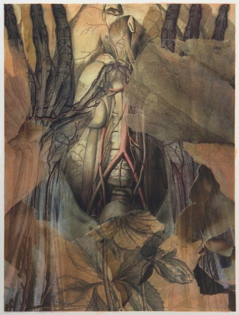
Torso with Hellebore by James Martin archival digital print with monotype.
Nancy Campbell
I absolutely adored my time spent in the Drs. Barri and Bobbi Coller Rare Book Reading Room at the New York Academy of Medicine. Handling objects so old, delicate, and precious was a rare treat, indeed.
While I enjoyed studying an array of different volumes in the Academy Library, Okamoto Ippo’s Jūshi kei ryaki wago (1693; 3 vol. book of Moxa-cautery) was a perfect match for me. Medieval Japanese picture scrolls have been a long fascination, and I have studied them in museum exhibitions in Japan and the USA. Of course, I have never held an actual medieval scroll and experienced the sequential unfolding of its story (scrolls being so incredibly fragile). Therefore, handling a 17th century Japanese book during my residence, with its ultra-thin, semi-transparent printed paper, was an amazing first-time experience for me and one that will surely affect my work for years to come.
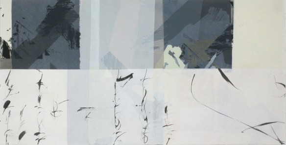
Artemisia by Nancy Campbell.
In my artwork I strive to evoke an Eastern sense of balance between fragility and strength while using a system of highly structured, intricate abstraction. My methods are slow and measured, but I work for a spontaneous result that inhabits an ambiguous realm between the visible and invisible, the logical and the intuitive, the representational and the abstract. Echoed in all of my work is a continuous play of opposites – often found at the heart of Japanese aesthetics.

Meridian by Nancy Campbell.
My work for the Plant Cure exhibition references text and diagrams that appear to be layered on top of one another. Each page in the Japanese books I viewed has hints of the previous page showing through the thin Japanese paper. I printed and painted on both sides of Japanese papers and used the method of collage (with Japanese glue) to layer multiple sheets together. A large screenprint based on a collage is still in process.

Pingback: Artist Inspiration: Plant Cure (Part 2) | Books, Health and History
Pingback: Artist Inspiration: Plant Cure (Part 3) | Books, Health and History