Jacques Gamelin’s Marauding Skeletons and Écorché Crucifixions: Guest Post by Morbid Anatomy

Gamelin 1

Gamelin 1

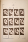
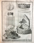
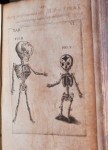
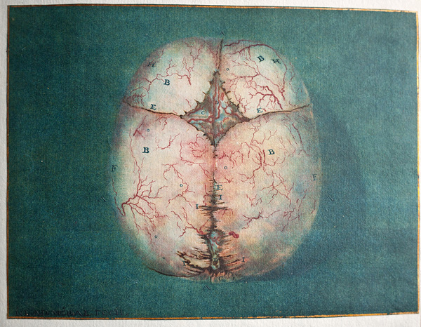
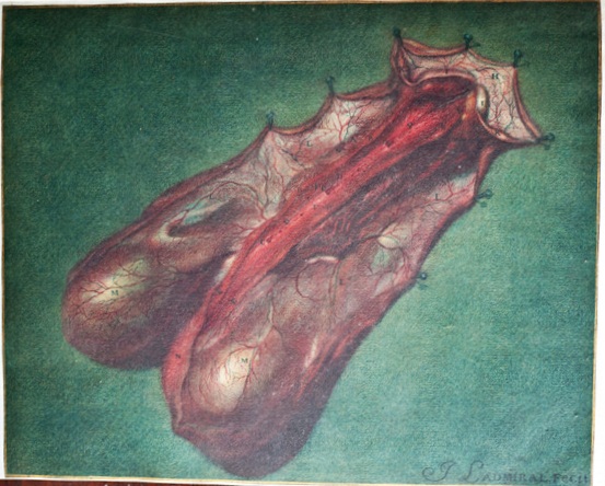
I was so excited to finally have the opportunity to pore over a book in the NYAM collection which I had long admired from afar: Jacques Gamelin‘s beautiful and lavish Nouveau recueil d’osteologie et de myologie (“New collection of osteology and mycology”) of 1779. This book, as explained to me by Arlene Shaner—acting curator and reference librarian for historical collections, at NYAM—was intended as a large scale, deluxe manual for artists interested in understanding human anatomy in order to create more convincing depictions of human figures.

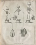
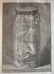
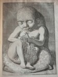
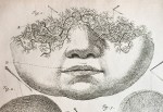
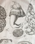
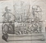
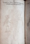
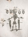
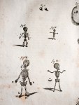
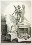
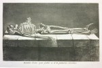
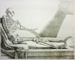
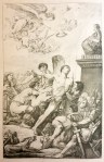
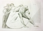
The meat of the book, as it were, is a collection of extremely virtuosic anatomical renderings (12-15) showing skinned—or écorché—human figures in a variety of poses. But what is much more interesting—at least to me—is the assortment of animated skeletons which fill the opening pages; these fanciful figures are engaging in such activities as waking up in a cemetery to the trumpet of the resurrection (text reading: surgite mortui venite ad judicium, or “Rise up, come to the judgment of the dead”; image 6); brandishing anatomical drawings in what looks to be a dissection room littered with bones (3); and raping and pillaging the parties of fashionably bewigged lords and ladies (9, 10). I am also very drawn images playing on biblical themes, such as a calm Saint Bartholomew being flayed alive (an old staple of anatomical illustration; image 11), and a skinned and anatomized Christ on the cross (12) which evokes this more literal rendition, cast from a convicted murderer just a few decades after this book was published.

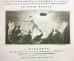
Interestingly, Gamelin is best remembered today not for this book, but as a painter and engraver of battle scenes, genre scenes, and portraits. He dedicated this book to his mentor, a certain Baron de Puymaurin (image 1 and 2), who had recognized Gamelin’s artistic abilities and funded his training when his father refused to do so. It is thought that Gamelin funded the book himself after inheriting a great deal of money upon his father’s death, which perhaps accounts for its delightful eccentricity.

This post was written by Joanna Ebenstein of the Morbid Anatomy blog, library and event series; click here to find out more.

Item of the Month: “Better Babies” on Things Which Are Bad For All Babies

By Johanna Goldberg, Information Services Librarian

When in the stacks recently, I came across two slim issues of “Better Babies: Infant Welfare and Race Progress,” one published in December 1921 and the other in April 1924.

BetterBabies_Cover-1921

BetterBabies_Cover-1924

While the title suggests an interest in eugenics, the two issues in our collection focus solely on ways to keep babies healthy, including articles on clothes for children, playgrounds and public health, the benefits of breast feeding, and disease prevention.

This last topic inspired the following list, published in the 1924 issue. As it is (coincidentally) Baby Safety Month, it seems appropriate to share it.

Which piece of advice is your favorite?

Identification Anthropométrique, Alphonse Bertillon, 1893; Guest Post by Morbid Anatomy

Title pageAnother fascinating book from the NYAM’s holdings is French criminologist and anthropologist Alphonse Bertillon’s Identification anthropométrique; instructions signalétiques of 1893. Alphonse Bertillon (1853–1914) invented a criminal identification system using photography and physical measurements which allowed police to document and convict repeat criminals. His system was called signaletics or bertillonage and involved recording certain traits, such as measurements of the head and body and the shape of the ear, and converting them into a unique formula. The formula was paired with a photo of the individual and filed. The system was successful and widely used, but errors were possible, since different officers might obtain different measurements and bodies can change, and eventually fingerprints became the preferred method of identification. Bertillon also introduced the systematic use of photography to document crime scenes. The reductionism of such medicalized views of the body went on to inspire the works of the surrealists, as seen in works such as Salvador Dali’s “The Phenomenon of Ecstasy” (see last image) of 1933.

This post was written by Joanna Ebenstein of the Morbid Anatomy blog, library and event series; click here to find out more.

“Artist of Death” Frederik Ruysch at NYAM: Guest Post by Morbid Anatomy’s Joanna Ebenstein

Ruysch1

1720 frontispiece to Opera Omnia, 1721.

My very favorite figure operating at the intersections of art and medicine–and probably the most bizarre to the modern eye–is Dutch anatomist, artist, preparator, and early museologist Frederik Ruysch (1638-1731). A pioneer in the art of preserving the human body, he was famed for his uncannily life-like and imaginative human preparations (i.e. bits of bodies preserved for study) which he achieved through a combination of injections of colored wax and a secret alcohol-based preservation formula. He is best remembered today for his lavish memento mori-themed tableaux utilizing real human fetal skeletons and other bits of human remains (see images 15-18) which are beautifully explained by Steven Jay Gould in his book Finders, Keepers: Eight Collectors:

Ruysch made about a dozen tableaux, constructed of human fetal skeletons with backgrounds of other body parts, on allegorical themes of death and the transiency of life… Ruysch built the ‘geological’ landscapes of these tableaux from gallstones and kidneystones, and ‘botanical’ backgrounds from injected and hardened major veins and arteries for “trees,” and more ramified tissue of lungs and smaller vessels for ‘bushes’ and ‘grass.’

The fetal skeletons, several per tableau, were ornamented with symbols of death and short life–hands may hold mayflies (which live but a day in their adult state); skulls bemoan their fate by weeping into ‘handkerchiefs’ made of elegantly injected mesentery or brain meninges; ‘snakes’ and ‘worms,’ symbols of corruption made of intestine, wind around pelvis and rib cage.

Quotations and moral exhortations, emphasizing the brevity of life and the vanity of earthly riches, festooned the compositions. One fetal skeleton holding a string of pearls in its hand proclaims, ‘Why should I long for the things of this world?’ Another, playing a violin with a bow made of a dried artery, sings, ‘Ah fate, ah bitter fate.’

Rusych showcased his thousands of human preparations in his own cabinet of curiosities visited by medics and philosophers, as well as members of the aristocracy and royalty. Here, one could see not only his fantastic tableaux, but also his imaginative human specimens in glass jars, preserved organs, exotic birds, butterflies and plants. Ruysch published several lavishly illustrated guides to his cabinet; in image 1, above, you can see an allegorical view of his museum as depicted in a frontispiece to his Opera Omnia, 1721.

Very sadly, none of Ruysch’s astonishing tableaux are known to exist today, and are only known to us through book illustrations. One can get a sense of what the real thing probably looked like, however, in image 19, a contemporaneous 17th fetal skeleton tableau emblazoned with a memento-more-themed Virgil quote; this photo was featured in my recent exhibition The Secret Museum, on which you can find out more here. You can also still see visit many of Ruysch’s wet preparations in collections such as the St. Petersburg Kunstkammer (which has 916 of them), Museum Bleulandinum in Utrecht, and the Anatomisch Museum LUMC in Leiden.

All of these images, save the photo, are drawn from the exceptionally rich Ruysch holdings of the NYAM Historical collections. Hear more about Frederick Ruysch at the October 5th Festival of Medical History & the Arts, when Daniel Margoscy will speak on “The Anatomy of the Corpse: Ruysch, Descartes, and the Problem of Wax.”

This post was written by Joanna Ebenstein of the Morbid Anatomy blog, library and event series; click here to find out more.

“Most Wonderful and Glorious Collection of Anatomical Matter in the World:” Popular Anatomy at NYAM; Guest Post by Morbid Anatomy

Grand Anatomical Museum

“Six splendid female figures, size of life… the EXQUISITE FORM in all its natural delicacy… and consummate BEAUTY which ever has and ever will captivate the heart of man.”

Above is a fantastic piece of ephemera housed in the NYAM Historical Collections which was recently brought to my attention by Arlene Shaner, Acting Curator of Rare Books & Manuscripts at NYAM.

This is a handbill advertising New York City’s Grand Anatomical Museum, one of the many for-profit, open to the pubic anatomical museums which were operating in New York and other European and American cities in the 19th and early 20th centuries. These collections were popular with the general public; they were both educational and spectacular, and often showcased objects of a titillating bent such as beautiful, unclothed wax women with real hair and glass eyes called Florentine, Parisian or anatomical Venuses (more on these fabulous creatures here), human freaks and–at a time when syphilis was both widespread and incurable–lurid wax depictions of genitalia deformed by venereal disease. These last could be found, more often then not, in a special “gentleman’s only” chamber.

Such museums were initially lauded by the medical establishment as excellent for laymen and medics alike. However, by the late 19th century, they became increasing associated with “quack” medical practitioners, who would use them as an kind of advertisement for their often mercury-based cures for sexuality transmitted diseases. Eventually, most of these museums were closed down–or even destroyed–under anti-obscenity laws.

Grand Anatomical Museum 2

You can find out more about popular anatomical museums in this article and book by Michael Sappol, who will be participating in NYAM’s upcoming Festival of Medical History and Arts on October 5th.  They were also explored in The Wellcome Collection’s 2009 exhibition Exquisite Bodies (for which I acted as curatorial consultant), and Maritha Rene Burmeister’s wonderful dissertation on the topic.

This post was written by Joanna Ebenstein of the Morbid Anatomy blog, library and event series; click here to find out more.

Theatrum Anatomicum, by Caspar Bauhin and Theodor de Bry, 1605 : Guest Post by Morbid Anatomy’s Joanna Ebenstein

Bauhin1Here are some wonderful images from another of my favorite books in the NYAM historical collections, Theatrum Anatomicum (1605), by Caspar Bauhin and Theodor de Bry. This edition also has some especially lovely image ghosting going on.

This post was written by Joanna Ebenstein of the Morbid Anatomy blog, library and event series; click here to find out more.

Icon durae matris in concavâ superficie visae…, Jan Ladmiral and Frederik Ruysch, 1738: Guest Post by Morbid Anatomy’s Joanna Ebenstein

Here is a lovely small frontispiece etching and some lush and wonderful mezzotints by Jan Ladmiral, pioneer of early color printing, drawn from the NYAM copy of Icon durae matris in concavâ superficie visae, ex capite foetus humani octò circiter à conceptione mensium, desumtae; ad objectum artificiosissimè praeparatum à clarissimo viro Fred. Ruyschio by Jan Ladmiral and Frederik Ruysch from 1738.

Ladmiral1

Ladmiral3 Ladmiral2

Ladmiral4

This post was written by Joanna Ebenstein of the Morbid Anatomy blog, library and event series; click here to find out more.

Item of the Month: The Medical Museum, Mythology and Medicine

By Arlene Shaner, Acting Curator and Reference Librarian for Historical Collections

Medical Museum title page 1763

Recently, while looking for something in the rare periodicals collection at NYAM, I came across a charming allegorical frontispiece that appeared in the first volume of The Medical Museum, a short-lived journal that appeared in three volumes published in London 1763 and 1764.  The editors of the journal, who described themselves as “Gentlemen of the Faculty,” remain unknown.  The subtitle and the introduction make clear that they viewed themselves primarily as the collectors and disseminators of already published works from a variety of sources. 

Even 18th century people struggled to make sense of an overwhelming amount of information.  “Many physical people very justly complain of the great expense attending the purchase of medical treatises, especially foreign ones… the pains and time to select and examine the matters that may concern their profession, are with many too much to be dispensed with,” (ix) they noted, while explaining the task they had chosen for themselves, that of serving as the selectors of the most useful materials from disciplines ranging from anatomy, medicine, chemistry, botany and other assorted sciences.  Works from a remarkable range of publications appear in the Museum, many of them translated from their original languages into English to make them more accessible, as the compilers hoped their journal would find an audience among the public, not just among medical men.

Medical Museum frontispiece 1763

The first volume contains a specially engraved frontispiece that shows Apollo bringing his son Asclepius to the centaur Chiron to learn about the art of medicine.  Coronis, Asclepius’ mother, was either killed by Apollo for being unfaithful to him or died in childbirth, and Apollo rescued the unborn baby from her womb.  Needing someone to raise the boy, Apollo handed him over to Chiron, who taught him the healing arts.  Asclepius went on to father many daughters, some of whom are also remembered for their connections to medicine and health.  One of his daughters, Hygeia, is the goddess of health, while another, Panacea, is the goddess of universal remedies.

If you visit The New York Academy of Medicine’s building, you will see that Asclepius and Hygeia were important touchstones for NYAM and for the building’s designers as well.  An ornamental frieze above our front door depicts the two of them together, attended by their snakes and dogs, a visual reminder of the classical heritage of medicine.

A&H

Eighty Years and Counting

Gallery

This gallery contains 4 photos.

By Arlene Shaner, Acting Curator and Reference Librarian for Historical Collections Many of you are aware that the Malloch Suite of rare book rooms (the Coller Rare Book Reading Room and the Seminar Room) has been under renovation since early … Continue reading

Festival of Medical History & the Arts

We are excited to announce our first all-day extravaganza, co-curated by Lawrence Weschler, Morbid Anatomy, and the Center, and featuring esteemed speakers, artists workshops, behind-the-scenes tours, and more. Please check the Festival of Medical History & the Arts page and schedule for more information.

save the date Oct 5