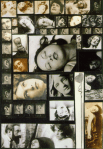
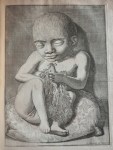
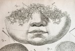
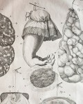
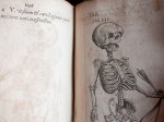
My very favorite figure operating at the intersections of art and medicine–and probably the most bizarre to the modern eye–is Dutch anatomist, artist, preparator, and early museologist Frederik Ruysch (1638-1731). A pioneer in the art of preserving the human body, he was famed for his uncannily life-like and imaginative human preparations (i.e. bits of bodies preserved for study) which he achieved through a combination of injections of colored wax and a secret alcohol-based preservation formula. He is best remembered today for his lavish memento mori-themed tableaux utilizing real human fetal skeletons and other bits of human remains (see images 15-18) which are beautifully explained by Steven Jay Gould in his book Finders, Keepers: Eight Collectors:
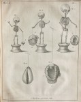
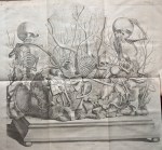
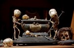
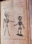
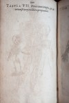
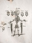
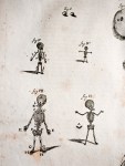
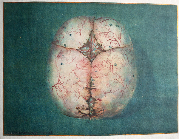
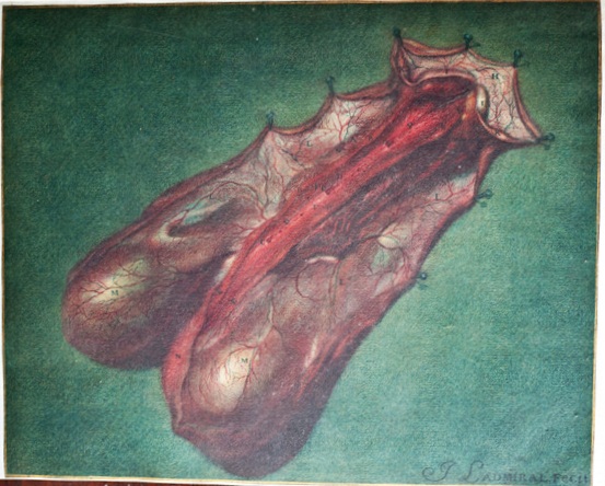
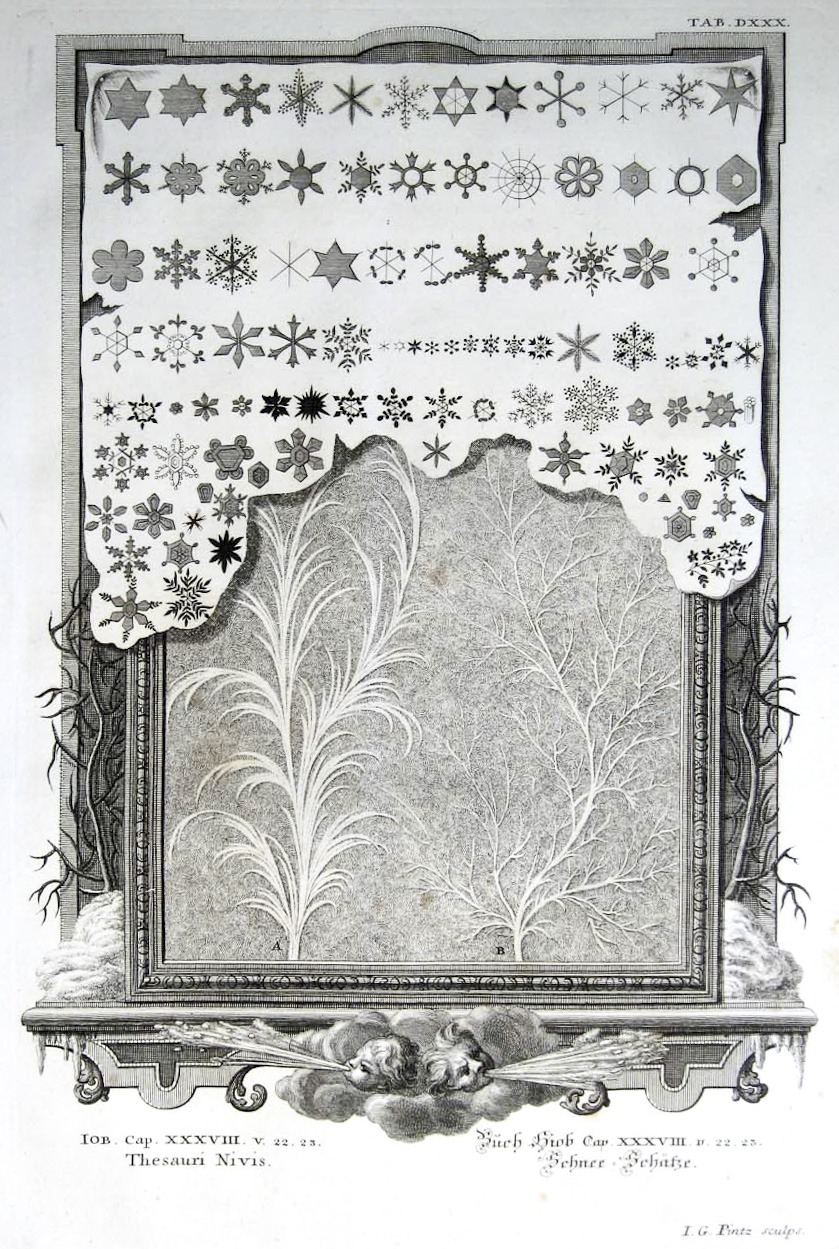
Ruysch made about a dozen tableaux, constructed of human fetal skeletons with backgrounds of other body parts, on allegorical themes of death and the transiency of life… Ruysch built the ‘geological’ landscapes of these tableaux from gallstones and kidneystones, and ‘botanical’ backgrounds from injected and hardened major veins and arteries for “trees,” and more ramified tissue of lungs and smaller vessels for ‘bushes’ and ‘grass.’
The fetal skeletons, several per tableau, were ornamented with symbols of death and short life–hands may hold mayflies (which live but a day in their adult state); skulls bemoan their fate by weeping into ‘handkerchiefs’ made of elegantly injected mesentery or brain meninges; ‘snakes’ and ‘worms,’ symbols of corruption made of intestine, wind around pelvis and rib cage.
Quotations and moral exhortations, emphasizing the brevity of life and the vanity of earthly riches, festooned the compositions. One fetal skeleton holding a string of pearls in its hand proclaims, ‘Why should I long for the things of this world?’ Another, playing a violin with a bow made of a dried artery, sings, ‘Ah fate, ah bitter fate.’
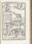
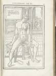
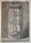
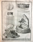
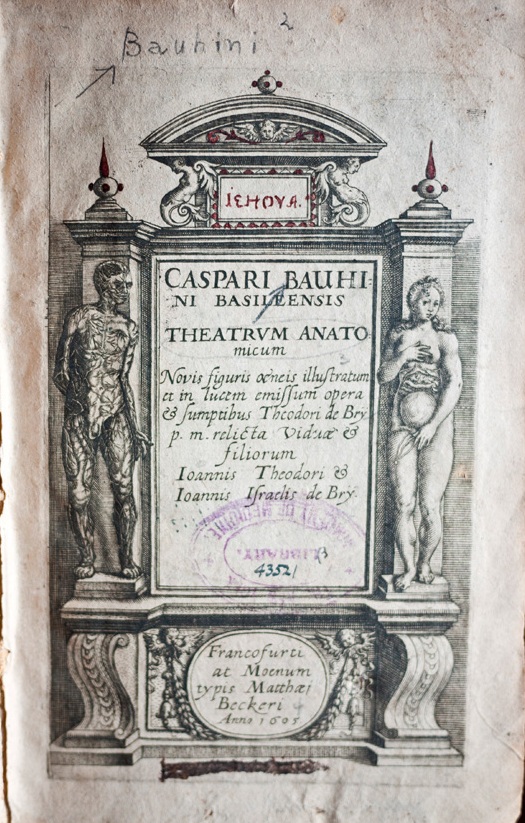
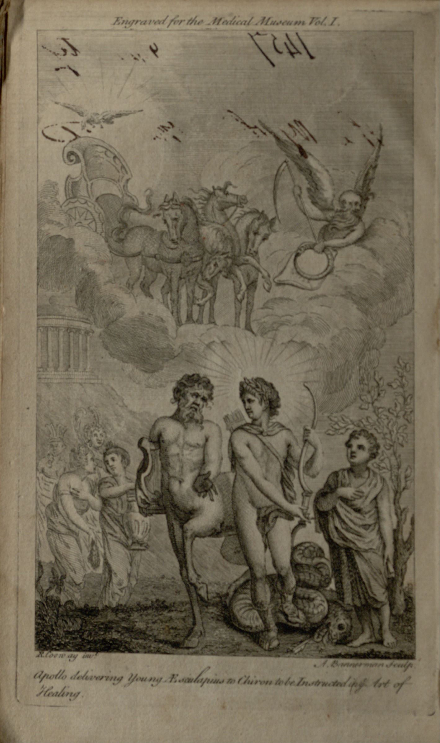
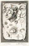
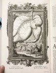
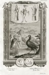
Rusych showcased his thousands of human preparations in his own cabinet of curiosities visited by medics and philosophers, as well as members of the aristocracy and royalty. Here, one could see not only his fantastic tableaux, but also his imaginative human specimens in glass jars, preserved organs, exotic birds, butterflies and plants. Ruysch published several lavishly illustrated guides to his cabinet; in image 1, above, you can see an allegorical view of his museum as depicted in a frontispiece to his Opera Omnia, 1721.
Very sadly, none of Ruysch’s astonishing tableaux are known to exist today, and are only known to us through book illustrations. One can get a sense of what the real thing probably looked like, however, in image 19, a contemporaneous 17th fetal skeleton tableau emblazoned with a memento-more-themed Virgil quote; this photo was featured in my recent exhibition The Secret Museum, on which you can find out more here. You can also still see visit many of Ruysch’s wet preparations in collections such as the St. Petersburg Kunstkammer (which has 916 of them), Museum Bleulandinum in Utrecht, and the Anatomisch Museum LUMC in Leiden.
All of these images, save the photo, are drawn from the exceptionally rich Ruysch holdings of the NYAM Historical collections. Hear more about Frederick Ruysch at the October 5th Festival of Medical History & the Arts, when Daniel Margoscy will speak on “The Anatomy of the Corpse: Ruysch, Descartes, and the Problem of Wax.”
This post was written by Joanna Ebenstein of the Morbid Anatomy blog, library and event series; click here to find out more.


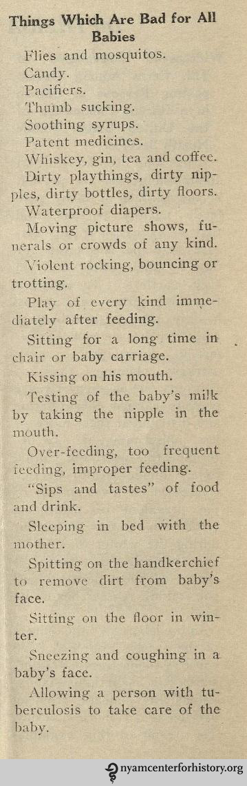 Which piece of advice is your favorite?


