Today’s post was written by the 2013 Gladys Brooks Conservation Intern, Caroline Evans.
The diploma collection at The New York Academy of Medicine contains over eight hundred certificates, diplomas, seals and proclamations granted by universities, professional societies and institutions across a wide geographical span. The items in the collection range from the mid-eighteenth century up to the late twentieth century. The diplomas were the subjects of a major collections care project carried out in the Gladys Brooks Book and Paper Conservation Lab by Caroline Evans (summer intern), with the assistance of Emily Moyer (Collections Care Assistant) and Allie Rosenthal (volunteer).

Piles of diplomas to be sorted, cleaned, and housed.
While most of the earlier diplomas are printed or written on parchment and display elaborate calligraphy, many of the later items in the collection are printed on paper. The diplomas can provide a glimpse into the changing methods of printing during this period, as well as into the preservation needs of flat paper—in some cases, for instance, some of the ink in the signatures had begun to flake, and seals on parchment were cracked. In addition to dry cleaning the diplomas and making the appropriate efforts to stabilize each of these items, we constructed folders and housing for each diploma or seal before sorting them by size, date, and granting institution.
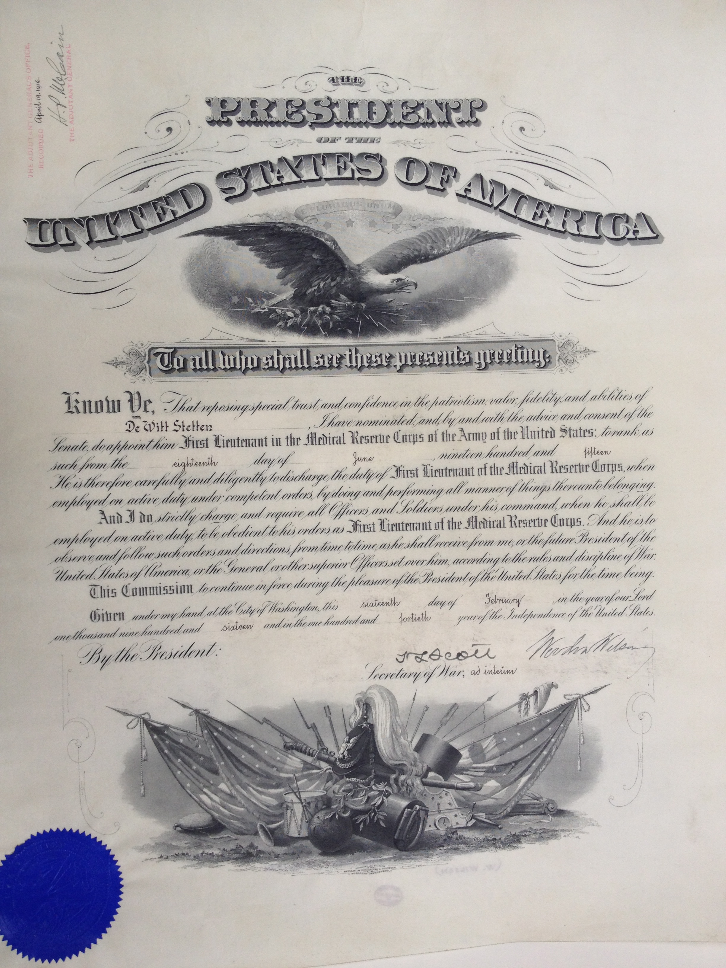
Over the course of this undertaking, some gems emerged—documents significant to the history of the Academy and to the history of medicine. Among these are certificates nominating and appointing military ranks to fellows of the Academy and other doctors serving in wartime. In addition to signatures from the “Secretary of War”, many of these documents boast signatures from various Presidents of the United States. Indeed, while sorting through the collection, we encountered wartime documents—appointments or commendations thanking military doctors for their service—with signatures from Presidents Abraham Lincoln, Andrew Jackson, Andrew Johnson, Herbert Hoover, William Howard Taft, Woodrow Wilson, and Warren G. Harding, to name a few.

Certificate signed by Woodrow Wilson.

Certificate signed by Andrew Johnson.

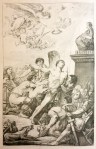
Certificate signed by Abraham Lincoln.
Occasionally found tacked onto the back of certificates and acknowledgments of service were documents indicating the intersection of military service and medical research—for example, a letter from Walter Reed Hospital to a soldier encouraging him to participate in a study on the effect of injections of yellow fever. There are also a significant number of female medical professionals whose successes and contributions to the field of medicine and women’s health are commemorated in the collection. Some of these awards and diplomas are dated as early as the nineteenth century.

Photographs of Howard and Edith Lilienthal attached to a passport.
The diploma collection contains items printed in French, Portuguese, Hungarian, Latin, Hebrew and Arabic that display a variety of design styles. One particularly beautiful certificate from 1945 was granted by the Société Impériale de Médecine de Constantinople, written in Arabic on thin paper with gold leaf.

Certificate in Arabic with gold ornament

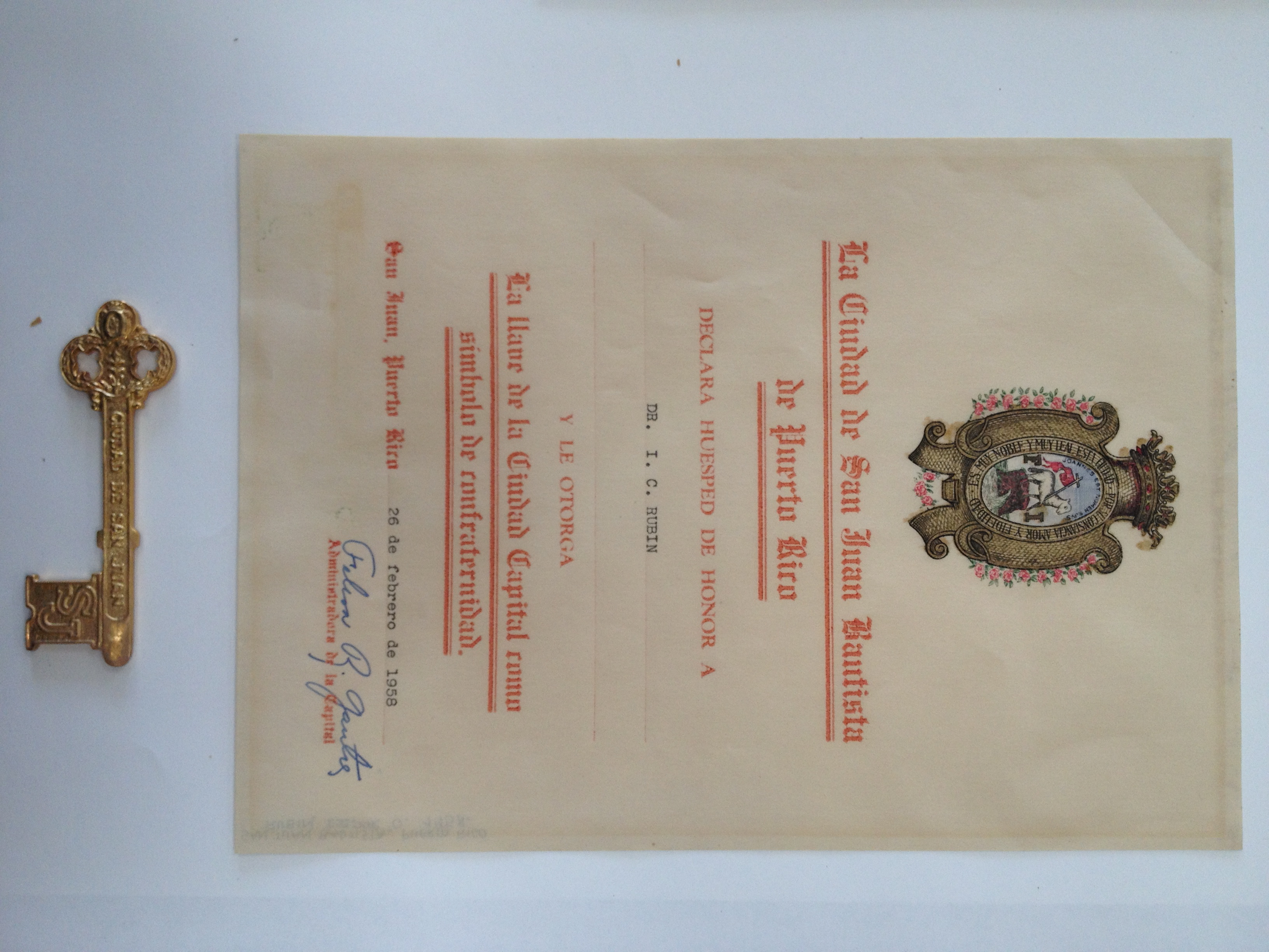
Key to the City of San Juan Batista, granted to Isidor Rubin.
Some more recent certificates are printed in color, with hand-colored borders and modern, stylized type. The diplomas on paper were a special challenge to clean and house, as many of the papers had become brittle or were adhered to acidic backings. This allowed the aspiring conservators interning and volunteering in the lab ample practice with paper repair. Diplomas printed on vellum provided their own challenges, however, as humidity fluctuations over time caused some of the works to curl and stretch, obscuring and fading labels and printed text.

Repairing paper certificates in the conservation lab.
These challenges, in addition to the diverse languages present in the collection, necessitated some additional investigation for the creation of new labels for each item. In the end, though, the lab was able to create a location guide with the identifying information for each sorted, cleaned, and re-housed object, so that the diploma collection will be accessible well into the future.

A certificate and seal, re-housed.