The Talented Dr. Knox

Lisa Rosner, PhD, author of today’s guest blog, will present “The True and Horrid Story of the Burke and Hare Anatomy Murders” at our October 18th festival, Art, Anatomy, and the Body: Vesalius 500.

Engraving of Dr. Robert Knox. From our online collection The Resurrectionists.

Engraving of Dr. Robert Knox. From our online collection The Resurrectionists.

Dr. Robert Knox, the anatomist whose cadaver purchases kept William Burke and William Hare in the murder business, has always been an enigma. Born in Edinburgh, Scotland, he served in the army and studied in Paris before returning home to set up a private anatomical school. He taught hundreds of students, lecturing twice a day in addition to holding separate dissection classes. He was curator of the surgical museum, wrote articles on human and comparative anatomy for scientific societies, and was in the process of seeing several books on anatomy through publication. His supporters claimed he knew nothing about the murders; his detractors argued that he simply turned his blind eye—for he had lost an eye to smallpox as a child.

Plate II in Knox's Man: His Structure and Physiology, shown flat and with lifted parts. Click to enlarge.

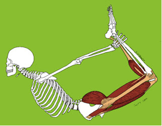
Plate II in Knox’s Man: His Structure and Physiology, shown flat and with lifted parts. Click to enlarge.

What we can see, using the extensive collection of Robert Knox materials in the New York Academy of Medicine Library, is just how talented an anatomist Robert Knox was. His edition of Hippolyte Cloquet’s A System of Anatomy is more than just a translation: it is instead a critical analysis of contemporary anatomical knowledge, enriched by examples from Knox’s own research and teaching. The same is true of his edition of Friedrich Tiedemann’s The Plates of the Human Arteries, prepared with two of his students, Thomas Wharton Jones and Edward Mitchell. The catalogue he prepared for the anatomical and pathological museum of the Royal College of Surgeons of Edinburgh is filled with his detailed insights: on anomalies of the biceps flexor cubiti, on the precise position relative of a fatal brain tumor, and on popliteal aneurism. Knox discussed the implications of these, and many more of his anatomical and surgical observations, in several series of articles for the London Medical Gazette. We can follow his teaching methods in The Edinburgh Dissector, the handbook he wrote for the use of his dissecting classes. “Nobody could ever say that he gave a dry lecture, or one that was not specially instructive,” reported his former student, Henry Lonsdale. Even in the midst of the detailed description that makes up most of the Edinburgh Dissector, Knox’s love of his subject shines through, as in his description of the bones of the foot, which “when well formed yields in beauty and perfection to no part in the human body.”

Could such a passionate observer of all subjects anatomical really have missed the fact that sixteen of his own “subjects” had been murdered? Contemporaries from Home Secretary Sir Robert Peel (founder of the London Metropolitan Police) to the Edinburgh evening papers refused to believe it and called for wider investigation. On the advice of legal counsel, Knox refused to answer any questions—just as he had refused to ask any, his professional rivals muttered darkly, when presented with Burke’s and Hare’s murder victims. There was no real case against him, and there are no records of any deliberations by the prosecuting attorneys. We will probably never know what Knox knew or when he knew it.

"Execution of the notorious William Burke the murderer, who supplied Dr. Knox with subjects." Engraved print in The Resurrectionists collection. Click to enlarge.

“Execution of the notorious William Burke the murderer, who supplied Dr. Knox with subjects.” Engraved print in The Resurrectionists collection. Click to enlarge.

The anatomical career of the talented Dr. Knox survived the Burke and Hare scandal, but it did not long survive the change in medical teaching and practices that followed it. He had a second career as a public teacher and lecturer: his books A Manual of Artistic Anatomy and Great Artists and Great Anatomists: A Biographical and Philosophical Study sold very well. But he never achieved the academic position he had striven for, and his research agenda, like his sixteen most famous subjects, died at the hands of Burke and Hare.

For more on Robert Knox and the Burke and Hare murders, visit our online collection, The Resurrectionists.

On Presenting Resisterectomy

Chase Joynt, co-author of today’s guest blog with Dr. M. K. Bryson, will present Resisterectomy at our October 18th festival, Art, Anatomy, and the Body: Vesalius 500.

Chase Joynt, left, and Dr. M. K. Bryson, right.

Chase Joynt, left, and Dr. M. K. Bryson, right.

Chase Joynt:

I announced my desire to find a collaborator for my then-still-hypothetical project Resisterectomy at every available opportunity. Lulls in dinner party conversation were filled with the always laughter-stopping question: “Does anyone know someone who has had a mastectomy and a hysterectomy who might be willing to talk about their experiences?” Anecdotes shared about eccentric distant relatives who happened to be both cancer survivors and watercolor painters were followed up with: “Do you think that person might be interested in working on a project with me?” And friends unfamiliar with the artistic process of starting a project from a place of utter-not-knowing (and/or perhaps at best “a hunch”) continued to entertain my quest suspiciously, albeit with sympathy. The most frequent reactions to my casual inquiries were blank stares and occasional bursts of conversational sarcasm directed at the seemingly impossible identifactory requirements of the project’s specificity. One day however, after lobbing the question into a blue-couch-filled Toronto living room, I was met with an animated, sarcasm-free answer: “You need to talk to my friend Mary Bryson.” Within hours of sending Mary the initial “Hello, how are you, might you be interested in chatting about these things?” e-mail, I was met with some necessary and critical resistance.

Dr. M. K. Bryson:

When I first heard from Chase (“I am looking for a woman who has had both a mastectomy and a hysterectomy.”), I was simultaneously deeply skeptical and intensely interested in his project. And even though I really did not, and do not, “feel like a woman” I assumed that because of Chase’s up-front trans* alliances, the complexities of our potential dialogue would find plausible vocabularies if not any shared experiences. I don’t in any case expect shared experiences no matter how self-evident they may appear to be. And besides, I had by then had at least a year’s worth of experiences interviewing participants in the Cancer’s Margins research project—Canada’s first ever nationally funded research project focused on LGBT experiences of breast and gynecologic cancer.

I knew several things by then about bodies, cancer, and the impact of mastectomy and hysterectomy. For one thing, my research interviews confirmed what I learned from my own cancer experiences—that for people with histories that overlap in minor or major ways with trans* health, the simple “fact” of the double-duty these surgeries take up—that mastectomy and hysterectomy are both cancer surgeries and also surgeries related to trans* health—means that these surgeries are already much more culturally complex than is typically within healthcare providers’ understanding and training. I knew that gender is very definitely implicated in how cancer patients experience cancer-related treatments and surgeries generally, and very specifically, that cancer patients’ histories of gender will shape what is meaningful about mastectomy and hysterectomy in ways that reveal the impact of trans* culture in the larger world of gender. I have always been very fond of exploring both/and relationships that organize how people located in precarious communities experience our lives and therefore, how organizations and institutions that create systems of care need to think about caring for marginalized people.

CJ:

It has been two years since our first meeting, and Resisterectomy continues to tour galleries, festivals, and schools internationally. In May 2014, we were invited to present the work as a part of the Sexuality Studies Summer School at the University of Manchester. Unbeknownst to the organizers, the occasion marked the first time since its creation that we were able to talk about the work together in public. As a result of living and working on opposite coasts, we rely on Skype and DropBox for our project-related intimacies, and I often tour and speak alone. Presenting a collaborative work alone is a complicated and precarious endeavor. How can I speak to the specificities of the project without problematically narrating (and therefore truncating) the experiences of someone else? And yet simultaneously, how can I protect that person by speaking to the assumptions so easily made about their experiences on account of their physical absence from these encounters? After our presentation in Manchester, Mary approached me at the reception with a smile, “I didn’t know you talked about the fact that this project was hard!” I smiled, “If there is one thing that every person in every room has thus far agreed upon, it is that talking about this project is hard,” I said.

JOYNT_RESIST_POSTER

Resisterectomy poster.

MKB:

I have been thinking for a while about the academic work that I am doing concerning cancer, gender, and marginalization under the general umbrella of “An Archive of the Talking Dead.” There is something absolutely unique in my experience of talking about cancer research and cancer experiences compared to talking about any other difficult, painful, or harrowing experience. North America is in many ways a culture obsessed with cancer and with mortality—and specifically, with avoiding cancer despite the fact that almost everything we do, like aging or driving a car, is something over which we have almost no control, and which increases our risk of cancer. In Resisterectomy, there is for most people who view the multimedia installation, a story of a trans* person (Chase) and a story of a cancer patient (Mary), both of whom have had a mastectomy and a hysterectomy. But that’s not how I see it at all. I am a trans* person for whom, having a mastectomy did double-duty as breast cancer surgery. However, when Chase and I are in the same space – either because our photographs are hanging on the wall, or our faces broadcast on a screen where the Resisterectomy video feed is playing, then the inevitable assumptions about Who-is-What overwrite what can be made visible in those spaces, and the play with what might be possible is cloaked by conventions. And so there we are.

What is a residual for me, every time I hear about one of Chase’s adventures installing Resisterectomy, or talking about the art with folks, is that he and I have enacted a mode of caring for each other’s responses. Resisterectomy then acts as a kind of Live Case History where a very diverse group of people gets to think, again, about things—about stories—that might benefit from a hell of a lot more energy and creativity. Chase and I took a huge risk in just saying, “Hello. Let’s compare notes. And actually, let’s mix up these stories we think we already know how to tell.” Let’s take great care in the curation of difficult stories—from the Archive of the Talking Dead… Any doctors or nurses in the house? Pay special attention. How could you talk to your patients as if you might be very surprised to learn who they are, and how their life stories are impacted by the changes health inevitably brings? And most of all, learn to enjoy how hard it needs to be. Learn to love what you don’t yet know about me.

A Lifetime of Healthiness? The Golden Health Library’s “Seven Ages of Man” (Item of the Month)

Cara Kiernan Fallon, this post’s guest author, is a history of science PhD candidate at Harvard University.

"The seven ages of man." From The Golden Health Library.

“The seven ages of man.” From The Golden Health Library. Click to enlarge.

Childhood can be full of “vigor and zest” but “Middle age is the time when our sins against the laws of health find us out,” warned physicians writing for The Golden Health Library’s inaugural volume. Published in the late 1930s, The Golden Health Library offered readers five volumes of advice on the “principles of right living” so they could secure health throughout their lifespans.1 Authors included physicians, nurses, professors, and even birth control advocates like Margaret Sanger. September, Healthy Aging Month, is the perfect time to revisit this publication.

Part of the New York Academy of Medicine’s extensive collection of health guides, public health pamphlets, and medical magazines, The Golden Health Library highlights the growing health concerns associated with longer lives and an emerging notion of the elasticity of health in later life. Although originally published in the United Kingdom, people on both sides of the Atlantic expressed concerns over health into old age as they were living longer than ever before. Between 1901 and 1931, the population over age 65 nearly doubled from 1.8 to 3.5 million in the United Kingdom, and went from 3 million to 6.6 million in the United States.2 Life expectancy at birth, a figure largely affected by infant and childhood mortality, grew dramatically along with the expanding older population. With more people surviving childhood and living decades longer, a new wave of health concerns—and health advice—came with it.

"Healthy womanhood." From The Golden Health Library. Click to enlarge.

“Healthy womanhood.” From The Golden Health Library. Click to enlarge.

"The wrestlers." From The Golden Health Library.

“The wrestlers.” From The Golden Health Library. Click to enlarge.

In a section directly addressing health throughout the life course—“The Seven Ages of Man”—The Golden Health Library provided a series of articles on maintaining health in each of seven stages of life: infancy, childhood, adolescence, maturity, middle age, elderly age, and old age. Physicians identified “the elderly age” as a “very elastic” time between middle age and old age (87). Rather than following an arc of growth to decline, “The Seven Ages of Man” presented the elderly age as an expandable period of potential health, one determined by physical condition rather than a particular chronological period. Men who followed the rules of health and hygiene, and who had “lived wisely…may feel justified in expecting to live for the full period of life free from disease… and to die of old age” (88). Moderate diet, exercise, rest, and regular medical examinations would also ensure a “healthy elderly age for all women—the best antidote to old age” (91).

"On skis at 63." From The Golden Health Library.

“On skis at 63.” From The Golden Health Library. Click to enlarge.

The idea of a healthy and elastic elderly age reflected important new concepts emerging in the 20th century. As people around the globe reached sixty, seventy, and eighty years of age in quantities never before seen, later life became a period of great diversity in physical, mental, social, and economic conditions. Readers were told that the “vigour and ability to do physical or mental work efficiently varies enormously in different people” but the “idea that advanced age in man must necessarily involve an arm-chair existence…is obsolete” (87, 89). Instead, it argued that “men are now never too old to lead an active life” (89). To demonstrate this new ideal, images of athleticism filled the pages of the elderly age. Fitness guru J. P. Muller was shown skiing in his undergarments at 63, and Lord Balfour was shown swinging for a tennis ball at the age of 80, both depicting the possibilities of health and vigor.

"Lord Balfour at eighty." From The Golden Health Library.

“Lord Balfour at eighty.” From The Golden Health Library. Click to enlarge.

Yet, the mid-century concept of a healthy elderly age also narrowly imagined health through a masculine body with physical freedom and strength. Despite women’s greater longevity—the article reminded readers that women lived on average five years longer than men at the time—the article offered no images of women living actively in the elderly age. Could no women be found to depict an ideal of healthy aging? Or did notions of health and age have different meanings for women than for men? Women may have been told they could achieve a healthy elderly age, but none could be found in these pages.

While the idea of healthy habits leading to a healthy older age offered a new optimism for the aging process, it also overlooked the powerful social and cultural influences on the biology, ability, and mobility of individuals. Recommendations throughout the lifespan for clean milk, sunny outdoor play, access to healthy foods, exercise, and regular physical exams reflected not merely physiological processes but more complex social and economic opportunities. Although the authors indicated that health throughout life was a matter of willpower, they acknowledged that few reached a disease-free old age. Were the ideals too lofty or were the challenges too great? Had their model failed to account for the complexities of health beyond a controllable regimen?

"A fine old age." From The Golden Health Library.

“A fine old age.” From The Golden Health Library. Click to enlarge.

“The Seven Ages of Man” offers an intriguing look into the early notions of healthy aging in the mid-20th century. While it responded to the growing population of older individuals, offering opportunities for self-determination and responsibility, it also reduced healthy aging to a matter of knowledge, willpower, and habit.

Decades later, efforts to improve the quality of life of older individuals continue to grow with the expanding population. Through its healthy aging initiatives and participation in Age-friendly NYC, the New York Academy of Medicine aims to address not only the physical components of aging but also issues of employment, housing, social inclusion, community health services, and many other social, psychological, and economic concerns for seniors. Looking back to The Golden Health Library allows us to explore the formative stages of important themes today – the growing belief in the elasticity of later life, the new emphasis on “healthy” and “active” aging, and the changing understandings of the powerful social and cultural influences on biology.

References

1. Browning, E., Stanford Read, C., Williams, L. L.B., Crawford, B. G. R., Arbuthnot Lane, W., Somerville, G. (193?). The seven ages of man. In W. Arbuthnot Lane (Ed.), The golden health library (pp. 48–96). London: William Collins Sons & Co. All parenthetical page numbers refer to this publication.

2. For the United Kingdom, see the Office for National Vital Statistics, Chapter 15 Population: Age distribution of the resident population, 15.3(a). For the United States, see the Center for Disease Control and Prevention, National Vital Statistics System, Population by Age 1900 to 2002, No. HS-3.

Introducing Graphic Medicine

Ian Williams and MK Czerwiec, authors of today’s guest post, co-run the website GraphicMedicine.org. They will present “Graphic Medicine and the Multiplanar Body” at our October 18th festival, Art, Anatomy, and the Body: Vesalius 500.

The 2010 Comics & Medicine gathering before Senate House.

The 2010 Comics & Medicine gathering before Senate House.

In the summer of 2010 a group of scholars, health care professionals, and comics artists gathered in Senate House, London. This brutal-looking art deco building, said to have inspired George Orwell’s “Ministry of Truth,” represented Gotham City Courts in the films Batman Begins and The Dark Knight. Those gathered, however, were not particularly interested in superheroes. They focused on graphic memoirs of illness, a modern phenomenon born of the counterculture in the 60s and 70s that has gathered momentum over the last 20 years.

Among the 75 delegates from around the world were the authors of this blog entry. The lead organizer of the conference was Ian Williams, a doctor and comics artist, creator of The Bad Doctor (2014, Myriad Editions). MK Czerwiec (pronounced sir-wick), aka Comic Nurse, has been making comics about her work in HIV/AIDS and hospice care since the late 1990s as a way of processing these caregiving experiences. We have now worked together for four years, talking and writing about the interplay between the comics and health care. We make comics, collaboratively and separately, and will give a talk on October 18th at “Art, Anatomy, and the Body: Vesalius 500” about Graphic Medicine, the field we helped pioneer.

The Bad Doctor. Cover by Ian Williams.

The Bad Doctor. Cover by Ian Williams.

MK Czerwiec teaching at Northwestern Feinberg Medical School. Still from BBC story by Katie Watson.

MK Czerwiec teaching at Northwestern Feinberg Medical School. Still from BBC story by Katie Watson.


Often when we describe Graphic Medicine, people say that comics must make an excellent educational medium for patients, especially those with poor literary skills and marginalized groups such as drug addicts, teenage mothers, or the mentally ill. While comics have certainly been used to reach these audiences, the idea behind this response is freighted with assumptions about comics, their target demographics, and the literacy skills or aesthetic proclivities of the social groups so named.

Stack of medically-themed graphic novels. Photo by Ian Williams.

Stack of medically-themed graphic novels. Photo by Ian Williams.

We regard comics as a sophisticated, rich, and adaptable system through which to explore the complex issues of health care. Our primary interest has been the use of graphic illness narratives to provide new knowledge about the illness experience and commentary on the pervading cultural conceptions of disease and health care. We are also interested in the psychological process of making comics. We have also been teaching using comics—both making them and reading them—in medical schools in the US and UK.

Binky Brown Meets The Holy Virgin Mary cover by Justin Green

Binky Brown Meets The Holy Virgin Mary cover by Justin Green

In 1972 Justin Green became the first comics artist to unburden his psychological troubles onto the page, creating Binky Brown Meets the Holy Virgin Mary. This inspired subsequent generations of artists to articulate their corporeal experiences in words and pictures, a process that Elisabeth El Refaie refers to as “pictorial embodiment.”1 More than 40 years later, the myriad comics titles that appear each year include stories of disease or trauma, known as “graphic pathographies,”2 in which the authors give highly subjective accounts of their own illnesses or caregiving experiences. The production of these works involves the repeated drawing of the author’s or subject’s body over a prolonged period, which may have interesting effects on how the artist perceives the body. The relentless decision-making process forces the artist to examine fears, suffering, anger, disgust, disappointment, and grief and distill the whole into a succinct series of sequential panels through which to transfer the narrative to the reader.

2014 Comics & Medicine poster. Art by Lydia Gregg.

2014 Comics & Medicine poster. Art by Lydia Gregg.

Since the London gathering, we have held international conferences in Chicago, Toronto, Brighton, and Baltimore. The movement is growing and what was initially viewed by some as a novelty interest is gaining respect in academia. As the nature of literacy changes, moving from the textual towards the image, comics is once again in ascendance, gaining new readers who might have previously dismissed the medium.


References

1. El Refaie, E. (2012). Autobiographical comics: Life writing in pictures. Jackson: University Press of Mississippi.

2. Green, M. J., & Myers, K. R. (2010). Graphic medicine: Use of comics in medical education and patient care. BMJ, 340, c863.

Postures of Childhood: A Conversation

This blog post presents a discussion between Riva Lehrer, artist and anatomist and our “Art, Anatomy, and the Body: Vesalius 500” festival guest curator, and Sander Gilman, distinguished professor of the liberal arts and sciences and professor of psychiatry at Emory University. Dr. Gilman will present “STAND UP STRAIGHT: Toward a History of the Science of Posture” at our October 18th festival. Register here.

Riva Lehrer:

Sander, when reading a scholar’s work, I often wonder whether it relates to personal experiences that set them on the path of intellectual obsession. For me, your work is so empathetic on the subject of difference it’s as if you’ve lived in the bodies of those you’ve written about. It’s fascinating to find out where that path started for you.

Riva Lehrer as a young child. Photo courtesy of Riva Lehrer.

Riva Lehrer as a young child. Photo courtesy of Riva Lehrer.

It seems that both of us were confronted with the problem of difference beginning in elementary school. Mine began right away. From kindergarten through eighth grade, it didn’t matter whether I was in math class, or English, or social studies; I knew at some point an aide would show up at the classroom door and call my name. All us kids knew this; twice an hour, someone would get pulled out of class and sent to the big open room on the third floor. There, we’d get down on one of the vinyl mats on the floor and start following orders.

These were our daily physical therapy sessions. Almost every student at Randall J. Condon School for Handicapped Children went through this same routine. Most of us had some variety of orthopedic impairment. Condon punctuated our academics with treatments for these perceived aberrations. My brothers were not disabled. They went to regular schools, where their growing bodies were exercised in gym class. This may have been wretched on its own terms but was at least somewhat communal, being arranged around games and team sports. Here, in PT, it was isolating.

Sander Gilman:

Why is gym always the horror! When you are in third grade gym is a horror in most cases any way—except for the two guys you always get chose first for ALL the teams — but when you wear high boots with VERY long laces that had to be cross tied all the way to the top and those boots had metal braces in them, even going into the locker room was a horror. Last one in (on purpose) and last one dressed. And then gym itself—jump, climb, run. But you run like a duck, the gym teacher shouted: STAND UP STRAIGHT!

RL:

A class at the Condon School. Photo courtesy of Riva Lehrer.

A class at the Condon School, with Riva Lehrer kneeling, second from left. Photo courtesy of Riva Lehrer. Click to enlarge

Well, we never played any games together. Whenever I showed up, there’d be 8 or 10 kids already in the PT suite, mired in separate islands of exercise mats, or on high tables that put them at arm’s level with the physical therapists. We half-ignored each other, though if someone let out a loud enough squawk that faux-privacy would end. As a rule, we were an obedient lot; splaying like starfishes on huge medicine balls, lifting our knees, doing wobbly push-ups, clutching squishy objects to build up our hand strength. In the 1960s, most disabled children weren’t even given basic academic instruction; Condon was unusual in its goals to give us some kind of mainstream education. But it was clear that in the battle between teaching disabled children how to read and pushing our bodies towards normalcy, the toss would always land us back on a vinyl mat.

In that room, every weakness and failure of our bodies was brought to our attention, and then set upon by the therapists. I was told I walked as if I had a broken leg, dragging it a half-step behind me.

SG:

In truth, a duck was not wrong. I waddled without my shoes and indeed with them. Standing was hard, running was difficult and the worst part of it was being always the one who was different. I could never quite stand up the way the gym teacher or others wanted me to. Now I know that all third graders KNOW that they are too different, too visible, too comical, but somehow I knew I really was odd. STAND UP STRAIGHT! Still haunts me.

RL:

Kids at Condon used to be called abnormal. Condon was a refuge of sortsat least we weren’t called the brutal names people used outside of school. The PT suite was the only place when I ever saw some of my friends out of their wheelchairs. If a kid could stand at all, the PTs made us watch ourselves walk. (it turned our most of my friends were taller than me; my assumption that I was one of the taller kids in class was an illusion). The room was divided by long metal poles that formed a narrow corridor ending in a tall mirror. I’d start at the far end, clutching the steel poles and trying to get my legs to regulate themselves as instructed. My reflection swayed and bounced as if I were on a ship in my own personal storm.

Riva Lehrer teaching at the School of the Art Institute of Chicago, circa 2008. Photo courtesy of Riva Lehrer.

Riva Lehrer teaching at the School of the Art Institute of Chicago, circa 2008. Photo courtesy of Riva Lehrer.

Until I stopped growing (ending up at 4′ 9″), and my spine was less curved, my limp was most the obvious sign of my disability. Thing was, my limp didn’t hurt. I didn’t even feel it. I only saw it in that tall mirror, where I watched myself list and sway, buffeted by those invisible shipboard winds. I seldom thought about the way I walked at all, but my doctors did, and operated. Nothing made much difference. A year after surgery, my limp always came back, tenacious as malaria.

I am not one who thinks that impairments should not be treated, or that bodies should not be given the chance to experience individual interpretations of health. But health cannot take its bearings from the polestar of normal. Bodies should be supported and encouraged according to specific, idiosyncratic parameters. What was missing from those well-meaning treatments at Condon was any pleasure in the body itself. These were the bodies we’d had at birth. According to our parents, teachers, and doctors, we’d come ashore in broken vessels.

For us, posture regulation, gait repair, and physical therapy rested on a bedrock of shame. We were not given the option of simply loving our bodies as-is, and exercising those bodies out of delight and wonder for what our bodies could do.

SG:

The thing is that that sense of being odd never leaves you as, perhaps, we never stop being third graders when we look deep into our souls. When I started my new project on the history and meaning of posture, the title seemed obvious: STAND UP STRAIGHT! We all write autobiographies, even those who avoid writing autobiographies. That is true of artists as well as scholars.

RL:

Our early lives taught us both that crooked is a posture that tilts your head and gives you a most unexpected view of the world.

Patient Photographs and Medical Collecting

Heidi Knoblauch, the author of today’s guest post, is our 2014–2015 Klemperer Research Fellow. She is a Ph.D. candidate in the History of Science and Medicine Department at Yale University.

Tucked away in the New York Academy of Medicine’s special collections is a small green metal box, simply labeled “daguerreotypes.” The box contains twelve photographs and one painting. A few are images of doctors, but most are of patients.

The small green metal box, simply labeled “daguerreotypes.” Photo by Heidi Knoblauch.

The small green metal box, simply labeled “daguerreotypes.” Click to enlarge. Photo by Heidi Knoblauch.

You would not necessarily know these photographs were of patients unless you looked closely for a misshapen nose, outline of an excision, or nondescript facial scars. The subjects’ posing more closely resembles 19th-century photographic portraits circulated between family members than the poses we currently associate with a clinical image. These poses are accentuated by the fact that most of the photographs are housed in hinged frames with gold matting.

These photographs straddle the line between the medical and the personal that was becoming more defined during the 19th century. They blend intentional subjectivity with a new technology used to make what contemporary physicians described as a “more perfect record.”

During the 19th century, medical men collected photographs of patients and pasted them into personal scrapbooks, case records, and put them on display. These personal collections of notable cases represent not only the use of photographic technologies in consultation, but also the continuation of an engrained practice of collecting that began long before the advent of the daguerreotype. Like all archives and collections, they highlight the inclusion of things meant to be remembered and exclusion of things meant to be forgotten.

Another view of the special collection. Photo by Heidi Knoblauch.

Another view of the special collection. Click to enlarge. Photo by Heidi Knoblauch.

Tracking the social practices associated with amassing medical collections is crucial for understanding this small box of photographs, almost all of which lack identifying information. These photographs have the potential to help us sketch out the formation of communities of collecting and exchange during the middle of the 19th century and to think about how doctors interpreted their relationships with their patients.

The famed surgeon Valentine Mott was one of many physicians who collected surgical and pathological specimens—including the images in the small green box. His museum, which was located at the University Medical College, was composed mainly of pathological specimens from surgical operations, collected in part from his students, who submitted dissections through an annual competition. Like many of his contemporaries, Mott thought collecting would advance the surgical art. In 1858, he declared that his collection was “believed to be the largest that any American surgeon had the occasion to form.”

Mott also sought photographs from his students. Although most of the examples in the small box are unmarked, one of Mott’s students, Edward Archelaus Flewellen, labeled a photograph he sent Mott: “A.P Jackson, Thomaston, Georgia. A supposed case of subcutaneous aneurism by anastomosis. Referred to Dr. Mott by E.A. Flewellen.”

In 1856, Flewellen sent a letter with this daguerreotype to his instructor to obtain a consultation for his patient. Flewellen told Mott that he “did this reluctantly” because he was sure that Mott was “taxed by frequent consultation by many of the thousands of students who have had the pleasure and benefits of [his] instruction.” But, Flewellen added, he believed that Mott would find this an interesting and rare case.

Dr. Edward Archelaus Flewellen's note and photograph, sent to Dr. Valentine Mott. Photo by Heidi Knoblauch.

The note and daguerrotype Dr. Flewellen sent to Dr. Mott. Click to enlarge. Photo by Heidi Knoblauch.

Flewellen’s patient, A.P. Jackson, was a 33-year-old mechanic from Georgia who developed a tumor over his right eye when he was very young. Flewellen described the case in great detail, saying that he had watched the tumor grow for the past five years. Flewellen asked Mott what surgical treatment he would recommend to “rid this poor young man of this hideous deformity” and then promised to send Mott another daguerreotype of Jackson if the surgery was successful so Mott could contrast the before and after photographs. There is no record of Mott replying to Flewellen.

Patient photographs began to represent a new type of scientific aesthetic practice, aligned with graphs and charts, during the 1870s. Patients contributed photographs to their case records during the 19th century, but by the 1890s patients became less willing to actively participate in creating a photographic record of their disease. Today, many patients—especially in genetics, plastic surgery, and dermatology departments—have their photographs taken by a physician or technician (with a digital camera of course) to include in their electronic medical record. Yet employing a professional photographer to take a photograph with the express purpose of mailing it to a physician would seem odd to most people today.

Concerns about privacy surfaced at the end of the 19th century, which changed the way patients thought about photography in the clinic. Standards for clinical photography emerged during the 1920s and, because of this, we would find it strange to have a clinical photograph taken with a piece of bone or a bullet. Photographs are now more sterilized than they were in the 19th century and, unlike in the case of Flewellen, patients are rarely told to dress up before being photographed. The culture of photography has changed and, with it, the way physicians use photographs has shifted.

Virtual Dissection

Kriota Willberg, the author of today’s guest post, explores the intersection of body sciences with creative practice through drawing, writing, performance, and needlework. She will present at our October 18th festival, Art, Anatomy, and the Body: Vesalius 500.

Artistic and clinical examinations of the body share many of the same processes. The artist and the clinician study the body’s mass, look for irregularities in its shape and color, locate bones, joints, and muscles, take notice of the breath. They watch the body and its parts move through space, assess joint alignment, and determine if their subject’s physical parts and relationships are assembled or functioning in a desirable form. The languages for and techniques of analysis vary by discipline but the object of exploration is the same.

"Dhanurasana illustration" by Kriota Willberg

“Dhanurasana illustration” by Kriota Willberg. Click to enlarge.

My careers are grounded in the exploration of the body. There was a time when I would take a morning ballet class, teach anatomy in the afternoon, and in the evening either work a shift as a massage therapist or go to a dance rehearsal. To relax on the weekends I would draw musculoskeletal anatomy illustrations for my class handouts.

Drawing, dancing, and massage all require skills in postural assessment. As a massage therapist I also palpate deeper structures, locating them under skin, fat, and other layers of muscle. As a dancer I learned to feel my musculoskeletal structures via movement exercises that isolate muscle groups or coordinate the body as a whole. Through years of building experiential and objective understanding of the body, physical assessment has become second nature to me.

Friction “…Bundle of Fibers” by Kriota Willberg.

“Friction” by Kriota Willberg. Click to enlarge.

Anatomy entertains and delights me everywhere I go. I study the foot and ankle alignment of strangers as they climb the subway steps. I monitor my two amputee cats for the development of functional scoliosis. I measure and palpate the skin and adipose of my husband, or myself, or the cats, as we sit on the couch and watch the Bond film Tomorrow Never Dies. I comment on Pierce Brosnan’s resemblance in the film to a dissected human subject illustrated in an Albinus anatomy text from 1749.

Tomorrow Never Dies from “The Anatomy of 007” by Kriota Willberg. Click to enlarge

Tomorrow Never Dies from “The Anatomy of 007” by Kriota Willberg. Click to enlarge

The world is an anatomical wonderland. Anatomy is all around us and all we have to do is see it and feel it.

"Pictorial Anatomy of the Cute" by Kriota Willberg. Click to enlarge.

“Pictorial Anatomy of the Cute” by Kriota Willberg. Click to enlarge.

I’m not unique in my perspective of the world of anatomy. There are many people in arts, sciences, and health professions who are skilled at virtual dissection. We can look at you, through your clothing, and through your skin and fat to see the muscle and bone beneath. We share the same skills and sometimes we share the same sense of humor. But our cohort is somewhat rarified. I intend to bring more people into the knowledge and skills that will enable them to join our “club.”

I train others in methods of seeing the body as a clinical or artistic tool. As a part of this instruction, I draw the body on a body. Using a live model, I locate bones and joints, tracing bony landmarks in rinseable ink. Then I locate a muscle’s attachment sites, connect them, and “flesh out” the muscle’s contours and fiber direction.

Willberg anatomy drawing. Model: Wendy Chu

Willberg anatomy drawing. Model: Wendy Chu

Willberg anatomy drawing. Model: Wendy Chu

Willberg anatomy drawing. Model: Wendy Chu

We watch levator scapula lengthen with upward rotation of the scapula. Or the hamstring elongate to seemingly impossible length as the model moves through deep hip flexion. The upper pectoralis major shortens as the lower part lengthens when the model brings her arms overhead. After 27 years of teaching, I am still entranced by these simple movements.

At the Vesalius 500 celebration on October 18, we will look at the body with the double vision of the anatomist. Part live-drawing performance, part slide show/lecture, part conversation, we will explore the (kin)esthetic relationships of our anatomy. I’ll present a narrated slideshow of artworks from A(lbinus) to V(esalius) to enhance and define actual and fanciful relationships of our parts to our whole. A live model and I will create associations between these illustrations and the living body by tracing superficial and deep connections of muscle to movement. The presentation will include opportunities for you (the audience) to ask questions and comment on your own experiences with the study of anatomy.

See you there!

Naissance Macabre: Birth, Death, and Female Anatomy

Brandy Schillace, PhD, the author of today’s guest post, is the research associate and guest curator for the Dittrick Museum of Medical History. She will speak at our October 18th festival, Art, Anatomy, and the Body: Vesalius 500.

The dance of death: Death emerges from the ground and is greeted by a group of allegorical women, symbolizing the vices. Woodcut after Alfred Rethel, 1848. Credit: Wellcome Library, London

The dance of death: Death emerges from the ground and is greeted by a group of allegorical women, symbolizing the vices. Woodcut after Alfred Rethel, 1848. Credit: Wellcome Library, London. Click to enlarge.

The danse macabre, or dance of death, features whirling skeletons and other personifications of death stalking the living. These images appeared regularly in the medieval period, particularly after outbreaks of bubonic plague. One of the salient features was death and life pictured together, frequently in the form of a young and beautiful woman. The juxtaposition symbolized how fleeting life could be, and served as a warning against vice and vanity. While death and the maiden might remind viewers of their own mortality, another set of images became far more instructive to the preservation of life: death and the mother—the anatomy of the pregnant womb.

From Jacob Reuff’s The Expert Midwife. Image courtesy of the Dittrick Museum.

From Jacob Reuff’s The Expert Midwife. Image courtesy of the Dittrick Museum.

The 1500s saw the proliferation of full-figure anatomy. Jacob Reuff’s The Expert Midwife (and other texts like it) displayed women with their torsos peeled back, daintily displaying their inner organs. Plenty of scholarship has focused on the near-wanton and sexualized poses of these and of the “wax Venus” figures, some of whom appear to be in raptures despite being disemboweled. Male figures also appeared in full and sometimes opened—many of Vesalius’ plates in On the Fabric of the Human Body provide these interior views. The male gaze is often directed at the viewer or at the anatomy, while female figures tend to look askance (perhaps with modesty or shame at the revelation of their innards). By the 18th century, however, the whole had been replaced by sectioned and partial anatomies. No longer were the figures walking, dancing, or—in the case of women—curtseying. Instead, only the relevant bits appear in the pages of the atlas, which meant (in pregnant women) only the womb.

Easily the most famous works on pregnant anatomy in the 18th century, William Smellie’s A Sett of Anatomical Tables and William Hunter’s Gravid Uterus provide a portal for viewing key developments in the practice of 18th-century midwifery. In Tables, Smellie set out to demonstrate technique, but, as historian Lucy Inglis explained in a recent talk at the Dittrick Museum, Hunter was more interested in ensuring his fame by making scientific discoveries on the causes of maternal death in childbirth. In fact, the title Gravid Uterus suggests just how primary the womb had become; the women to whom they belonged are depicted headless, limbless, with bloodied cross-sections of stumped legs.

From Hunter’s Gravid Uterus. Image courtesy of the Dittrick Museum.

From Hunter’s Gravid Uterus. Image courtesy of the Dittrick Museum. Click to enlarge.

Neither anatomist provided entire forms—there was no expectation that they should. But Smellie’s models often included sheets of cloth to hide, but also to suggest, extremities. There is some debate about whether Hunter deliberately tried to achieve artistic or visceral impact,1 but unlike the birthing sheet, which hid the woman’s body from the midwife, the atlas rendered the female form more than denuded: It was naked of flesh, severed in places, the internal matter laid open for observation. At the same time, these female anatomies, like silent muses, were invaluable to the practice of midwifery, particularly as it pertained to difficult and dangerous cases. So what was gained—or lost—by these piecemeal renderings?

In February 2013, I worked with Lucy Inglis on a temporary gallery at the Dittrick that showcased both atlases, not for the sake of their authors, but to exhibit the work of the artist. Jan Van Rymsdyk—the artist behind the majority of figures in both atlases—had a “forensic eye.” He attended when Hunter obtained a new corpse and sketched as the dissections took place. Once, he watched a stillborn baby, more suited to the illustration, substituted within a dead woman’s womb. Lucy and I pondered the ramifications of this, the strange artificial quality of these posed cadavers. Enlightenment ideals required strict adherence to evidence, to the “real.” And yet, even here, anatomies were constructed by doctor and artist, a “dance” that renders plain the problems and process of birth at the moment of death.

In Dream Anatomy, historian Michael Sappol suggests that mastery over the dead body was akin to mastery over oneself, and even a kind of mastery over death.2 He notes, too, the attempts of early anatomy texts to shock the reader, and even the pleasure of shock; the sense that anatomists and anatomy artists wielded an erotic power in undressing the body.2 The detachment necessary to the task (and feared by a public concerned that dissection rendered doctors inhuman) cannot be universally applied to all, however. Van Rymsdyk suffered something akin to a breakdown from the hours spent hovering over dead women and their children with his palette of chalks—and Smellie turned his anatomical information into instruction for saving the lives of women and children. Even so, in the naissance macabre, artist and author reduce female anatomy to constituent parts: woman becomes womb, objectified as teaching tool…a mute muse, but a muse none the less.

References

1. McCulloch, N.A., D. Russell, S.W. McDonald. “William Hunter’s casts of the gravid uterus at the University of Glasgow.” Clinical Anatomy 14, no. 3 (2001): 210-217.

2. Sappol, M. (2006). Dream Anatomy. Washington, D.C.: Government Printing Office, 34.

Guest curator Riva Lehrer on Vesalius 500

Our “Art, Anatomy, and the Body: Vesalius 500” festival guest curator, artist and anatomist Riva Lehrer, describes some of her thinking about bodies, anatomy and art.

In 1543, when Andreas Vesalius published his De humani corporis fabrica (On the fabric of the human body) many contemporaries refused to accept his results. They contradicted canonical texts passed down over millennia: belief and expectation trumped direct experience and observation.

It’s easy to smile condescendingly at such pig-headedness. Yet we can scarcely look in the mirror without being caught in a fog of distortion. Every day we’re overloaded with information about how we should look and how our bodies should work. There are still plenty of ways in which our biases form medicine, and medicine, in turn, forms us.

"Circle Stories #4: Riva Lehrer" 1998  self portrait

“Circle Stories #4: Riva Lehrer” (1998).

I was born with visible disabilities. My body has always been seen as lacking, in need of correction, and medically unacceptable. My parents and doctors pushed me to have countless procedures to render it more “normal” as well as more systemically functional. These were two different streams of anxiety—how I worked and how I looked— yet they became inextricably woven together. My life in the hospital gave me a tremendously intimate view of medicine, as does the fact that I come from a family of doctors, nurses, and pharmacists. It gave me an acute awareness of how medical choices control and shape our bodies.

I first studied anatomy at the School of the Art Institute of Chicago and later at the University of Illinois at Chicago, as a visiting artist in the cadaver lab. I often think about what my first anatomy professor told me, many years ago. She remarked that when she was a child, people grew into their original faces. Whatever oddities they were born with formed what they looked like, year after year. Faces were hard-won and unique. But modern dentistry, nutrition, grooming—all the large and small interventions of medicine—made people look much more alike than they did sixty years ago.

In the 21st century, medicine is not just about the “correction” of significant impairments; personal perfectibility is as much the point of modern medicine as the curing of significant diseases. We view our bodies as lifetime fixer-upper projects.

Yet, it’s that very fluidity that opens profound questions about the identities our bodies express. Technologies such as radical cosmetic surgery, cyborgian interfaces, and gender reassignment procedures raise and complicate our expectations. Medicine offers new options if the inside of our bodies does not match the appearance of the outside. We live in a state of wild restlessness, trying to see and feel who we are. We see chimeras of possibility.

"At 54" Riva Lehrer 2012 self portrait

“At 54” by Riva Lehrer (2012).

My body was not normalized through all my surgeries; yet the original body I had would not have lived. It’s been changed so many times that I can’t even guess at what it would have been. My own mutability has given me a deep interest in the two-way relationship between one’s body and the course of a life.

I teach anatomy for artists at the School of the Art Institute and am a visiting artist in Medical Humanities at Northwestern University. My studio practice focuses on the intersection of the physical self and biography. I interview people in depth about the interweaving of their bodies and their stories. These interviews become narrative portraits, as I try to understand what can be known about a life in a single portrait image.

Join us as we explore the role of anatomy in identity formation through our celebration of the 500th anniversary of Vesalius’ birth. We’ve invited artists, performers, scholars, and historians to help us ask how our imaginations form our living flesh. Let’s all look in the mirror and ask, what are we really seeing, and what do we believe we see?

Some of the issues our speakers will explore include:

""Chase Joynt" by Riva Lehrer and Chase Joynt 2014

“”Chase Joynt” by Riva Lehrer and Chase Joynt (2014).

—How do we decide what is “lifesaving” and what is “elective” surgery when it comes to identity? Transgender performer Chase Joynt questions what it means to save a life, and how his dealings with the medical establishment led him to question such choices.

—How many of us were raised with the constant imprecation to stand up straight? Sander Gilman peers into the use of posture lessons in public schools to control the American body.

—Artist Steven Assael creates dramatic portraits of New Yorkers, from street performers to elderly eccentrics. His work shows us how identity travels from the inner self to the outer shell.  Assael is a long-time professor at New York’s School of Visual Arts, one of the last bastions of serious anatomical study in the U.S.

—Famed choreographer Heidi Latsky will discuss GIMP and how she creates dance for performers with a range of movements and morphologies. A performance and film excerpt bring us into the innovative strategies used by the GIMP collective.

—Many contemporary artists use anatomy in investigations of identity and formal exploration. Curator Ann Fox will present images from an international roster of artists. She will be joined by Taiwanese artist Sandie Yi, who will show work that deals with the intense difficulties of having a physically different body in China.

"Coloring Book" Riva Lehrer 2012

“Coloring Book” by Riva Lehrer (2012).

Graphic Medicine is a consortium of comics artists who explore medicine from the standpoint of doctor, nurse, patient and family member. The founders of Graphic Medicine, MK Czerwiec and Ian Williams, will discuss how the vulnerable body is rendered in comics form. Comics allow artists to move from the inside of the body to the outside in seamless transitions, to weave together objective perspectives and highly personal, subjective experiences.

Tracking the History of Cancer Drug Development

Lourdes Sosa, today’s guest blogger, is an associate professor in the department of management at the London School of Economics and Political Science.

Have new cancer drugs entered the market targeting ever-smaller portions of the total cancer patient population? If so, is this a symptom of a high-tech market phenomenon known to economists as submarket fragmentation?1 If we accurately answer these questions, we will better understand oncology drug discovery competition and thus will offer better strategic recommendations to enhance drug discovery efficiency.

My co-authors, Prof. Roberto Fernandez (MIT Work and Organization Studies), Prof. Andrew Lo (MIT Finance), and myself, Prof. Lourdes Sosa (LSE Department of Management), set about to answer these questions more than a year ago. As we began our research, our most important first step was to identify the anticancer drugs available in the US market since the birth of chemotherapy in the 1940s. A perfect data source became the Physicians’ Desk Reference (PDR®), an annual directory of approved drugs and full prescribing information that began publication in 1947.

Our next challenge came about immediately: where could we locate an accessible repository that held the entire collection to date? Although key local libraries offered us access to a large portion of the collection in print, we found in the New York Academy of Medicine Library full access to the entire collection. Furthermore, NYAM holds the collection in microfiche format, making it easy to browse from one year to another.

Starting a year ago, we began collecting data from the NYAM Library. We are now happy to report how our study is taking shape (we are also delighted to have an avenue to thank the support of Ms. Danielle Aloia and the team of expert librarians at NYAM).

The title page and an entry in from the 1949 Physician's Desk Reference.

The title page and an entry from the 1949 Physicians’ Desk Reference.

The figure below shows the oncology drugs available in the US market from 1947 until 2001 (data entry is still in progress). The process to identify these drugs started with the Product Category Index of the PDR®, where all cancer-related drugs can be found. We then read the full prescription information included in the product information section of the PDR® to extract the actual indications approved per drug. This latter step allowed us to make a precise decision on whether the drug was a treatment for cancer (as opposed to a treatment for a side effect or complication), and if so, to define for which cancer indications the drug was approved.

Courtesy of Roberto Fernandez, Andrew Lo, and Lourdes Sosa.

Courtesy of Roberto Fernandez, Andrew Lo, and Lourdes Sosa.

As can be seen in the figure, there is a big change in reporting in 1970. Starting that year the Product Category Index of the PDR® reported a category titled antineoplastics that made it straightforward to identify relevant drugs. In contrast, the categorization used in 1947–1969 has categories such as multiple myeloma and breast carcinoma listed separately. More importantly, during those earlier years a vast majority of drugs listed as cancer-related were in fact general-purpose drugs such as steroids, analgesics, and diuretics, which just happened to be novelties in the market.

As mentioned, we used the full prescription information to discern between the cancer-treating drugs that constitute the core of our study and those of either general application (e.g., steroids) or symptom-relief purpose (e.g., anemia treatments). The actual population of cancer-treating drugs for us to use is the black portion of the above figure shown with the legend “treating drugs.”

Our next step (after completing this exercise to year 2013) will be to calculate an index of coverage that proxies for the percentage of all cancer patients that each drug can treat. We will eagerly report on our progress as soon as we have preliminary results to share.

Reference

1. Sutton, J. 1998. Technology and Market Structure: Theory and Structure. MIT Press, Cambridge, MA.