About nyamhistmed

Editor Allison Piazza, MLIS, Reference Services and Outreach Librarian

10 Gifts for the [Anyone] in Your Life

By Emily Miranker, Project Manager

How do you choose the perfect gift to symbolize the relationship you have with another person? Daunting task; no wonder the holidays get stressful!

Never fear, the images from our library’s collections offers up scores of ways to express any sentiment. Find some new products and old favorites below. As our gift to you, use code ZTHANKS15OFF for 15% off at check out.

Shop Entire Store

  1. We’re a Library, so we have to encourage getting wrapped up in a good book. In this case, a cozy blanket showing a beautiful illuminated manuscript from the sixteenth-century.

Book Blanket

  1. Warm people from the inside too with coffee or tea in this china mug with a caduceus. In the late 19th century the caduceus began to be widely accepted as a symbol of medicine. Perfect for the surgeons, nurses, doctors, and all the healers in your life.

Caduceus mug

  1. A thing of beauty, a tool of healing, a feat of design: the stethoscope –stocking stuffer sized!

Stethoscope bookmark

    1. Brilliant thoughts deserve an extraordinary notebook. The thinking skeleton on this journal has been brainstorming since he was first printed by woodcut in the 16th century.

Thinking skeleton notebook

  1. Our botanical images adorn items from stationery to home goods. These engravings, illuminations, and woodcuts from our herbals, encyclopedias and recipe books reveal the age-old human fascination and affection for plants as things of beauty, medicine and meaning.

Clove pillow

  1. Librarians never judge a book by their cover; but we definitely judge a present by its packaging.

Amarylis gift tag

  1. Send warm wishes to loved ones on chilly winter days. This stunning card of plants from 17th-century botanist, astrologer and physician Nicholas Culpeper is just one of hundreds of cards and stamps in our stationery collection.

Culpeper card

  1. Only prize winning cats are fit to tote your belongings around! These feline beauties come from a pet project of British Naval doctor, W. Gordon Stables, a guide to keeping cats.

Longhaired black cat tote

  1. Our sunflower earrings from the longest running botanical magazine – Curtis’ Botanical Magazine, in publication since 1787- brighten the dreariest day.

sunflower earrings

  1. The iconic Brooklyn Bridge is always a stylish choice.

Brooklyn bridge pencil case

Grand finale: for that impossible-to-shop-for-person adopt a book from our collections in their honor. With your donation, you or an honoree of your choice become a permanent part of a book’s story through a beautiful bookplate inserted into the book. Alternately, adopt a card catalog drawer, that iconic symbol of libraries, learning and love of reading.

Bookplate -YourName

All adoptions are fully tax deductible and all Library Shop sale proceeds support the growth and care of the collection; for which we are very grateful to you and all our supporters.

Happy Holidays!

Historical “Cures” Uncovered During Cataloging of Rare Pamphlets

By Becky Filner, Head of Cataloging

The New York Academy of Medicine Library is currently undertaking the cataloging of a collection of rare pamphlets from the 17th century through the early 20th century. Through this process, we’ve come across many fascinating (and sometimes perplexing) items. Here I will highlight four pamphlets from the 18th and 19th centuries that prescribe surprising cures for ailments.

In 1704, Nicolas Andry de Bois-Regard (1658-1742)[1], a French physician and writer, published a pamphlet in Paris, Le thé de l’Europe, in which he enumerates the health benefits of “Europe tea,” or Veronica officinalis (also known as speedwell or gypsyweed.) Our pamphlet, titled Preservatif contre les Fièvres Malignes, ou Le Thé de l’Europe et les Proprietez de la Veronique, is a 1710 edition of this work printed in Lyon. Andry recommends Veronica officinalis for treating headaches, sore throats, dry coughs, fevers, asthma, dysentery, and many other ailments. Speedwell is still used as a dietary supplement today, however its efficacy in treating any of these conditions is inconclusive.

andry_titlepage_1710_watermark

The title page to Andry’s 1710 Lyon edition of Le Thé de l’Europe.

andry_frontispiece_1710_watermark

Engraving of Veronica officinalis from Andry’s Le Thé de l’Europe.

The second pamphlet, by John Andree (1697/8-1785)[2], a physician and co-founder of the London Hospital, recommends against using hemlock to treat cancer. Andree is responding to the Viennese physician Anton von Störck’s 1760 publication on hemlock, An Essay on the Medicinal Nature of Hemlock. In his Observations upon a Treatise on the Virtues of Hemlock, in the Cure of Cancers, Andree warns that when he tried to replicate Störck’s claims about hemlock, he found that “some curable Scirrhuses [tumors] were, during the Use of the Extract of Hemlock, instead of mending, brought to the State of deplorable Cancers.”[3] Not only is hemlock not a cure for cancer, it is “detrimental.”[4] Throughout the pamphlet, Andree provides specific details of the negative side effects his patients encountered while trying hemlock as a treatment. Andree is hopeful that new cures will be found for cancer, but he warns that “we should be cautious not to recommend any Thing before we are well assured of the Certainty of its Effects; nor publish Cases glossed over with a Design, perhaps, to raise our Reputation.”[5]

andree_observationstitlepage_1761_watermark

Title page from John Andree’s 1761 work, Observations upon a Treatise on the Virtues of Hemlock, in the Cure of Cancers.

The third pamphlet, Paul Bolmer’s New Receipts and Cures for Man & Beast (1831), is by far the strangest of the four pamphlets. Printed on cheap paper and apparently self-published (the cover reads, “Published by Paul Bolmer,” and the title page reads, “Printed for the Purchaser” where we would expect to see a printer or publisher’s name), this compilation of folk remedies is strikingly odd.[6] Bolmer’s cures include tea made of catnip for treating morning sickness, pills made of brown sugar and pepper for treating toothache, garlic juice mixed with whiskey and breast milk for treating colic in children, and a concoction of white oak bark, pennyroyal, knotgrass, whortleberries, and French brandy for curing dysentery. Many of the recipes have specific directions involving times of day or phases of the moon: a “certain cure for the tooth-ache” is to:

take a goose quill and cut it off where it begins to be hollow, then scrape off a little from each nail of the hands and feet, put it into the quill & stop it up, after which bore a hole towards the rise of the sun, into a tree that bears no fruit, put the quill with the scrapings of the nails into the hole and with three strokes close up the hole with a bung made of pine wood. It must be done on the first Friday in New moon in the morning.[7]

bolmer_newreceipts_cover_1831_watermark

Paul Bolmer, in his New Receipts and Cures for Man & Beast (1831), compiles folk remedies that seem outlandish today.

bolmer_newreceipts_cureforthefelon_1831_watermark

Bolmer’s “A cure for the felon” is perhaps the strangest moment in this weird pamphlet.

The most bizarre cure in this pamphlet is Bolmer’s “A cure for the felon, if used directly in the beginning” (see image of the page above.)[8] After writing the word “Javsvsra” (with the J backwards and dots specifically placed between and under the letters) and the phrase, “Now I rely on the name of God, that this word will destroy the seed of the Felon,” Bolmer promises that by “tie[ing] the side of the paper with the writing over the felon and leav[ing] it on for 24 hours . . . the felon will be killed.” It is hard to imagine that anyone ever believed this would work. Someone must have been buying what Bolmer was selling, however, because this book went through three German editions (1831, Philadelphia 1838, and Harrisburg, PA 1842), the 1831 English translation discussed here, and an English reprinting in 1853 in Greencastle, PA.[9]

The fourth pamphlet, The Boston Cooking School, 372 Boylston Street: Invalid Cookery: Nurses’ Course, was published in 1898 to accompany a Boston Cooking School class for nurses.  Divided into six lessons, it covers beverages; beef tea, gruels, and mushes; eggs, toast, sandwiches, etc.; fish, jellies, etc.; soups; and desserts. Many of the recipes are familiar (oatmeal, omelets, toast), but others (Irish moss blanc mange, Junket custard, ivory jelly) use unfamiliar ingredients or simply do not suit our taste. The Library holds many related pamphlets and cookbooks about invalid cookery, a popular topic in the 19th and early 20th centuries.

The front cover and the first page of The Boston Cooking School’s Nurses’ Course in Invalid Cookery.


As we continue to catalog our extensive rare pamphlet collection, we expect to uncover many more fascinating, illuminating, and downright weird pamphlets. Stay tuned!

References:
[1] For more information about Andry, see Remi Kohler. “Nicolas Andry de Bois-Regard (Lyon 1658-Paris 1742): the inventor of the word “orthopaedics” and the father of parasitology.” Journal of Children’s Orthopaedics. 2010 Aug.; 4(4): 349-355. Available online: https://www.ncbi.nlm.nih.gov/pmc/articles/PMC2908340/
[2] For more information about Andree, see D.D. Gibbs. “Andree, John (1697/8-1785).“ Oxford Dictionary of National Biography. Oxford University Press; 2004. http://www.oxforddnb.com/view/article/514, accessed 2 Nov 2017.
[3] John Andree. Observations upon a Treatise on the Virtues of Hemlock, in the Cure of Cancers. London: J. Meres; 1761, p. iv.
[4] Ibid., p. vii.
[5] Ibid., p. vii.
[6] This pamphlet was apparently published in southern Pennsylvania, not Germany (as the title page notes.) This is an English translation of a pamphlet originally printed in German as Eine Sammlung von neuen Rezepten und erprobten Kuren fur Menschen und Thiere (Deustchland [i.e. Pennsylvania]: Gedruct fur den Kaufer, 1831.) Bolmer’s work may be plagiarized from Daniel Ballmer’s Eine Sammlung von neuen Recepten und bewahrten Curen fur Menschen und Vieh (Schellsburg, PA: Friedrich Goeb; 1827.)  For further discussion of these pamphlets, see Christopher Hoolihan. An Annotated Catalogue of the Edward C. Atwater Collection of American Popular Medicine and Health Reform. Volume III. Rochester: University of Rochester Press; 2008, p. 79-80. Available through Google Books.
[7] Paul Bolmer. New Receipts and Cures for Man & Beast. Germany: Paul Bolmer; 1831, p. 12.
[8] Ibid., p. 27.
[9] This information about the various editions of this work is taken from Christopher Hoolihan’s An Annotated Catalogue of the Edward C. Atwater Collection of American Popular Medicine and Health Reform. Volume III. Rochester: University of Rochester Press; 2008, p. 80.

Asthma and the Civil Rights Movement

Today’s guest post is written by Ijeoma Kola, a PhD candidate in Sociomedical Sciences at Columbia University Mailman School of Public Health and a former National Science Foundation graduate fellow. Her dissertation examines the history of asthma in urban African Americans in the 20th century, with special attention to medical history, environmental racism, and community activism. On Tuesday, November 14 at 6pm, Ijeoma will give the talk “Unable to Breathe: Race, Asthma, and the Environment in Civil Rights Era New Orleans and New York.” Click HERE to register for this event.

In July 1965, several months after the assassination of Malcolm X and the freedom marches from Selma to Montgomery, the New York Times ran a story about “an emotional epidemic” of asthma sweeping across New York City.[1] Although the writer focused on psychosomatic explanations to link asthma symptoms to the hostility of the Civil Rights Movement, it prompted me to explore the significance of asthma’s emergence as a racial problem during the 1960s.

Asthma Linked to Rights Drive

Osmundsen, John A. “Asthma Linked to Rights Drive.” New York Times. 1965.

Before the 1960s, little was written about asthma in African Americans. For much of the early twentieth century, doctors debated whether black people could have asthma, as they understood the disease to afflict middle and upper-class whites, who were believed to have more civilized lifestyles and delicate constitutions than poor blacks.[2]

However, in the 1960s, several “outbreaks” of asthma made national news headlines. In the fall of 1960, nearly 150 patients from adjoining neighborhoods were treated for asthma at Charity Hospital in New Orleans. One patient, a 73-year-old man, died.[3] After several years of seasonal asthma admission spikes in the same hospital, researchers at Tulane University found that asthma related visits to the emergency room correlated with fire department calls from spontaneous fires at the base of garbage heaps, some five to twenty years old, around the city. Smoke containing silica particles would drift downwind to where the majority of people who visited Charity Hospital, triggering asthma attacks.[4]

Air Pollution and NO Asthma

Lewis, Robert, Murray M. Gilkeson, and Roy O. McCaldin. “Air Pollution and New Orleans Asthma.” Public Health Reports 77, no. 11 (November 1962): 953.

Air Pollution and NO Asthma 2

Lewis, Robert, Murray M. Gilkeson, and Roy O. McCaldin. “Air Pollution and New Orleans Asthma.” Public Health Reports 77, no. 11 (November 1962): 948. with modifications.

At the time, however, the New Orleans asthma epidemic of November 1960 was quickly forgotten, as events over the course of the next few days would quickly turn attention away from asthma to something more urgent. A week after Dennis Knight’s death, on November 14, 1960 – four black 6-year old girls – Leona Tate, Tessie Provost, Gaile Etienne, and Ruby Bridges – began the school integration process at two elementary schools in New Orleans. Violent protests broke out across the city, and only 13 of the usual 1,000 students at the two schools attended on integration day.[5]

In New Orleans in 1960, and in several other American cities with a large concentrated black community over the next decade, asthma appeared to present itself alongside moments of racial tension. Although the New York Times connects these two phenomena with a psychosomatic explanation of emotional distress, I view the relationship differently. Neighborhoods where African Americans lived – often restricted to due to segregation and redlining – were more exposed to both indoor and outdoor particles that triggered asthma symptoms. While struggling to breathe, black people simultaneously fought for the right to live as equals. Rather than think of Civil Rights as a cause of asthma, I see asthma outbreaks in black urban America and subsequent efforts to reduce the asthma disparity as both a symptom and a symbol of the Civil Rights movement.

References:
[1] John A. Osmundsen, “Asthma Linked to Rights Drive: Authorities Note Sharp Rise in Ailment Among Negroes and Puerto Ricans in City CAUSE STILL UNCERTAIN Tensions of Fight for Gains Play at Least Some Role, Many Experts Contend,” New York Times, 1965.
[2] Horace F. Ivins, “Pollen Catarrh-Hay Fever,” in Proceedings of the Fourth Quinquennial Session of the International Homoeopathic Congress, Held at Atlantic City, N.J., U.S.A., June 16 to 22, 1891 (Philadelphia: Sherman & Co., 1891), 732–43.
[3] “Medics Puzzled:: Asthma Epidemic Hits New Orleans; 149 Seized, 1 Dead,” Philadelphia Tribune (1912-2001); Philadelphia, Penn., November 12, 1960, sec. 2.
[4] Robert Lewis, Murray M. Gilkeson, and Roy O. McCaldin, “Air Pollution and New Orleans Asthma,” Public Health Reports 77, no. 11 (November 1962): 947–54.
[5] John G. Warner, “Mob of 5000 Is Hosed By New Orleans Police: Police Hose New Orleans Segregation Rioters,” The Washington Post, Times Herald  (1959-1973); Washington, D.C., November 17, 1960.

Caring for a Collection of Seventeenth Century Ivory Manikins

By Scott W. Devine, Head of Preservation

The Gladys Brooks Book and Paper Conservation Laboratory recently completed the rehousing of a fascinating collection of seventeenth century ivory manikins (small sculptures which open to reveal details of human anatomy). As with most items that are treated in the conservation lab, recent consultation and study of the collection by a researcher provided the starting point for conservation assessment and a review of the current housing.

2_Number27_Closed_watermark

Each manikin includes delicately carved features and is often attached to a support of carved wood. Finely detailed pillows are a common feature on items in the collection. Webster Anatomical Manikin Collection #27.

1_Number27_Open_watermark

In most female manikins, the abdominal wall removes to reveal tiny painted organs and a small fetus connected by a linen cord. Webster Anatomical Manikin Collection #27.

History of Ivory Manikins

The renewed interest in human anatomy following the publication by Andreas Vesalius of De humani corporis fabrica in 1543 resulted in a growing demand for écorché drawings which depicted anatomical cross sections of the human body. In addition to drawings, sculptors in France, Italy and Germany began to specialize in detailed cross sections of specific organs which could be used for anatomical study. Out of this tradition of producing three-dimensional study models, either molded from wax or sculpted from wood or ivory, grew the art of carving ivory manikins:

Quite apart from the écorché figures, the ivory eyes, ears and skeletons, yet another product of the carver’s skill was produced in considerable numbers during the seventeenth and eighteenth centuries. This was a small manikin of a man or a woman measuring from 12 to 24 centimeters in length with the anterior thoracic and abdominal wall removable to reveal the viscera. By far the greater number of these lie supine on a stand or in a fitted case and are carved in ivory; some stand on a small pedestal. Although they do occur in pairs, male and female, it is more common for single female figures to be found and in almost every case the figure is represented in an advanced state of pregnancy; the foetus being attached to the uterus by a red cord or else loose within the cavity.[1]

The term manikin is preferred as it denotes a figure with articulated limbs, the moveable arms being essential for allowing the removal of the abdominal wall.

The New York Academy of Medicine Library holds seven manikins, including a rare male and female pair. The manikins do not contain physical markings to indicate artist or date of creation. We do know that one of the largest producers of ivory manikins was Stephan Zick (1639-1715) of Nürnberg and that the Zick workshop produced possibly more manikins than any other workshop in Germany.[2]

Significance and Use

Unlike the detailed écorché figures designed for study purposes, it is unlikely that the manikins were used for teaching or instruction. The lack of detail on the internal organs would limit their function in this capacity. Le Roy Crummer (1872-1934) describes a female patient who remembers learning about pregnancy in 1865 with the aid of an ivory manikin, although such instruction does not seem to be the intended use of the manikins.[3] It is possible that the manikins were considered objects of curiosity, collector’s items that perhaps represented a growing interest in women’s health and the physiology of pregnancy. It is also conceivable that the manikins were given as gifts to newly married couples as good luck tokens intended to signify a future of healthy childbirth. Regardless of the original purpose, as art form the manikins represent an intriguing merger of Baroque art and science.

Designing a New Enclosure

Maintaining complex three-dimensional moveable objects such as the manikins is similar to the work required to preserve rare books in good working condition. In both cases, proper storage and housing are critical for long term preservation.  Enclosures designed for the delicate manikins must account for many moving parts, including fragile ivory fingers and tiny internal organs. The previous temporary housing consisted of wrapping the manikins in acid-free tissue and tying labels to each manikin, stacking them in a Coroplast® polypropylene box.  While this solution protected the manikins during storage, it did not allow for easy viewing and required a complex unwrapping and re-wrapping procedure to access each manikin.

3_OriginalHousing_watermark

The previous temporary housing did not facilitate easy access and introduced the possibility of damaging the delicate manikins during the unwrapping process.

The new enclosure takes into consideration the needs of each manikin by creating a small custom designed tray with two types of polyethylene foam to make sure that each manikin fits securely inside each tray: dense Ethafoam® provides basic support and is lined with softer Volara® foam in areas where the foam directly touches the manikin. The trays are fitted with handles of linen tape that allow the tray to be removed from a larger housing without touching the manikin. The trays are designed to fit into pre-made archival boxes purchased from Gaylord Brothers. The pre-made boxes were retrofitted with Ethafoam® supports lined with Volara® foam. The addition of the Ethafoam® allows the boxes to be easily transported from the environmentally controlled stacks to the Rare Book Room, minimizing vibration and movement within the box.

4_BoxDesign_1_watermark

Yungjin Shin, Collections Care Assistant, designed the interior of the storage boxes, taking advantage of the box depth to fit as many trays in each box as possible. In this case, the manikin’s tortoise shell bed and pillow rest in a tray above the actual manikin, pictured in the next image. Webster Anatomical Manikin Collection #23.

5_BoxDesign_2_watermark

Chloe Williams, 2017 Pre-Program Intern, designed customized trays for each manikin, taking into consideration the contours of each object. Webster Anatomical Manikin Collection #23.

As an additional support, each tray includes a custom fitted pillow of Tyvek® filled with polyester batting that rests on top of each manikin. The pillows further minimize shifting within the box without introducing a rigid support that could damage the fragile ivory features of each manikin. Typical of most artifact housings, each box is labeled with a photograph of the contents so that there is no confusion about which manikin is inside.

6_BoxExterior_watermark

Boxes labeled with photographs allow for easy identification of contents without having to check inventory numbers or search for less obvious identification marks.

Gloves are used when the manikins need to be handled to reveal the intricate internal organs. In situations where the manikin needs to be removed from the tray, the placement of supports within each tray is intentional and designed to encourage the use of two hands when removing the manikin.

7_Gloves_watermark

The use of gloves when handling the manikins protects the item and allows for better control when handling the smooth ivory surface.

Working with this extraordinary collection has allowed the conservation staff to refine our skills in objects housing and to begin designing similar projects to preserve the rich collection of artifacts that complement the Academy Library’s rare book collection.

References:
[1] K.F. Russell. Ivory Anatomical Manikins. Medical History 1972; 16(2): 131-142.
[2] Eugene von Philippovich. Elfenbein. Munich: Klinkhardt und Biermann, 1981.
[3] Le Roy Crummer. Visceral Manikins in Carved Ivory.  American Journal of Obstetrics and Gynecology 1927; 13: 26-29.

Birds of New York

By Rebecca Pou, Archivist

The Academy sits directly across Fifth Avenue from one of the loveliest areas in Central Park: the Conservatory Garden. It’s a favorite lunchtime spot for Academy librarians, and lately we’ve been enjoying an array of avian visitors. We’re in the midst of fall migration and New York City is on the Atlantic Flyaway bird migration route, with hundreds of species of birds flying through.

Inspired by our feathered friends, I want to highlight a series of reports on New York state wildlife published in the 19th century that also features hundreds of species of birds, along with other animals, plants, minerals, fossils, and more. In 1836, the New York State Legislature approved a plan to conduct a geological and natural history survey of the state. This survey took place from 1836 through 1840 and resulted in an epic 22-volume set of reports on New York’s zoology, botany, mineralogy, geology, agriculture, and paleontology, published from 1842 through 1867.

Left: Plate 81, Great Blue Heron, Black-crowned Night Heron, & Great White Heron; Right: Plate 118, Ruddy Duck & Buffle-headed Duck.

The survey was part of a larger trend of state sponsored geological and natural history surveys, beginning with North Carolina’s geological survey in 1823. For the most part, these surveys focused on geological resources. As F. C. Newcombe points out in a 1913 Science article, the earliest surveys were comprehensive, writing “the prevailing idea in these early surveys in the various states seems to have been what we may designate by the word recognizance, including geology, physiography, botany, and zoology.” Over time, the scope of the surveys narrowed. Newcombe laments the decline in zoological and botanical efforts, though he singles out New York as “the only example known to the writer that from the first has continued its natural history studies.”[1]*

Left: Plate 137, Red-throated Loon & Great Loon; Right: Plate 40, Red-bellied Nuthatch & Red-throated Hummingbird.

Economic interests figured highly in New York’s survey, but it was also prompted by a desire for scientific knowledge. Curiously, the work begins with a far-reaching, 178 page introduction by Governor William H. Seward, detailing such varied topics as “a geographical and political description of the state;” “a notice of the theological profession;” “an account of the formation and establishment of the constitution of the United States;” and “notices of the application of the steam engine to navigation.” It is not until page 174 that Seward begins to discuss the survey, writing of geological studies that preceded it and the surveys of other states. Seward briefly describes some of the geological and mineralogical discoveries (including the disappointment of not finding coal) and makes little mention of other kinds of natural resources.

Left: Plate 50, Spotted Warbler, Blackburnian Warbler, & Black-throated Green Warbler; Right: Plate 79, American Ring Plover, Black-breasted Snipe, Killdeer.

Still, the survey did include zoology, botany, and even paleontology. The zoological study was led by the zoologist James E. De Kay and the reports were published in 5 parts, covering mammals, birds, amphibians and reptiles, fish, and mollusks and crustaceans.  All are beautifully illustrated, but the volume on birds is the largest, with 141 hand-colored lithograph plates. As De Kay explains in his preface, the illustrations are by the artist J. W. Hill and most were based on a living animal or mounted specimens. Initially, the illustrations were meant to be engravings and most of the mammals are engraved, but due to the expense and time involved, they switched to lithographs. There was some anxiety over this change, and De Kay writes “We hope that in the lithographies furnished by Mr. G. Endicott, the naturalist will not regret a departure from the original plan.” I can’t claim to be a naturalist, but I don’t think he should have worried; the plates are almost as striking as the birds we see in the park.

Left: Plate 18, Yellow-bellied Woodpecker & Crested Woodpecker; Right: Plate 4, Pigeon Hawk & Cooper’s Hawk.

Reference:
[1] Newcombe, F. C. “The Scope and Method of State Natural History Surveys.” Science, New Series, No. 956 (Apr. 25, 1913), pp. 615-622. Retrieved 9/21/2017.

* The work of the New York State Natural History Survey continues today in the New York State Museum; the Museum was established as part of the survey in 1836.

Wound Ballistics: The Science of Injury and the Mystery of Exploding Bullets

1018Johnkinder-FBToday’s guest post is written by John Kinder, Associate Professor of History and American Studies at Oklahoma State University. He is the author of Paying with Their Bodies: American War and the Problem of the Disabled Veteran (University of Chicago Press, 2015). On Tuesday, October 17, Kinder will give his talk, “A History of American War in Five Bodies.” To read more about this lecture and to register, go HERE.

On March 11, 1944, an American soldier in the 182d Infantry was digging a foxhole on the island of Bougainville when a Japanese bullet ricocheted and hit him in the ankle. The wound didn’t look that serious. There was almost no blood. Still, it was better to be safe than sorry. Medics bandaged the wound, loaded the soldier onto a litter, and started down the hill to the aid station. He was dead before they reached the bottom.

I recently discovered this story in a volume on wound ballistics published by the US Army Medical Department in the early 1960s. Wound ballistics is the study of the physiological trauma produced by modern projectile weapons. It achieved quasi-scientific status in the late nineteenth century, as military physicians and other self-proclaimed wound experts carried out experiments to measure and ultimately predict what happened when chemically-projected metal collided with living human tissue.

Early on, much of their research involved shooting ammunition into pine boards or the carcasses of  animals to estimate the casualty-causing potential of various armaments. Over time, however, wound ballisticians developed increasingly sophisticated techniques for mapping the body’s vulnerability to different weapons and fine-tuning the production of physiological trauma.

Dog

Microsecond X-ray of the femur of a dog after it has been shot by an 8/32-inch steel ball travelling at 4,000 feet per second. The bone has been shattered despite the fact that it was not actually hit by the steel ball. In order to understand the mechanisms of human injury, World War II-era scientists carried out ballistics experiments on a variety of “model” targets including living dogs, cats, pigs, and horses, as well as blocks of gelatin and tanks of water. 

In the process, they also managed to solve one of the most head-scratching mysteries in nineteenth-century military medicine. The mystery emerged in the mid-century, when growing numbers of observers began to notice a peculiar phenomenon: soldiers were dying from what initially appeared to be relatively minor “through-and-through” wounds. High-velocity bullets seemed to enter and exit the body with only minimal damage. Upon autopsy, however, surgeons discovered extensive internal trauma—pulped tissue, ruptured veins, shattered bones—far outside of the track of the bullet. How was this possible? As early as the 1840s, critics charged that the wounds must be the product of “exploding bullets,” which were subsequently banned by international treaty in 1868. In later years, physicians speculated that the internal explosions were caused by compressed air or heat, but nothing could be proven.

Cat

Microsecond X-ray of a thigh of a cat that has been shot by a 4/32-inch steel ball at an impact velocity of 3,000 feet per second. The dark area is the temporary cavity formed as the ball passes through the muscle tissues. X-rays like this one helped wound ballisticians explain the “explosive effect” that mystified nineteenth-century military physicians. 

By the 1940s, scientists were able to use X-rays and high-speed cameras to solve the mystery once and for all. They discovered that, around 200-400 microseconds after a high-speed bullet strikes a human body, a temporary cavity begins to form around the bullet path. This cavity, which expands and contracts in a fraction of a second, can be more than 20 times the volume of the permanent wound track, resulting in the explosive damage to nearby tissue and bone. And, thanks to the elasticity of human skin, the bullet’s entrance and exit wounds might be nearly closed over by the time the patient reaches medical attention. It was remarkable discovery—not least because it affirmed wound ballisticians’ belief that, when it came to understanding injury, the human eye was no match for a scientist and a machine.

To this day, practitioners of wound ballistics like to justify their work in humanitarian terms. The goal of their research, they often say, is to help military surgeons and body armor manufacturers cut down on unnecessary deaths. All of this is true—to a certain extent. From the very start, however, the field of wound ballistics has played a more ominous role in military history. If wound ballistics is the science of injury, it is also the science of injuring others. Understanding the body’s vulnerabilities has allowed warring nations to develop deadlier antipersonnel weapons: armaments designed to pulverize, poison, burn, shred, emulsify, and eviscerate the bodies of one’s enemies.

No doubt, some readers might be wondering about the soldier at Bougainville, the one who died after a light wound to the ankle. Was he too a victim of the “exploding bullet” phenomenon? As it turns out, his death can be chalked up to a more quotidian threat: human error. Today, we can only speculate about the medics’ actions: perhaps they were in a hurry, or perhaps they were exhausted after a brutal day of fighting, or perhaps—and this is my guess—they were so used to seeing war’s butchery that this soldier’s injury appeared inconsequential by comparison. Whatever the reason, they failed to apply a tourniquet to the wounded man’s leg.

Shortly after the litter party started down the hill, the soldier’s ankle began to hemorrhage. As blood drained from his body, he said that he felt cold. Within minutes, he was dead.

References:
1. International Committee of the Red Cross. Wound Ballistics: an Introduction for Health, Legal, Forensic, Military and Law Enforcement Professionals (film). 2008.
2. Kinder, John. Paying with Their Bodies: American War and the Problem of the Disabled Veteran. Chicago: University of Chicago Press, 2015.
3. Saint Petersburg Declaration of 1868 (full title: Declaration Renouncing the Use, in Time of War, of Explosive Projectiles Under 400 Grammes Weight”). November 29-December 11, 1868.
4. United States Army Medical Department. Wound Ballistics. Washington DC: Office of the Surgeon General, Department of the Army, 1962.

Images:
Dog X-ray: Newton Harvey, J. Howard McMillan, Elmer G. Butler, and William O. Puckett, “Mechanism of Wounding,” in United States Army Medical Department, Wound Ballistics (Washington DC: Office of the Surgeon General, Department of the Army, 1962), 204.
Cat X-ray: Ibid, 176.

Open Access to Your State Medical Society Journals

By Robin Naughton, Head of Digital

In 2015, The New York Academy of Medicine Library embarked on a mass digitization project with the Medical Heritage Library (MHL), a digital curation consortium.  Over the course of two years, the Academy Library along with MHL collaborators digitized state society medical journals from 48 states, the District of Columbia and Puerto Rico.  The Academy Library contributed state medical journals from 37 states, which accounted for 716 volumes of the digitized content now available.   Today, you can find, 97 titles, 3,816 volumes and almost 3 million pages of digitized journals on the Internet Archive.

Digitizing the medical journals of state societies has been an amazing experience for the Library and it is a significant contribution to preserving our cultural heritage and making it accessible to anyone with an internet connection.  Researchers and the general public now have access to a major resource on medical history that includes journals from the 19th and the 20th centuries that would not otherwise be available to the public.  “One of the great values of having the state medical journals online is the willingness to provide full-text digital content for materials that would normally be available only with limited content because they are still in copyright,” says Arlene Shaner, Historical Collections Librarian.

Dr. Daniel Goldberg, Associate Professor at University of Colorado, Denver and 2016 Academy Library Helfand Fellow, agrees:

“As an intellectual historian, medical journals in general are really important for my work because they can reveal much about significant ideas and concepts circulating in medical discourse.  I am working on several projects where the specific local and state histories are crucial to the story I am trying to tell, so having full access to digitized state medical journals will be enormously helpful.  I continue to be so grateful for the important work of the MHL and its partners!”

A quick exploration of the journals can be the catalyst for a deeper research project across many disciplines.  For example, what style and design trends can be identified from the covers of the Illinois Medical Journal?

SMJournals
Illinois Medical Journal through the years.

We invite you to explore the journals, use them, and share with us how they’ve impacted your work: https://archive.org/details/nyamlibrary

Charles Terry Butler and the “War before the War”

By Paul Theerman, Associate Director

The centenary of the United States entry into World War I was this past April. But wars—even those having such sharp cease-fires as this one did, on November 11, 1918—rarely have well-defined beginnings and endings. Even before the official American entry, Americans served in France from the outbreak of the war in 1914. Expats in Paris formed the American Ambulance (the term then meant field hospital), which spun off the American Field Service, charged with transporting wounded soldiers from the front line and providing immediate care. In direct combat, the famed Lafayette Escadrille was founded in 1916, made up of volunteer American air fighters under French command, who battled the Germans up until actual American military deployment two years later. And in the realm of battlefield medicine and surgery, Americans served as volunteers in France from 1914 up to 1917. One of the most noted was Dr. Joseph A. Blake (1864–1937) who, at the outbreak of war, resigned from his prominent surgical positions at Presbyterian Hospital and Columbia College of Physicians and Surgeons, and went to France. There he successively headed up three volunteer hospitals in Neuilly, Ris-Orangis, and Paris, up until his induction to the American military medical corps in August 1917 where he continued his work.

christmas_watermark

“Merry Christmas to J.A.B” [Joseph A. Blake, chief surgeon and hospital director], December 1916. Image: Charles Terry Butler papers, New York Academy of Medicine Library.

Blake had an outstanding reputation, so much so that he readily attracted both funds and workers. One such surgeon was Charles Terry Butler (1889–1980) whose memoir, A Civilian in Uniform (1975), and personal papers are held in the Academy Library. Butler was born in Yonkers, New York, to a prominent family. He was the son of lawyer William Allen Butler, Jr., whose father, William Allen Butler, Sr., both lawyer and author, was himself the son of Benjamin Franklin Butler, U.S. attorney general in the Andrew Jackson and Martin Van Buren administrations. Charles Butler led a life among the New York elite. As one example, he remembers that his family hosted William Howard Taft to dinner during his presidency.[1] Butler went to Princeton University, where he graduated in 1912, and then to medical school at Columbia University College of Physicians and Surgeons. After his graduation in 1916, he was due to take up an internship at Presbyterian Hospital that July. He postponed it to January in order to serve under Blake, then at the Anglo-French volunteer hospital in Ris-Orangis, France, some 25 miles southeast of Paris. As Butler put it:

My two year internship would be put off six months, but here was the opportunity to learn the treatment of serious war wounds under a great surgeon, perhaps my only chance to have such training, and if the United States were forced into the war, I would be much more useful to the Army.[2]

Blake promised Butler scant remuneration, 400 francs travel expenses each way, and 100 francs a month salary, relying on his “contribution” to aid the cause.[3]

Butler_watermark

Charles Terry Butler identity card for Ris-Orangis hospital, June 1916. Image: Charles Terry Butler papers, New York Academy of Medicine Library.

Butler left for Liverpool on May 27, and—after a long period of negotiating his credentials to enter France, as authorities were concerned about German infiltrators—he arrived at the Ris-Orangis hospital on June 10. A converted college, long empty before its refitting, the hospital was organized by two English patrons and operated by private donations and support from the French military. The hospital held about 200 beds, with a surgical theater and supporting radiology and bacteriological facilities, as well as, of course, kitchens and laundries.

with patient_watermark

Charles Terry Butler dressing a wound with the aid of two nurses, 1916. Image: Charles Terry Butler papers, New York Academy of Medicine Library.

ward_watermark

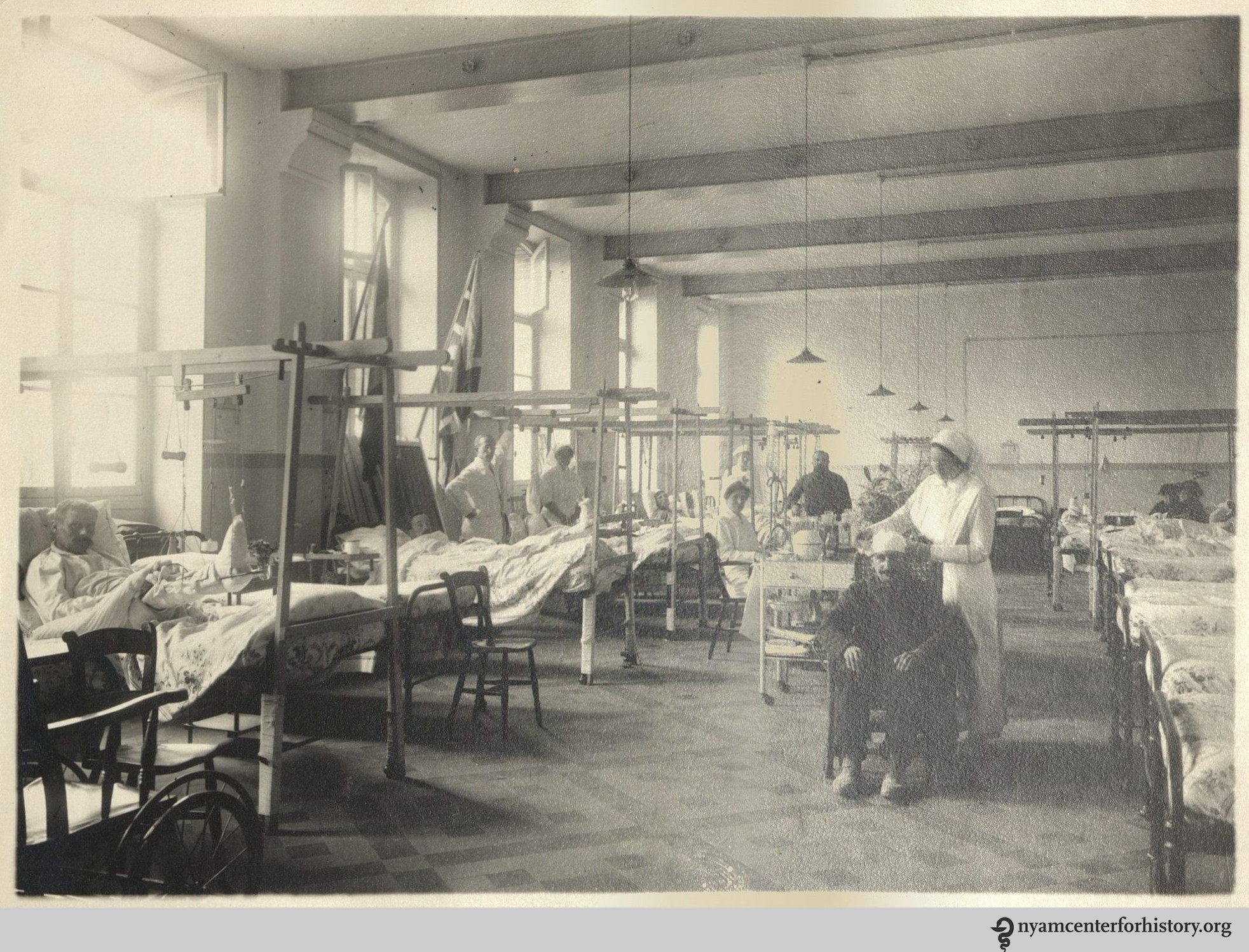
A recovery ward, 1916. The flags of Britain and France are mounted at the window, as this hospital was a joint effort: operated within the French military hospital system, sponsored by private British philanthropy, and staffed by American surgeons. Image: Charles Terry Butler papers, New York Academy of Medicine Library.

Butler’s letters home trace his awakening to war and medicine. Within a week, he wrote to his uncle Clare:

The hospital has about 200 beds, and on my arrival I was put in charge of two wards with over 90 beds and some 80-odd patients. It was some contract to start with, and for two or three days I hardly knew whether I was coming or going. I did about forty dressings a morning with three nurses to help me, and two getting their patients ready for dressing ahead of me and bandaging up when I was through. It took over three hours of hard, steady work.[4]

After a month, to his mother:

Last Sunday, 65 new blessés arrive—the majority of them frightfully wounded. They come by ambulance from a distributing railroad station some 6–7 kilometers away. Arriving in bunches of four or eight, they are sent immediately to their beds. Most of the orderlies had been given leave that day, so we doctors had to turn to and help carry them to the wards. (It isn’t particularly easy carrying a large man on a heavy stretcher with his trappings up three flights of stairs.) There they are undressed; their clothes put in a bag, tagged, and sent to be sterilized and cleaned; and then bathed. . . . The next thing is food. Many have not had anything for 24 hours or more while en route from the front or the last hospital. Then the surgeon comes along. Dressings, casts, splints, etc. are removed so as to see the condition and nature of the injury. It would be impossible to describe the state of some of the wounds—many not having been dressed for several days, some even for 10 or 14 days. A hasty and rather superficial cleansing must suffice for the time being, until the patient comes back from the X-ray room. … All the wounds are terribly infected, and a large percentage have foreign bodies (balls, pieces of shell, clothing, stones, dirt, etc., etc.) lodged…. [Surgery followed, aided by X-ray and fluoroscopy.] The recoveries are wonderful. Men whom no one would expect to live, ordinarily, in a civil hospital, hang by a hair for days and come around O.K.[5]

Butler noted that the average length of stay at the hospital was almost 50 days.

lg group_watermark

The staff of the Ris-Orangis Hospital, 1916. Dr. Joseph A. Blake, director, is the central figure (second row, seated); Charles Terry Butler is the third man to his left. Image: Charles Terry Butler papers, New York Academy of Medicine Library.

Ris-Orangis was considered one of the most successful hospitals in the war. [One of the founders, Harold J. Reckitt, wrote a detailed history of the hospital, V.R. 76: A French Military Hospital (1921)]. Butler spent most of his time dressing wounds, with little occasion for actual surgery. He returned to New York in January 1917 to take up his internship at Presbyterian. But upon the American entry into the war in April 1917, he was commissioned a first lieutenant with the United States Medical Corps, serving into 1919—the topic of a future blogpost. Butler’s experience at Ris-Orangis was crucial to his surgical accomplishments in this second phase of war service. After the war, he entered private practice, but by 1923 ill health—apparently resulting from wartime conditions—led Butler to retire. Moving to the Ojai Valley of Ventura County, California, he became a prominent civic and cultural leader up to his death in 1980.

References:
[1] Butler, Charles Terry. A Civilian in Uniform. Butler, 1975, p. 28.
[2] A Civilian in Uniform, p. 49.
[3] Blake to Butler, 29 April 1916, A Civilian in Uniform, p. 49.
[4] Butler to “Uncle Clare” [Clarence Lyman Collins (1848–1922)], 17 June 1916, A Civilian in Uniform, p. 57.
[5] Butler to “mother” [Louise Terry Collins (1855–1922)], 7 July 1916, A Civilian in Uniform, p. 62–64.

Images:
Charles Terry Butler, “Ris-Orangis, France, 1916,” photographic album. Charles Terry Butler papers. New York Academy of Medicine Library.

More Than Medicine: Social Justice and Feminist Movements for Health

COS-Series-Twitter-R1 Event 1005Today’s guest post is written by Jennifer Nelson, Professor at University of Redlands, specializing in women’s history, the history of feminism in the United States, and medical histories associated with social justice movements. She is the author of More Than Medicine (NYUPress, 2015).

On Thursday, October 5, Nelson will give her talk, “More Than Medicine: Social Justice and Feminist Movements for Health.” To read more about this lecture and to register, go HERE.

I begin my story of social justice and feminist movements for health with the Mound Bayou demonstration clinic—located in the Mississippi Delta. The clinic was founded by medical doctors who had been part of the Medical Committee for Human Rights (MCHR). Most had come to Mississippi to volunteer during the 1964 Freedom Summer, although several others were locals active in the Delta Ministry, a Mississippi based Civil Rights organization.

Dr. H. Jack Geiger and the other founders of the Delta Health Center clinic worked with the Mound Bayou community to prioritize health needs. They quickly discovered that community members needed more than traditional medicine: they demanded food, jobs, and housing—linking these to the promotion of health. The clinic included a cooperative farm that grew vegetables for the community, since most of what was grown in the Delta was for commercial consumption. Click HERE to listen to Dr. Geiger talk about his experiences in Mississippi at the National Library of Medicine exhibit on the Delta Health Center.

Many historians of social movements have emphasized that the women’s liberation movement emerged from the Civil Rights movement. I also pay attention to this connection, but focus on the use of medicine to achieve social justice goals in both the Civil Rights and women’s liberation movements. In both of these contexts, activists expanded the meaning of medicine in the process.

In the 1980s women’s health movement feminists were also grappling with conversations about race and racism. Since the early 1970s, women of color had been demanding that feminists pay more attention to issues raised by women of color. In the 1970s much of the focus was on sterilization abuse. In the 1980s attention shifted to HIV transmission, safer sex, and AIDS. Dazon Dixon Diallo, one of the only women of color working at the Atlanta Feminist Women’s Health Center in the 1980s, focused on developing a program called the Women with AIDS Partnership Project.

Here is a clip from a talk given by Diallo about her organization SisterLove, which she formed when she left the Atlanta FWHC to more directly address HIV/AIDS:

Clip

Dazon Dixon Diallo. (Click image to watch video).

My book also focuses on Loretta Ross, one of the most important founders of the Reproductive Justice Movement, which sought to broaden the feminist discourse around “choice” to address the systemic problems associated with poverty and discrimination that prevented many women of color from simply choosing to have or not have children. Ross’s work connects back to the Civil Rights efforts in Mound Bayou among MCHR activists and local organizers with the Delta Health Center. Ross, by forging a reproductive justice framework, maintained that health promotion for poor women could not rest on medicine alone.

More Than Medicine: Social Justice and Feminist Movements for Health is the third event in the three part event series, Who Controls Women’s Health?: A Century of Struggle. The series examines key battles over women’s ability to control their bodies, health choices, and fertility. It is developed in collaboration with the Museum of the City of New York and supported by a grant from Humanities New York.

So, You Want to Build a Digital Program? (Part 2)

By Robin Naughton, Head of Digital and Audrey Sage Lorberfeld, Digital Technical Specialist

This is the second in a two-part series on the creation of the Academy Library’s Digital Collections and Exhibits website. Part 1 is here.

Sometimes opportunities arise that you just can’t pass up. In late May 2017, our Curator of Rare Books and Manuscripts, Anne Garner, suggested a digital exhibit of items from our collection that would showcase the history behind many of the magical elements from J.K. Rowling’s beloved Harry Potter series; and suggested its launch coincide with Harry Potter and the Philosopher’s Stone‘s 20th anniversary on June 26. While we were a new digital team of two with new digitization equipment, we were up for the challenge. That is how we wound up creating the online exhibit, “From Basilisks to Bezoars: The Surprising History of Harry Potter’s Magical World,” in a mere 6 weeks.

Leo, Astronomicae Veteres_watermark

Leo from Aldus Manutius’ Astronomici veteres (1499).

Our first stop was to Garner, who, within a week and a half, handpicked two objects (including our infamous bezoar) and 34 images from over 20 different books  for us to digitize. While we were hustling to photograph these items in the lab, Garner was busy creating robust image metadata for us to ingest into Islandora. Next, we got to work churning out XML records through the use of OpenRefine and an Apple script, and then we were in the quality assurance phase. Once everything was checked, it was time to launch!

Of course, there were bumps along the road, too. At one point, we realized we had digitized the wrong phoenix! At another point, we had to go into all of our XML records and manually add in a download button. There were also some late Friday and Sunday nights spent working on our laptops to make the collection as perfect as possible. Bumps notwithstanding, we launched on-time, and the collection received a lot of great attention.[1]

contentselection

Anne Garner takes us through the images she has selected for digitization for our Harry Potter-inspired collection.

So, what did we learn from the launch of our digital program? We quickly discovered that it takes time and skill to create metadata. Both Garner and our Head of Cataloging, Rebecca Filner, expertly provided us with extremely detailed metadata for our current collections. Even in its quickest iteration, metadata-creation takes weeks![2]

We also learned that no digitized image is wasted. As we mentioned in Part 1, many of the digitized rare books with which we launched our Digital Collections were photographed for a separate project the Library had completed a few years earlier. The photographs were impeccable, so why double the work? Instead, we used what the photographer, Ardon Bar-Hama, gave us and co-opted these previously-shot images for our Digital Collection site. (Some notable examples are the Apicius and the Guy de Chauliac.)

robin

Our Head of Digital, Robin Naughton, trying out the new equipment.

And finally, we learned that the easiest part of the digitization process is photographing the items. As long as your equipment works, you are good to go! We received training by our expert Conservation staff in how to handle rare items, and we hit the ground running.

For those wishing to start a digital lab in your library, we have some hard-earned advice, all of which focuses on communication and outreach (the tech is the easy part!):

First, reach out to local colleagues who have been doing this longer than you. We visited many digital labs before our launch, including the beautiful labs at Columbia University, The Frick Art Reference Library, and the Museum of the City of New York. Each lab had a different setup, different workflows, and different amounts of staff with unique backgrounds. Without this exploratory research, we have no doubts our lab would not have done as well as it has.

Second, join listservs. Without the listservs to which we subscribe, we would not have gained nearly as much knowledge as we did before launch. Two great listservs to join are ALA’s digipres listserv and the ImageMuse Yahoo group’s listserv. We even reached out to a wonderful colleague from Coastal Carolina University because we admired how he used OpenRefine and the command line to batch-create and –export XML metadata records. He shared his script with us, and the rest is history. We now use his workflow in our lab for nearly every project.

teachingothers

Now we are even sharing our knowledge with others!

Third: join or create a digitization-related community in your area. By way of using the free, open-source Islandora platform for our digital collections, we became part of a very active and supportive network of Islandora admins, managers, developers, and vendors around the world. While that is incredible, it is also nice to have a support system in your own backyard. So, we co-founded the New York City Islandora Working Group with some colleagues from other institutions. Our group is open to anyone, regardless of whether they currently use Islandora. We meet once per month and share skills, ask each other questions, and, of course, eat pizza and sip wine together. It’s one of the most worthwhile professional endeavors we have been a part of, and our members have been instrumental in getting our lab up and running.

The secret to success really is communication. Talk to people, and you will learn so much!

Footnotes:

[1] “Study for your O.W.L.s with Library’s Harry Potter-Themed Online Collection” (DNAInfo); “There’s a New Digital Harry Potter Book Collection from NYC’s New York Academy of Medicine Library” (untapped cities); “Celebrating 20 Years of the Philosopher’s Stone Inside the Mini-Hogwarts in New York City” (The Verge); “Attention Harry Potter Fans: There’s A Mini-Hogwarts In East Harlem” (Gothamist)
[2] For those library-science fans among you, why didn’t we just pull the metadata from our online catalog? We did! But we encountered a lot of library-speak that we did away with for our Digital Collections audience and wanted to add some new metadata.