Ambroise Paré on gunshot wounds (Item of the Month)

By Lisa O’Sullivan, Director, Center for the History of Medicine and Public Health

The August item of the month is Ambroise Paré’s (1510 –1590) Les Oeuvres, or Works. Published in 1575 in 26 sections or books, the folio volume has 295 illustrations and includes Paré’s writings on anatomy, surgery, obstetrics, instrumentation, and monsters. This post focuses on Paré’s military surgery and is the first in a series of occasional posts looking at the relationship between medicine and war.

Pare_Oeuvres_1575_tp_watermark

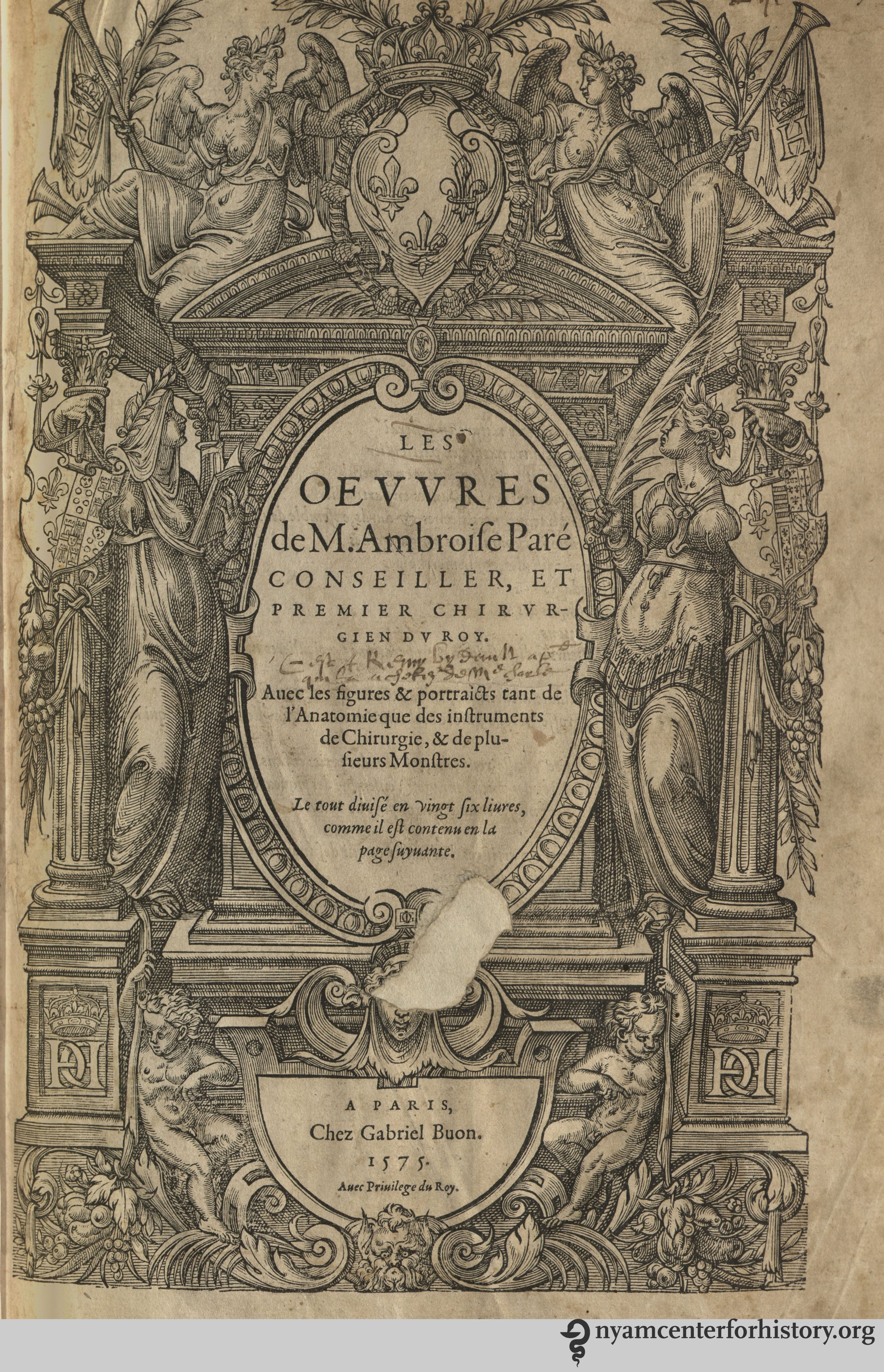
Frontispiece of the first (1575) edition of Les Oeuvres, dedicated to King Henri III. Click to enlarge.

Dedicated to Henri III, Paré presents Les Oeuvres as an accumulation of his life’s studies and experience, and it incorporates many of his earlier publications. The French barber surgeon spent much of his life at war, serving in over 40 campaigns, and published numerous highly influential books, many of them directly based on his practice of military surgery.i Paré’s career was a prestigious one, progressing from working as an apprentice barber surgeon to great prominence as surgeon to Henry II, and subsequently his successors Francois II, King Charles IX, and Henry III.

Like his contemporary Andreas Vesalius, Paré is now celebrated as an emblematic figure of Renaissance thinking, willing to look beyond the established authorities and instead rely on the evidence of his own experience. In the Oeuvres, for instance, he mocks the use of “mummy” or “mummia,” a popular remedy ostensibly created from Egyptian mummies and used extensively by physicians.ii Such a position was particularly provocative given Paré’s identity as a surgeon, rather than a university trained physician with a formal education and knowledge of Greek and Latin.

Despite Paré’s close connections with many of its members, the Parisian Faculty of Medicine attempted to block the publication of the Oeuvres, arguing that the Faculty needed to approve all publications relating to medicine and surgery. In addition, they objected to Paré’s use of French, as he was among a small but increasing number of practitioners writing in the vernacular rather than the more scholarly Latin, making such works vastly more accessible to students of surgery operating outside the universities and the lay public.iii

Pare_Oeuvres_1575_woundleg_watermark

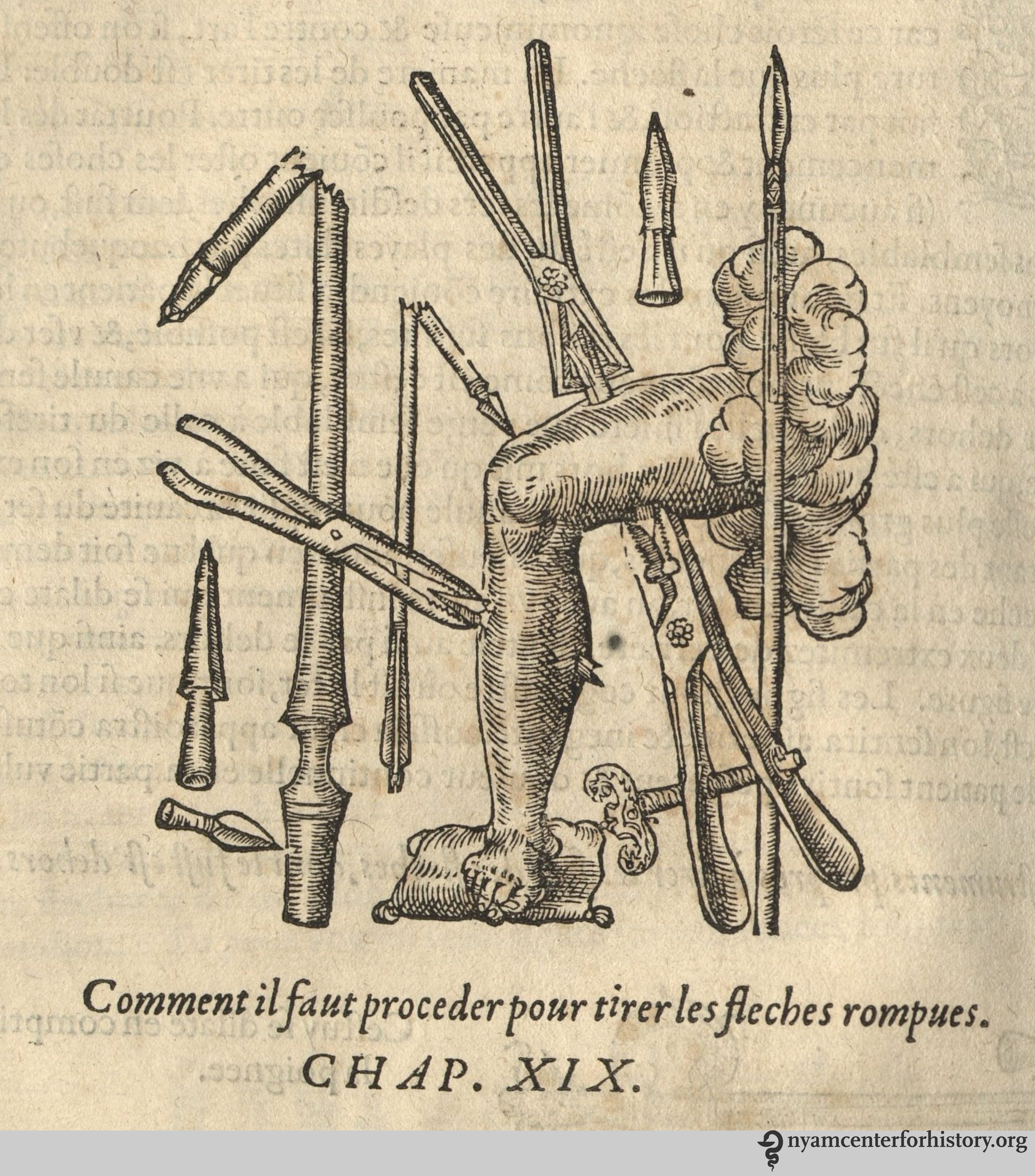
Reminiscent of a “wound man,” this illustration demonstrates techniques for extracting broken arrows from the body. Click to enlarge.

Much of Paré’s renown was based on his early work in the military context. Throughout the Oeuvres, he returns to examples of treating soldiers wounded during conflict. Perhaps the most famous vignette describes how, during his first campaign in 1536, Paré found that he had insufficient boiling oil to use in cauterizing gunshot wounds, and instead used a liniment made of egg yolk, rose oil, and turpentine. The following day, he discovered that those soldiers treated with the liniment were in a better condition than those whose wounds had been treated according to the prescribed manner. He subsequently argued for the treatment of gunshot wounds with liniments and bandaging, as well as removing affected tissue from the wound.iv

Gunpowder, whether projected from cannons or shot from firearms, had become a significant factor on European battlefields in the late 14th century. The use of gunpowder dramatically changed the practice of warfare. Increasingly numerous and accurate firearms contributed to the number of soldiers killed and wounded. These weapons produced new types of wounds that penetrated into the body, carrying foreign materials with them and leading to gangrene, while also deafening and blinding those near blasts.v

Descriptions of surgical tools, including a variety of tools for extracting bullets from wounds. On the top left, "crane bill" forceps for fragmented bullets; on right a shorter "duck bill" instrument designed for extracting whole bullets. At bottom, "lizard noses" for drawing out flattened bullets.

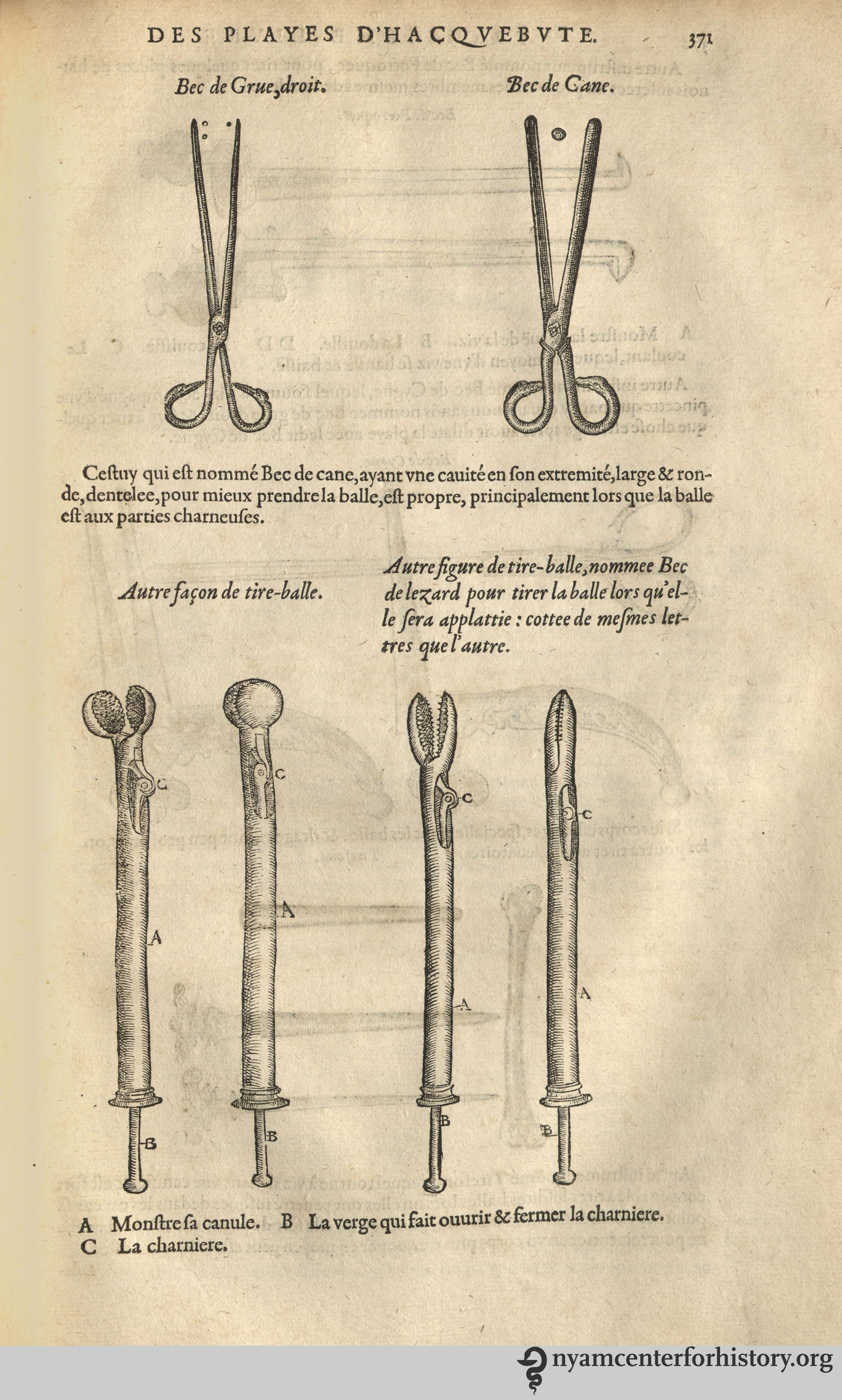
A variety of tools for extracting bullets from wounds. On the top left, “crane bill” forceps for fragmented bullets; on right a shorter “duck bill” instrument designed for extracting whole bullets. At bottom, “lizard noses” for drawing out flattened bullets. Click to enlarge.

Surgeons based their treatment of gunshot wounds on the belief that the gunpowder carried into the body by the bullets brought poison with it. This idea came from Giovanni da Vigo (1450–1525), an Italian surgeon whose 1514 Practica in arte chirurgica copiosa and 1517 Pratica in professione chirurgica were highly influential surgical texts. Rapidly translated into multiple European languages, these books include da Vigo’s suggestion to cauterize (burn) the wound with boiling oil in order to counteract the poisonous traces of gunpowder and to seal any severed arteries. This procedure became considered standard practice.viParé, after his experience with liniment rather than oil, experimented further, and recounts seeking advice from other surgeons and testing a folk remedy for onion poultices for burns suggested by an older local woman. Concluding that they were effective against blistering offered Paré another rhetorical opportunity to emphasize his commitment to observation and experimentation.vii

The evidence found in earlier surgical manuals suggests that medieval surgeons had made similar experiments, and that it was the popularity of the more recent ideas promulgated by da Vigo that led to treatments with cauterization and oil.viii While he was not the only surgeon to be working towards more humane and effective treatment of gunshot wounds, Paré became the most well-known and is often celebrated today as the “father” of modern military surgery.ix This reputation rests on not only his work around gunshot wounds but his broad interests, influence, and innovation. A future post will explore other aspects of Paré’s Oeuvres and its long-term impact on military surgery.

References

i.  A full bibliography of his works was produced by Academy librarian Janet Doe in 1937. See Janet Doe, A Bibliography of the Works of Ambroise Pare; Premier Chirurgien et Conseiller du Roy (Chicago: University of Chicago Press, 1937).

ii. Ambroise Paré, Les Oeuvres de m. Ambroise Paré … Avec les figures & portraicts tant de l’anatomie que des instruments de chirurgie, & de plusieurs monstres. Le tout diuisé en vingt six livres … (Paris : Chez G. Buon, 1575), p399.

iii. Paré defended his publication with a written defense and in the Parisian courts. While the verdict was not recorded, the book went on sale and sold out almost immediately. See Wallace B Hamby, Ambroise Paré, Surgeon of the Renaissance (St. Louis: W.H. Green, 1967), pp153-156.

iv. Ambroise Paré, Les Oeuvres de m. Ambroise Paré, pp357-359.

v. John Pearn, “Gunpowder, the Prince of Wales’s Feathers and the Origins of Modern Military Surgery,” ANZ Journal of Surgery 82 (2012): 240–244, 241; Kelly R DeVries, “Military Surgical Practice and the Advent of Gunpowder Weaponry,” The Canadian Bulletin of Medical History / Bulletin canadien d’histoire de la médecine 7(2) (1990):131-46, p135.

vi. DeVries, “Military Surgical Practice and the Advent of Gunpowder Weaponry,” pp141-142.

vii. Ambroise Paré, Les Oeuvres de m. Ambroise Paré, p359.

viii. DeVries, “Military Surgical Practice and the Advent of Gunpowder Weaponry,” p142.

ix. Frank Tallett, War in Context: War and Society in Early Modern Europe : 1495-1715 (London, US: Routledge, 2010), pp108-110.

The Origins of Automated Ice

By Danielle Aloia. Special Projects Librarian

This August, for most of us, ice is a second thought:  easily obtained for cooling drinks and chilling food, and usually only a few steps away.   An 1844 title in our collections offers an intriguing snapshot of a time when this was not always the case.

In 1844, a Londoner with a shop on Regent Street and an inventive mind published The Ice Book: Being a Compendious and Concise History of Everything Connected with Ice.  His name was Thomas Masters.   In this publication, Masters enumerates the practical uses–both culinary and medical– of his own patented ice machine.  In his introduction, Masters describes his obsession with the process of freezing:

The transformations narrated in the “Arabian Nights,” those gorgeous repositories of Eastern legendary lore, are not more marvelous or more speedy than the change of a liquid body to a block of solid ice.1

During the course of The Ice Book, Masters introduces his invention and its applications and takes readers on a whirlwind tour of ice through space and time.  Along the way, he also supplies some delectable frozen recipes–sign us up for the maraschino ice cream, the nectar ice and the punch a la Victoria, stat.

Masters reports that the Greeks and Romans were known to use snow from the surrounding mountains to cool their wine.2 Nero’s cooks flavored snow with “honey, juices, and pulp of fruits,” creating a precursor to the flavored ice of today, and eventually ice cream.3

nationaldairycouncil_icecreamyears_1946_watermark

Depiction of a runner delivering snow from the mountains to Nero. Published in the National Dairy Council’s Ice Cream Through the Years, 1946.

Masters also describes Indian methods of making artificial ice, reporting that during the winter months, ice was created by filling rows of small earthen pans with boiled water, which was then cooled and left overnight.  The thin ice was gathered up, thrown in a pit that was lined with straw and layered with blankets, and pressed into a solid mass.  The pit was closed up with straw, blankets and a thatched roof.

Masters devotes a significant portion of his narrative to the promotion of his portable “patent freezing machine.”  In his introduction he writes:

The preparation of one of the most delectable refections known to this advanced era of modern culinary civilization, has been hitherto left to the experienced confectioner, on whose skill, not always within reach, depended the supply.  By attending to the instructions contained in the following pages, ices may now be procured from the machine within five minutes.4

A review of the book in The Patent Journal and Inventors’ Magazine offers this glowing endorsement of The Ice Book:

The specification of Mr. Masters’ patent appeared in #53 of our journal…it will be seen that he invented a number of very ingenious apparatus, by means of which, the luxury of cold liquors, &c. may be the most readily supplied; his Ice safes and well are excellent, and his ready mode of freezing, astonishing.  It is really a disgrace to buttermen and other shopkeepers to vend their edibles in the nasty state they frequently do, and the public should demand the use by tradesmen of these safes…5

The benefits of Masters’ machine were not limited to food and drink preparation.  Ice was used in medicine to relieve headaches, fever, hemorrhaging, and, believe it or not, symptoms of rabies.6 Masters includes testimonials from MDs.  One Dr. John Ryan writes that Masters’ machine will “enable [doctors] at all seasons, whether in the crowded fever wards of the hospital, or in private practice, to obtain for the patient a necessary adjunct to medical treatment.”7

An elevation of a double-motion machine with pails (B), a2 (machinery), and P (flapdoor).  Some were made with a drawer underneath, which serves as a wine-cooler.  Plate 1 published in Thomas Masters' The Ice Book, published in 1844.

An elevation of a double-motion machine with pails (B), a2 (machinery), and P (flapdoor).  Some were made with a drawer underneath, which serves as a wine-cooler.  Plate 1 published in Thomas Masters’ The Ice Book, published in 1844.

A single-motion machine with a freezer that is rotated by turning the crank handle at the top.  Plate 3, published in Thomas Masters' The Ice Book, 1844.

A single-motion machine with a freezer that is rotated by turning the crank handle at the top.  Plate 3, published in Thomas Masters’ The Ice Book, 1844.

The machine had various interchangeable parts and could be setup for private use to make blocks of ice, flavored ice and ice cream, and to cool wine and drinks. In plate 6 below, Figures 1-3 depict the special churns needed to get the fineness and smoothness necessary to keep the flavored ice or ice cream from separating; “a proper beating-up, a process which never can be accomplished by the hand.”8 Figures 4-5, depict separate ice preserving containers for game, fish, butter, etc. Figures 6-8, depict the cold storage for beverages, such as wine and beer.

Plate 6 published in Thomas Masters' The Ice Book, 1844.

Plate 6 published in Thomas Masters’ The Ice Book, 1844.

Below, we’ve included a few tantalizing recipes from the book.  Masters supplies instructions for making plain and flavored ice creams:

Recipes for plain, pistachio, biscuit, maraschino, "nouveau" and cinnamon ice creams, from Thomas Masters' The Ice Book, 1844.

Recipes for plain, pistachio, biscuit, maraschino, “nouveau” and cinnamon ice creams, from Thomas Masters’ The Ice Book, 1844.

Recipes for pine-apple, ginger, and apricot ice cream, from Thomas Masters' The Ice Book, 1844.

Recipes for pine-apple, ginger, and apricot ice cream, from Thomas Masters’ The Ice Book, 1844.

Other recipes instruct on making flavored ices.

Wine ices, from Thomas Masters' The Ice Book, 1844.

Wine ices, from Thomas Masters’ The Ice Book, 1844.

Raspberry water ice et al., published in Thomas Masters' The Ice Book, 1844.

Raspberry water ice et al., published in Thomas Masters’ The Ice Book, 1844.

Apple water ice et al., published in Thomas Masters' The Ice Book, 1844.

Apple water ice et al., published in Thomas Masters’ The Ice Book, 1844.

masters_clarify sugar_1844_watermark

How to clarify sugar, from Thomas Masters’ The Ice Book 1844.

We found this errata slip laid in amusing:

Errata slip, Thomas Masters' The Ice Book, 1844.

Errata slip, Thomas Masters’ The Ice Book, 1844.

Another peculiar aspect of this work is the Appendix. Masters delights in supplying real-life anecdotes about ice.  Among the highlights are an ice storm in 1672 that destroyed numerous trees; an ice market in 19th-century St. Petersburg containing the bodies of thousands of frozen animals, captured inside ice; and in that same city, the Ice Palace of St. Petersburg built near the banks of the River Neva in 1739, which began to give way under its own weight before the last ice blocks were placed.9  We’ll be returning to this book again for these fascinating stories, and for the recipes within…particularly on hot summer days.

References

1.   Masters, Thomas. The Ice Book. London: Simpkin, Marshall, & Co., 1844.

2.  Masters, 6.

3.  National Dairy Council. Ice Cream through the Years.  Chicago: National Dairy Council, 1946.

4.  Masters, x.

5.  “Thomas Masters’ Ice Book:  The Ice Book: Being a Compendious and Concise History of Everything Connected with Ice.”  Patent Journal and Inventors’ Magazine, June 5, 1847, accessed online.

6.  Masters, pp. 180-187.

7.  Masters, pp. 185-187.

8.  Masters, pp. 194-196.

9. Masters, pp. 134-146.


Historical Advice on Breastfeeding in Honor of World Breastfeeding Week

By Becky Filner, Head of Cataloging

jewesbury_mothercraft_1932_53_watermark

“Mother nursing twins simultaneously.”  From Reginald Charles Jewesbury’s Mothercraft, antenatal and postnatal.

World Breastfeeding Week – August 1-7, 2016 – seeks to promote, protect, and support breastfeeding. How was breastfeeding regarded in the past? To answer this question, I consulted books on child rearing from the early 19th century to the mid-20th century.

buchan_advicetomothers_part1watermark

Title page, William Buchan’s 1804 Advice to Mothers.

The earliest book I looked at, Dr. William Buchan’s 1804 Advice to Mothers, on the Subject of Their Own Health; and on the Means of Promoting the Health, Strength, and Beauty, of Their Offspring, is extremely critical of women who do not breastfeed:

Unless the milk….finds the proper vent, it will not only distend and inflame the breasts, but excite a great degree of fever in the whole system… It may be said, that there are instances without number, of mothers who enjoy perfect health, though they never suckled their children. I positively deny the assertion; and maintain, on the contrary, that a mother, who is not prevented by any particular weakness or disease from discharging that duty, cannot neglect it without material injury to her constitution.1

At the end of the 19th century, Dr. Genevieve Tucker’s Mother, Baby, and Nursery: A Manual for Mothers (1896) also strongly advocates breastfeeding:

Every mother who has health sufficient to mature a living child ought, if possible, to nurse it from her own breast. Her own health requires it, as the efforts of the child to draw the milk causes the uterus to contract, and nothing else will take its place to her infant.2

Much of her other advice seems outdated now, including her claim that “nursing babies suffer from too frequent nursing” and her suggestion to nurse “as seldom as possible at night.” Perhaps strangest to modern ears is her analysis of a woman’s ability to nurse based on her physical and emotional state:

Different temperaments and constitutions in women have great influence in the quantity and quality of milk. The richest milk is secreted by brunettes with well developed muscles, fresh complexions, and moderate plumpness. Nervous, lymphatic, and fair-complexioned women, with light or auburn hair, flabby muscles, and sluggish movements, as a rule, secrete poor milk. Rheumatic women secrete acid milk, which causes colic, diarrhea, and marasmus in the child.3

Tucker also suggests that a nursing mother should be producing a whopping forty-four ounces of breast milk every twenty-four hours.

applebaum_babyamothersmanual_watermarkcrop

Breastfeeding baby, from Stella B. Applebaum’s Baby, A Mother’s Manual, published ca. 1946.

Dr. Charles Gilmore Kerley in his Short Talks with Young Mothers: On the Management of Infants and Young Children  wrote in the early 19th century that contemporary pressures on women hinder their breastfeeding abilities:

A mother, to nurse her child successfully, must be a happy, contented woman… The American women of our large cities assume the cares and responsibilities of life equally with men. Among the so-called higher classes, — those who have all that wealth and position can give, — there is a constant struggle for social pre-eminence. Among the majority of the so-called middle classes the contest for wealth and place never ceases from the moment the school days begin until death or infirmity closes the scene. Among the poor there are the ceaseless toil, the struggle for food and shelter, the care of the sick, and the frequent deaths of little ones in the family whom they are unable properly to care for. In all classes, therefore, the conditions of life are such as seriously to interfere with the normal function of nursing, no matter how excellent may be the mother’s physical condition.4

This emphasis on a woman’s mind being at rest is repeated in much of the early 20th- century literature on breastfeeding.

eldred_fortheyoungmother_part1_watermark

“Hungry!” from Myrtle M. Eldred and Helen Cowles Le Cron’s For the Young Mother, 1921. p. 31.

Most of the books from the first few decades of the 20th century contain a passage about keeping the breasts and nipples clean. Kerley and others recommend washing the nipples (and even the child’s mouth!) with a solution of boracic (boric) acid. Myrtle M. Eldred and Helen Cowles Le Cron write in For the Young Mother (1921) that “the breasts are tender and easily infected at first, so that the boric acid acts as a cleanser to protect the baby from possible germs and as a preventive of abscessed breasts.5”Boric acid, though it is sometimes used as an antiseptic, is toxic to humans if taken internally or inhaled in large quantities. Other books recommend rinsing the breasts with hot water prior to nursing.6

Many books also contain lists of foods the nursing mother should and should not eat. Dr. Anne Newton, in her Mother and Baby: Helpful Suggestions Concerning Motherhood and the Care of Children (1912), advises mothers to practice “sacrifice and self-denial” in eating meals, and to avoid rich and seasoned foods altogether.7 Newton specifies that mothers should eat “nothing about which there is any question of fermentation. Such vegetables as cabbage, turnips, cauliflower, and tomatoes should not be given until the baby is four months old at least, and even then certain things may cause discomfort and cannot be indulged until the child is weaned.8” Dr. Thomas Gray, in Common Sense and the Baby: A Book for Mothers, notes that the breastfeeding mother should “eat an abundance of wholesome, nutritious food; avoid indigestible pastries and salads. Take sparingly of tea and coffee. Drink freely cocoa and milk. Eat fruits – not acid.9”  Some more recent books are much less rigid about the mother’s diet. Dr. Dorothy Whipple, writing in 1944, is less cautious, and argues that there’s very little a mother can eat that harms a nursing baby, mentioning only certain foods like onions that may, in breast milk, deter babies with its “unusual taste.10

applebaum_babyamothersmanualcrop_watermark

A mother breastfeeding and a selection of foods recommended for the breastfeeding mother, taken from Stella B. Applebaum’s Baby:  A Mother’s Manual (1946).

nycdeptofhealth_careofbaby_part1cover_watermark

Front cover of the New York City Health Department’s The Care of Baby, 1932.

None of the books I consulted recommended breastfeeding to two years or beyond, the WHO’s current recommendation on breastfeeding. Most books recommend weaning the baby between eight and fourteen months of age.  The New York City Department of Health warns against weaning in summer because of the risk of spoiled cow’s milk:

If possible, do not wean your baby during the hot summer months…. If you are well, it will not harm you to nurse your child until the dangerous, hot weather is over. This precaution may mean saving your child’s life.”11

whipple_ouramericanbabies_part6_)watermark

“One of life’s richest experiences,” from Dorothy Whipple’s Our American Babies, published in 1944.

Another common thread in the literature about breastfeeding is an emphasis on the pleasure and health benefits experienced by the nursing mother.  According to Tucker, “under the right conditions of lactation, … the mother should thrive and even grow stout.12” Others emphasize that breastfeeding will help the mother “get her ‘good figure’ back much more quickly than the mother who doesn’t nurse” because “nursing causes the uterus or womb to contract.13” Stella Applebaum provides this summary of the mother-baby nursing relationship:

Mother’s milk is the perfect baby food. From a healthy mother’s clean nipples, this pure, fresh, warm, nourishing, digestible food is delivered, germ-free, directly into the baby’s mouth. At the same time mother’s milk protects him against certain diseases. Suckling at the breast makes the baby feel close to his mother, happy, and secure.

Nursing benefits you, too. It stimulates the uterus to contract to normal size and contributes to your personal enjoyment and contentment. Propped in a comfortable chair or bed, you share a uniquely satisfying experience with your baby.14

Other writers underscore the vital role nursing plays in strengthening the emotional bonds between mother and child.   Buchan writes in 1804 that “the act itself is attended with sweet, thrilling, and delightful sensations of which those only who have felt them can form any idea.15” Dorothy Whipple has the last word:

…to sit in a comfortable chair and hold a little snuggling baby in your arms, to watch him grab that nipple with all the fury of his tiny might and suck and work away until he reaches that complete satisfaction that comes to a baby with a full stomach is one of the pleasantest sensations in life.16

zabriskie_motherandbabycareinpictures_part1 0004_watermark

A mother and her baby breastfeeding while lying down, from Louise Zabriskie’s Mother and Baby Care in Pictures, published in 1941.

References

1.  Buchan, William. Advice to Mothers, on the Subject of Their Own Health; and on the Means of Promoting the Health, Strength, and Beauty, of Their Offspring. Philadelphia: John Bioren, 1804, p. 75-76.

2-3. Tucker, Genevieve. Mother, Baby, and Nursery: A Manual for Mothers. Boston: Roberts Brothers, 1896, p. 85-87.

4. Kerley, Chalres Gilmore. Short Talks with Young Mothers: On the Management of Infants and Young Children. New York: G.P. Putnam’s Sons, 1904, p. 13-15.

5.  Eldred, Myrtle M. For the Young Mother. Chicago: The Reilly & Lee Co., 1921, p. 37.

6.  Kenyon, Josephine Hemenway. Healthy Babies Are Happy Babies: A Complete Handbook for Modern Mothers. Boston: Little, Brown, and Company, 1934, p. 55-56; Zabriskie, Louise. Mother and Baby Care In Pictures. Philadelphia: J.B. Lippincott Company, 1941, p. 131.

7.  Newton, Anne B. Mother and Baby: Helpful Suggestions Concerning Motherhood and the Care of Children. Boston: Lothrop, Lee & Shepard Co., 1912, p. 74.

8.  Ibid., p. 78.

9.  Gray, Thomas N. Common Sense and the Baby: A Book for Mothers. New York: the Bewick Press, 1907, p. 39.

10. Whipple, Dorothy V. Our American Babies: The Art of Baby Care. New York: M. Barrows and Company, Inc., 1944, p. 139.

11.  New York City Department of Health. The Care of Baby. New York: Department of Health, 1932, p. 10.

12. Tucker, p. 86.

13. NYC Dept. of Health. The Care of Baby, p. 5.

14. Applebaum, Stella B. Baby: A Mother’s Manual. Chicago and New York: Ziff-Davis Publishing Company, 1946.

15. Buchan, p. 79.

16. Whipple, p. 122.

Godman’s mammals: An Illustrated Natural History

By Rebecca Pou, Archivist

I have our rare book cataloger, Tatyana Pakhladzhyan, to thank for introducing me to American Natural History, a delightful three-volume set by John D. Godman (1794-1830), a physician, lecturer, and naturalist. She initially came across it in our S.132 section, which comprises books on zoology, natural history, and mineralogy (The Academy library has a unique classification system – watch the blog for an upcoming series on our staffers’ favorite sections.)   After consulting with our curator, the decision was made to move the book into our Americana collection.

Godman, American Natural History, 1826-1828.

Engraved, added title page in Godman’s American Natural History, 1826-1828.

Though he is best known as a naturalist, Godman first made a name for himself as a medical man. Godman studied at the University of Maryland Medical School, graduating in 1818. He then moved around Pennsylvania and Maryland for a few years and succeeded in Philadelphia as a lecturer. Godman moved to Cincinnati in 1821, where he briefly taught at the Medical College of Ohio.1,2

In 1822, Godman moved back to Philadelphia. The next year he took over leadership of the Philadelphia School of Anatomy.1 Godman had a lifelong interest in nature, but it is in this period that he began to focus on his natural history studies. He became a member of the Academy of Natural Sciences of Philadelphia, the Franklin Institute, and American Philosophical Society.2 In 1826, he became the chair of anatomy at the Rutgers Medical College in New York City, but he was ill with tuberculosis and soon resigned. Too sick to lecture, he devoted himself to his literary pursuits and died in 1830. In his later years, he wrote a series of nature essays that were first published in a magazine and then posthumously as a collected work, Rambles of a naturalist.1,2 These essays are considered to be significant yet understudied American nature writings.2

Godman also contributed to medical literature, both as a writer and editor. He published a work on fasciae of the human body, Anatomical Investigations, in 1824. While living in Cincinnati, he edited the short-lived Western Quarterly Report of Medicine, Surgical, and Natural Science, which was the first medical journal published west of the Alleghanies.1,2 He later served on the editorial board of the Philadelphia Journal of the Medical and Physical Sciences, which was renamed the American Journal of Medical Sciences thanks to his efforts.1

American Natural History is Godman’s effort to document and classify North American mammals. The creatures include wolves, bears, seals, cats, weasels, the domestic dog, and the decidedly American bovine, the bison. The descriptions are accompanied by illustrations depicting the animals with remarkably expressive faces.

godman_americannaturalhistory_1826_v1_bats

Bats from Volume I of Godman’s American Natural History, 1826-1828.

As Godman details in the preface, he started on American Natural History in 1823 and anticipated a speedy year to year and a half of work to publish the first volume. Instead, the first two volumes were published in 1826 and the third followed in 1828. He explains the delay:

“It has been frequently necessary to suspend it for weeks and months, in order to procure certain animals, to observe their habits in captivity, or to make daily visits to the woods and fields for the sake of witnessing their actions in a state of nature. On other occasions we have undertaken considerable journies, in order to ascertain the correctness of statements, or to obtain sight of an individual subject of description.” (pp v-vi).

Godman’s emphasis on observation paid off; his work is noted for its accurate descriptions.2,3

Harlan_FaunaAmericana_tp_watermark

Title page of Harlan’s Fauna Americana, 1825. Click on enlarge.

Looking further down the same shelf, we found another early American book on mammals, Richard Harlan’s Fauna americana: being a description of the mammiferous animals inhabiting North America. This was published just a year before American Natural History. Harlan’s book was based on A. G. Demarest’s Mammologie (1820). Godman openly criticized Harlan for this reason and maintained the superiority of his work. A rivalry developed between the two, with Godman generally considered the victor.2 Wesley C. Coe corroborates this in his article “A Century of Zoology in America.” He regards Harlan’s text as “a compilation of work from European writers…[that] had little value,” while Godman’s is an “illustrated and creditable work.”4 Nevertheless, Fauna americana will soon join American Natural History on the shelves of our Americana collection.

Please enjoy this selection of illustrations from American Natural History:

godman_americannaturalhistory_1826_v1_commonwolf_duskywolf_watermark

Common wolf and dusky wolf in Volume I of Godman’s American Natural History, 1826-1828. Click to enlarge.

godman_americannaturalhistory_1826_v1_seals_watermark

Common and hooded seals from Volume I of Godman’s American Natural History, 1826-1828. Click to enlarge.

godman_americannaturalhistory_1826_v1_lynxandwildcat_watermark

Canada lynx and wild cat in Volume I of Godman’s American Natural History, 1826-1828. Click to enlarge.

godman_americannaturalhistory_1826_v1_harpseal_walrus_watermark

Harp seal and walrus in Volume I of Godman’s American Natural History, 1826-1828. Click to enlarge.

godman_americannaturalhistory_1826_v2_americangerbillus_watermark

American gerbillus in Volume II of Godman’s American Natural History, 1826-1828. Click to enlarge.

godman_americannaturalhistory_1826_v2_opossums_watermark

Opossums in Volume II of Godman’s American Natural History, 1826-1828.

godman_americannaturalhistory_1826_v2_goatandantilope_watermark

Mountain goat and prong-horned antilope in Volume II of Godman’s American Natural History, 1826-1828.

godman_americannaturalhistory_1828_v3_bison_watermark

Bison in Volume III of Godman’s American Natural History, 1826-1828. Click to enlarge.

godman_americannaturalhistory_1828_v3_dolphin_watermark

Dolphin in Volume III of Godman’s American Natural History, 1826-1828. Click to enlarge.

References

  1. “Godman, John Davidson.” In Dictionary of American biography, edited by Allen Johnson. New York : Charles Scribner’s Sons, 1931.
  1. Rosen, Susan A. C. “John D. Godman, MD.” In Early American nature writers: a biographical encyclopedia, edited by Daniel Patterson, Roger Thompson, and J. Scott Bryson. Greenwood Publishing Group, 2008. Retrieved from http://books.google.com, July 28, 2016.
  1. Faul, Carol. “Godman, John Davidson.” In Biographical Dictionary of American and Canadian Naturalists and Environmentalists, edited by Keir B Sterling. Westport, Conn: Greenwood Press, 1997. Retrieved from http://books.google.com, July 28, 2016.
  1. Coe, Wesley. “A Century of Zoology in America.” The American Journal of Science series 4, 46 (1918): 355-398. Retrieved from http://books.google.com, July 28, 2016.


The Influence of Sunshine and Pure Air: New York City Parks and Public Health

By Emily Miranker, Project Coordinator

My first picnic of the summer was picture-book perfect. Norman Rockwell would have approved: my friends and I clustered on blankets sipping lemonade, lightly toasted by the sun and gently cooled by a breeze, occasionally tossing a stray ball back to a child or sharing tidbit of our cold chicken lunch with an eager puppy.

Central Park - Harlem Meer_EM

Central Park’s Harlem Meer.  Photo:  Emily Miranker

The belief that public parks are “a fundamental need of city life,”1  goes very deep. The father of American landscape architecture, Frederick Law Olmsted ‑to whom (along with Calvert Vaux) New York City owes not only Central Park; but Prospect Park, Carroll Park, Fort Greene Park, the Parade Ground and Von King Park,2 commented that it was more than delight in nature that made parks so vital. There was a health benefit too. “The enjoyment of scenery employs the mind without fatigue and yet exercises it; tranquilizes it and yet enlivens it; and thus, through the influence of the mind over the body gives the effect of refreshing rest and reinvigoration to the whole system.”3

The Park Association of New York City (today New Yorkers for Parks) took up Olmsted’s charge after his death. Several small associations banded together in 1908 to form The Parks and Playgrounds Association of the City of New York; primarily concerned with advocating for children with no outdoor spaces in their neighborhoods. This organization merged with the Battery Park Association and the Central Park Association to become the Park Association in 1928. “Our purpose,” they declared, was to advocate park extension, defense and betterment, as parks were “essential to the mental, moral and physical well-being of city dwellers.”4  The starting point was that ever persistent New York City need: land.

Our collection boasts a wonderfully-designed pamphlet from this period soliciting support for the Park Association.  The pamphlet argues for the maintenance of city parkland, and the acquisition and development of more land dedicated to greenspace.

The pamphlet includes a colorful fold-out map. On the map, green illustrates the city’s parks as of 1927, yellow, the land purchased and intended for park use but not yet developed, and red, land recommended for purchase by the 1927 Metropolitan Conference on Parks but not yet purchased by the city.

parkassociation_toprotectandextend_map_192

Fold-out map published by the Park Association of New York City.  To Protect and Extend our City Parks for Posterity.  ca. 1927.

parkassociation_toprotectandextend_1_192

Inside of pamphlet with introductory letter from President Nathan Straus.  To Protect and Extend our City Parks for Posterity.  ca. 1927.

The pamphlet’s call to action is to “make the yellow and red green.” Indeed, many of those patches on the map have since become green.

The Trust for Public Land (TPL), whose mission to create and protect land for people ensuring healthy and livable communities is much like the Park Association’s just on the national scale, spends a fair amount of time bolstering their advocacy for parks with research on the health benefits they provide. In 2006, TPL released a white paper on the health benefits of parks, underscoring the argument that parks are a wise investment for communities.5 You can read the report online for percentages, statistics, financials, and citations of peer-reviewed work; but in brief: greenways enable people to exercise, improve mental health, offer vital space for child play, and contribute to the creation of stable communities

columbiau_builtenvironmentmap_2016

A 2011 geographic map of the distribution of parks and playgrounds done by the Built Environment and Health research team at Columbia University.6

trustforpubliclandsparkscore_map_2016

Map of New York City parkland (the dark green) created by The Trust for Public Land’s ParkScore rating system.7

A Fellow of The New York Academy of Medicine wrote on this very topic back in 1899. Dr. Orlando B. Douglas bemoans the lack of numbers to support his firm belief in the rejuvenating power of parks in The Relation of Public Parks to Public Health, written for the American Park and Outdoor Art Society. What he lacks in hard scientific data, he makes up for in poetic writing:

douglas,o_relationofpublicparks_1899

Orlando B. Douglas’ The Relation of Public Parks to Public Health, published in 1899.

While he didn’t have the same kind of data to fortify his arguments available to TPL more than a hundred years later, Douglas supports his claims that “the public park system in cities resulted in diminishing the rate of nervous disease [and] the improvement of the general health in cities”9 with testimony from twenty-one other doctors throughout the state of New York. I imagine that Dr. Douglas would have been thrilled at our ability today to quantify the beneficial effects of parks; though his pamphlet is more enjoyable reading than modern white papers.

References

1.  To Protect and Extend our City Parks for Prosperity. New York: Park Association of New York City; 1929.

2. “Olmsted-Designed New York City Parks,” NYC Parks. Accessed June 14, 2016. https://www.nycgovparks.org/about/history/olmsted-parks

3. Frederick Law Olmsted, “The Yosemite Valley and the Mariposa Grove of Big Trees,” The Saturday Evening Post, July 18, 1868.

4. To Protect and Extend our City Parks for Prosperity. New York: Park Association of New York City; 1929.

5. “Parks,” Built Environment and Health Research Group at Columbia University. Accessed June 14, 2016. https://beh.columbia.edu/parks/

6. “ParkScore: New York, NY,” The Trust for Public Land. Accessed June 14, 2016. http://parkscore.tpl.org/map.php?city=New%20York

7.  https://www.tpl.org/health-benefits-parks accessed June 14, 2016.

8.  The Relation of Public Parks to Public Health. Boston: Rockwell and Churchill Press; 1899.


Anatomical Illustrations: A Round-Up from our Visualizing Anatomy Workshop

Kriota Willberg, the author of today’s guest post, explores the intersection of body sciences with creative practice through drawing, writing, performance, and needlework.

On Mondays in June, I taught a drawing class in collaboration with staff at The New York Academy of Medicine Library.

The Visualizing and Drawing Anatomy workshop was open to artists as well as first time drawers willing to be challenged by the visual complexity of the human body in a short four-week course. Using the Academy’s historical collection as reference and instruction, artists and hobbyists learned to draw the body and found inspiration in the variety of illustrations.

Arlene

Historical Collections Librarian Arlene Shaner shared her knowledge about the collection with participating artists.

Working with rare books, a live model, and short presentations about the musculoskeletal system, workshop participants practiced looking through the skin to the model’s bony structures and large muscle groups.

In_Class2_NYAM2016

Drawing muscular anatomy on the model, we can compare a living body to images from historical texts.

Participants drew the model’s anatomy in class, and practiced during the week by doing various homework assignments.

In_Class4_NYAM2016

Artists drawing in our Hartwell Reading Room from our live model.

WhitTaylor2016anatomy1

Whit Taylor’s in class sketches of muscular anatomy from the live model.

WhitTaylor2016anatomy2

A second sketch by workshop participant Whit Taylor.

Debbie Rabina2016_4

Debbie Rabina’s in class sketch of the live model.

AllisonWhite4_2016

Allison White’s in class sketch of muscular anatomy from the live model.

Some homework used copied images from Vesalius and Dürer as subjects to anatomize with skeletal and muscular systems.

4Shaw_susan_Durer_Hmk2

Susan Shaw’s homework of anatomized Dürer images.

One of the participants proposed earning some extra credit, and anatomized two characters drawn by cartoonist Josh Bayer.

3Shaw_susan_Bayer2

Susan Shaw did a great job of re-configuring these skeletons to suit Josh Bayer’s iconoclastic drawing style.

Josh Bayer’s original cartoon can be viewed here.

Working with the historical collection as a teaching tool was very gratifying. I found new points of interest in familiar images, and developed a deeper appreciation for the artists and anatomists who generated so much rich material.

I love watching people draw.  As I watched this group work with the collection and the live model, I could observe and celebrate their growth during the course of the workshop. Witnessing the hard work, diligence, and growth of this group was truly inspiring!

Digitizing Medical Journals of State Societies

By Robin Naughton, Ph.D., Head of Digital

State medical journals digitized for the MHL collective project.

State medical journals digitized for the MHL consortium.

The New York Academy of Medicine Library is digitizing state society medical journals as part of a mass digitization project with the Medical Heritage Library (MHL), a digital curation consortium. The Academy Library is one of five collaborators on the project, along with the College of Physicians of Philadelphia; the Countway Library of Medicine at Harvard University; the Health Sciences and Human Services Library, University of Maryland; the Founding Campus; and the University of California at San Francisco.

Together, the MHL team is actively working to digitize 48 state society journals, more than 3,800 volumes that span much of the 20th century. Digitizing the state medical journals will provide open access to quality historical resources in medicine for researchers and the general public, letting them explore connections between medicine and society.

State medical journals digitized for the MHL collective project.

State medical journals digitized for the MHL collective project.

Evenly splitting the volumes among the MHL team makes the process of mass digitization more manageable and very collaborative. The Academy Library has already digitized almost 50% of the state medical journals assigned to it since Fall 2015. The journals are scanned by the Internet Archive (IA) and are publicly available as part of the Library’s and MHL’s collections on the IA site. Our digitized assets are open for anyone to access and use. Thus far, we have digitized journals representing 24 states and almost 238, 000 images.

The volumes are digitized in their entirety, showing the journals’ articles and  advertising. For example, in Alaska Medicine (vol. 29, 1987), as you read the article “Alaska State Hepatitis B Program – Past, Present and Future” by Elizabeth A. Tower, you can’t help but notice the advertisement for medical transcription. It is hard to resist the “Hello …. Museum of Primitive Civilizations and Hieroglyphs?”

Scan from Alaska Medicine, vol. 29, 1987.

Scan from Alaska Medicine, vol. 29, 1987.

State medical journals are valuable resources that should lead to many new and novel projects for researchers in the history of medicine. Look for more on the project as it progresses.

Explore our collection.

Get Crafty at the Museum Mile Festival on June 14

By Emily Miranker, Project Coordinator

When my office is perfumed by the smell of crayons and stocked with boxes of jumbo-sized sidewalk chalk, I know its Museum Mile Festival time. This year’s Museum Mile Festival takes place on Tuesday, June 14 from 6:00-9:00 pm, rain or shine.

Museum Mile (New York City’s Fifth Avenue from 82nd to 105th Street, which is technically three blocks longer than a mile) is one of the densest cultural stretches in the world.1 For the last 38 years, Fifth Avenue closes to traffic for a few hours on an early June evening. The eight major museums and their neighbors–that’s us!–throw open their doors and spill out onto the street in a block party.

Museum Mile at the New York Academy of Medicine. Courtesy of the Academy's Communications Office.

Museum Mile at the New York Academy of Medicine. Courtesy of the Academy’s Communications Office.

The first festival was held in 1979, the brainchild of the Museum Mile Association, to increase cultural audiences and garner support for the arts in time of great fiscal crisis in the city. The festival has since brought many New Yorkers and tourists to upper Fifth Avenue for the first time, and total attendance over the years has surpassed one million visitors.

Besides free admission to the museums along the mile, street performers, chalk drawing, live bands, balloons, and family-friendly activities abound. Dedicated to improving the health and well-being of people living in cities, the Academy has partners from the East Harlem Asthma Center of Excellence and Shape Up NYC joining us for the evening.

Getting physical with our community partners at Museum Mile. Courtesy of the Academy's Communications Office.

Getting physical with our community partners at Museum Mile. Courtesy of the Academy’s Communications Office.

The Library has planned some special crafts for the festival. We have the perennial favorite: coloring pages based on images from our collections. Feel free to download your own pages any time from #ColorOurCollections online.

Coloring sheets fro the New York Academy of Medicine Library. Photo: Emily Miranker.

Coloring sheets from the New York Academy of Medicine Library. Photo: Emily Miranker.

Among the treasures of our collection are the anatomical flap books. These are detailed anatomical illustrations superimposed so that lifting the sheets reveals the anatomy and systems of the body as they would appear during dissection. We created a simple DIY version of a flapbook inspired by these remarkable figures from the 1559 edition of Geminus’ Compendiosa totius anatomiae delineatio, aere exarata. The sheets are quite delicate, so it’s rare to see intact versions like this 400 years after they were made. Make your own flapbook with us during the festival.

Male flap anatomy from The Academy's copy of the 1559 English edition of Geminus’ Compendiosa.

Male flap anatomy from The Academy’s copy of the 1559 English edition of Geminus’ Compendiosa.

Female flap anatomy from The Academy's copy of the 1559 English edition of Geminus’ Compendiosa.

Female flap anatomy from The Academy’s copy of the 1559 English edition of Geminus’ Compendiosa.

Make this flap anatomy craft with us at Museum Mile! Photo: Emily Miranker.

Make this flap anatomy craft with us at Museum Mile! Photo: Emily Miranker.

And there’s nothing like using your own body to create art—finger print art!2

Make fingerprint art with us at Museum Mile! Photo: Emily Miranker.

Make fingerprint art with us at Museum Mile! Photo and artwork: Emily Miranker.

We look forward to seeing you at Museum Mile!

References

1. “Museums on the Mile.” Internet Archive Wayback Machine (June 2011). Accessed June 3, 2016. https://web.archive.org/web/20120101013336/http://www.museummilefestival.org/museums/

2. “Fingerprint Fun.” Bookmaking with Kids (June 2010). Accessed June 6, 2016. http://www.bookmakingwithkids.com/?p=1826

The Foresight of Trans-vision: An Innovative Anatomy of the Eye

By Anne Garner, Curator of Rare Books and Manuscripts

Early European anatomical lift-the-flap books made use of technologies available during the 16th century: woodcut and engraving, combined with manual cutting and pasting.1 Flap anatomies like Geminus’ Compendiosa (1559) allowed readers to peel away the layers of the body to reveal different organs, but these flaps were made of paper and opaque, and didn’t allow the reader to view the strata of the body simultaneously.

Female flap anatomy from The Academy's copy of the 1559 English edition of Geminus’ Compendiosa.

Female flap anatomy from The Academy’s copy of the 1559 English edition of Geminus’ Compendiosa.

Fast forward four centuries, when an innovation in printing technology let readers take a deep dive through the layers of the body all at once.

Anatomical illustrators used transparencies to show the layers of the body as early as the 1920s. J.E. Cheesman published Baillière’s Synthetic Anatomy, a series of 14 booklets, in London from 1926 to 1936. The series used a set of glassine sheets to show what lay beneath the surface of the skin.

Forearm in Cheesman, Baillière's Synthetic anatomy, 1926-1936.

Forearm in Cheesman, Baillière’s Synthetic Anatomy, 1926-1936. Click to enlarge.

Hand in Cheesman, Baillière's Synthetic anatomy, 1926-1936.

Hand in Cheesman, Baillière’s Synthetic Anatomy, 1926-1936. Click to enlarge.

In 1942, Richard Lasker patented a new printing process for Milprint Inc., a Milwaukee-based company. He called this new method trans-vision. Trans-vision allowed for the printing of images on the inner surfaces of folded sheets of transparent acetate. These sheets could then be piled on top of one another so that they overlapped, enabling a multi-layered view with the top sheets depicting the most superficial layers of an object and the bottom sheets the deepest level.2

The patent application for Lasker’s trans-vision process used a cutaway illustration of a mattress, with different layers of acetate offering views of the mattress’ filling.3 Trans-vision’s medical applications proved significantly more useful: it made possible the representation of complex anatomical relationships to health professionals and public audiences alike.

In 1943, Peter C. Kronfeld, an ophthalmology professor at the University of Illinois, published The Human Eye in Anatomical Transparencies. The book contains 34 color anatomical paintings printed using trans-vision.4 Each page offers a frontal and temporal view of the eye and its area, with transparent layers that can be peeled away by turning the pages. The paintings are printed on the inner side of the acetate to minimize damage from handling. The parts of the eye are numbered. Readers can use a bookmark key laid-in to identify the different parts by name.5

Figure I in Kronfeld, The Human Eye in Anatomical Transparencies, 1943. Click to enlarge.

Figure 1 in Kronfeld, The Human Eye in Anatomical Transparencies, 1943. Click to enlarge.

Kronfeld describes the project in The Human Eye’s preface:

It was at once obvious that the eye could advantageously be represented by this means, for it is a three-dimensional object in which great structural intricacy is combined with relatively small size. Ordinary drawings of its separate parts tend to isolate them too much from each other in the mind of the observer….The text has been so organized as to present not only a systematic account of ocular anatomy—taking up the various structures in a functionally logical order—but also a topographic treatment of the anatomy…which necessarily reveal the structures layer by layer in an order determined somewhat by the layers of dissection techniques.6

The paintings were made at twice the actual size by Gladys McHugh, an illustrator at the University of Chicago. The pioneering medical illustrator Max Brödel had been McHugh’s teacher and mentor at Johns Hopkins University, where she studied. It was Brödel who influenced McHugh to make her own dissections.

Figure II in Kronfeld, The Human Eye in Anatomical Transparencies, 1943. Click to enlarge.

Figure 2 in Kronfeld, The Human Eye in Anatomical Transparencies, 1943. Click to enlarge.

McHugh augmented her dissections of human eyes with specimens from pigs and monkeys. She describes her extensive dissection work in her introduction:

Over a course of time I obtained from baby autopsies ten good cases, making a total of twenty eyes and orbits. These I dissected layer by layer, making color notes and drawings from the fresh specimens. To develop a technique for separating the layers of the eyeball as intact semispheres, pigs’ eyes were employed. Also, to supplement my observation of the muscles and other structures not fully developed in the infant, monkey orbits were dissected.

As Professor Shelley Wall has argued, turning the pages in The Human Eye mimics the dissection process. As the layers on the recto side of the book are more deeply revealed with each page turn, the layers build on the opposite verso, allowing for the eye’s reconstruction.7 With The Human Eye, the union of format, technology, and material is harmoniously in sync.

Figues 3-6 in Kronfeld, The Human Eye in Anatomical Transparencies, 1943. Click to enlarge.

Figues 3-6 in Kronfeld, The Human Eye in Anatomical Transparencies, 1943. Click to enlarge.

Figure 14 in Kronfeld, The Human Eye in Anatomical Transparencies, 1943. Click to enlarge.

Figure 14 in Kronfeld, The Human Eye in Anatomical Transparencies, 1943. Click to enlarge.

Figures 19-22 in Kronfeld, The Human Eye in Anatomical Transparencies, 1943. Click to enlarge.

Figures 19-22 with bookmark key in Kronfeld, The Human Eye in Anatomical Transparencies, 1943. Click to enlarge.

By 1958, the book had enjoyed five editions.8 Students and educators embraced the text and its ingenious illustrations. In 1946, the trans-vision process was applied again to McHugh’s paintings in for The Human Ear in Anatomical Transparencies. Initially conceived as a wartime project useful to the aviation industry, the book’s value, as with The Human Eye, was in its power to demonstrate to both lay and specialized audiences the inner workings of organs not easily seen.

The finest examples of trans-vision printing occurred when the coffers of the pharmaceutical companies who published them were at their fullest. After The Human Eye and The Human Ear, medical illustrator Ernest Beck used trans-vision technology to produce more than 30 anatomical transparency projects published by Milprint for encyclopedias, pharmaceutical companies and other commercial concerns.9 A decade after Kronfeld, Ciba Pharmaceuticals published a 13-volume collection of anatomical illustrations using anatomical transparencies between 1953 and 1989. The illustrator of these, was a native New Yorker, fellow of the Academy, and former member of the Art Students League of New York. His name was Frank Netter.10

References

1. For more on this, see Andrea Carlino’s excellent Paper bodies: A catalogue of anatomical fugitive sheets, 1538-1687.

2. Wall, Shelley. “Mid-twentieth-century anatomical transparencies and the depiction of three-dimensional form.” Clinical Anatomy 23 (2010) 915-921. Accessed December 2015.

3. Wall, 917.

4. David Templeman, review of The Human Eye in Anatomical Transparencies by Peter C. Kronfeld. Optometry & Vision Science, 35 7 (1958). 388.

5. Wall, 919.

6. Kronfeld, Peter. The Human Eye In Anatomical Transparencies. Rochester, New York: Bausch & Lomb Press, 1943. iii.

7. Wall, 919.

8. David Templeman, review of The Human Eye in Anatomical Transparencies by Peter C. Kronfeld. Optometry & Vision Science, 35 7 (1958). 388-389.

9. Brierley, Meghan. Dialogue and Dissemination: The Social Practices of Medical Illustrators in the Pharmaceutical Context. Dissertation, University of Alberta. 2013. Accessed online May 31, 1916.

10. Frank Netter was the most prolific medical illustrator of the twentieth-century. During his more than sixty-years as a medical illustrator he produced more than 4,000 illustrations of the entire anatomic and pathologic character of the human body, system by system. His comprehensive and detailed illustrations, published in The Ciba Collection of Medical Illustrations, Clinical Symposia, and The Atlas of Human Anatomy educated generations of medical professionals and students all over the world.

20th-Century Teeth: Dentistry at the Turn of the Century

By Johanna Goldberg, Information Services Librarian

This is part of an intermittent series of blogs featuring advertisements from medical journals. You can find the entire series here.

“How did you learn to be a dentist? Did you go to a college?”

“I went along with a fellow who came to the mine once. My mother sent me. We used to go from one camp to another. I sharpened his excavators for him, and put up his notices in the towns–stuck them up in the post-offices and on the doors of the Odd Fellows’ halls. He had a wagon.”

“But didn’t you never go to a college?”

“Huh? What? College? No, I never went. I learned from the fellow.”

Trina rolled down her sleeves. She was a little paler than usual. She fastened the buttons into the cuffs and said:

“But do you know you can’t practise unless you’re graduated from a college? You haven’t the right to call yourself, ‘doctor.'”1

In Frank Norris’ 1899 novel McTeague: A Story of San Francisco—better known for its depiction of greed than the professionalization of dentistry—the title character loses his 12-year-old dental practice after California requires practitioners to hold a degree in the field. The timing couldn’t be worse for McTeague: he’d only just fulfilled a long-held dream, obtaining and hanging an enormous golden tooth outside his dental parlor.

McTeague’s fictionalized struggle was based in reality: until the mid to late 1800s, dentistry in the United States was not a regulated profession. Alabama became the first state to regulate dentists in 1841, and other states followed suit through the end of the century.2 In 1885, California passed a law requiring practicing dentists to register with a board, which could call up registrants for examination. Diplomas from a licensed dentistry school—the University of California College of Dentistry opened in San Francisco in 1882—also qualified registered dentists to practice. In 1901, a new law made practicing dentistry in California even more restrictive, part of a nationwide move to tighter regulation.3,4 In the novel as it would have been in real life, McTeague’s practice was toast.

Advertisements in dental journals from the era depict the trend toward professionalization, along with other technological advances. In 1840, the Baltimore College of Dental Surgery opened its doors as the first dental school in the world; by 1895, it had some local competition, including the Dental Department of the Baltimore Medical College.4 This school advertised heavily in journals like the American Journal of Dental Science.

Ad for the Dental Department of the Baltimore Medical College in the American Journal of Dental Science, vol. 33, no. 10, February 1900.

Ad for the Dental Department of the Baltimore Medical College in the American Journal of Dental Science, vol. 33, no. 10, February 1900.

Intriguingly, not only dental schools advertised in dentistry journals: The February 1901 volume of Dental Hints includes an ad encouraging dentists to take up a correspondence course in optometry, “on account of the intimate relationship between the eye and the teeth.” Huh?

Advertisement for the Philadelphia Optical College in Dental Hints, vol. 3, no. 2, February 1901.

Advertisement for the Philadelphia Optical College in Dental Hints, vol. 3, no. 2, February 1901.

Dental journal advertisements also reflect anesthetic advances. William Morton, a dentist, performed the first public demonstration of ether as a surgical anesthetic in 1846.2 A similar demonstration of nitrous oxide in 1845 did not go so well: dentist Horace Wells extracted a tooth before administering the proper dosage, and the patient cried out in pain. The drug was tabled for about 20 years; by 1869, it was commonly used either on its own or in conjunction with ether for dental procedures.2,5 Dental surgeries held less risk than other medical procedures, as they were commonly performed either in the patient’s or dentist’s home, locations less teeming with deadly microbes than operating theaters. After advances in antiseptic surgery by people like Joseph Lister, dental surgery became even safer—and Dr. Joseph Lawrence named an antiseptic mouthwash in his honor.5,6

Codman & Shurtleff's Inhaler for Gas or Ether advertisement in Dental and Oral Science Magazine, vol. 1, no. 2, May 1878.

Codman & Shurtleff’s Inhaler for Gas or Ether advertisement in Dental and Oral Science Magazine, vol. 1, no. 2, May 1878.

Listerine advertisement in the American Journal of Dental Science, vol. 33, no. 10, February 1900.

Listerine advertisement in the American Journal of Dental Science, vol. 33, no. 10, February 1900.

Local anesthetics also entered the market around the turn of the century. Some, like Mylocal, contained cocaine—though in the case of Mylocal, that cocaine was to be added by the practitioner prior to use. Perhaps unsurprisingly, the amount of cocaine used in local anesthetics was often poorly controlled, with sometimes dire results.5 Another local anesthetic, Eureka, proudly advertised that it “[avoids] that most dangerous drug that is known to the profession as COCAINE.” A third, Wilson’s Local Anaesthetic, notes that it is “non-secret and positively guaranteed.” Unfortunately, its ads don’t state what these non-secret ingredients are.

Advertisement for Mylocal anaesthetic in the American Journal of Dental Science, vol. 39, no. 1, January 1908.

Advertisement for Mylocal anaesthetic in the American Journal of Dental Science, vol. 39, no. 1, January 1908.

Advertisement for Eureka Local Anaesthetic in Dental Hints, vol. 3, no. 2, February 1901.

Advertisement for Eureka Local Anaesthetic in Dental Hints, vol. 3, no. 2, February 1901.

Advertisement for Wilson's Local Anesthetic in Dental Clippings, vol. 3, no. 6, April 1901.

Advertisement for Wilson’s Local Anesthetic in Dental Clippings, vol. 3, no. 6, April 1901.

Other turn-of-the-century advances include the development of tube toothpaste in the 1880s (previously, toothpaste had only been available in powdered form); awareness of microbial causes of tooth decay, leading to the promotion of flossing and brushing in the 1890s; and the use of gold foil as a cavity filling in the 1850s.2 The ads below reflect these advances and others, and were selected to show the relatively pain-free side of dentistry.

R. S. Williams Toothbrushes advertisement in Dental and Oral Science Magazine, vol. 1, no. 2, May 1878

R. S. Williams Toothbrushes advertisement in Dental and Oral Science Magazine, vol. 1, no. 2, May 1878.

Ney's Gold Plates advertisement in the American Journal of Dental Science, vol. 33, no. 1, May 1899.

Ney’s Gold Plates advertisement in the American Journal of Dental Science, vol. 33, no. 1, May 1899.

Dental Floss Silk advertisement in advertisements in the American Journal of Dental Science, vol. 33, no. 10, February 1900.

Dental Floss Silk advertisement in advertisements in the American Journal of Dental Science, vol. 33, no. 10, February 1900.

McConnell Dental Chair advertisement in Dental Hints, vol. 3, no. 4, April 1901.

McConnell Dental Chair advertisement in Dental Hints, vol. 3, no. 4, April 1901.

Standard Dental Manufacturing Co. advertisement in Dental Hints, vol. 3, no. 5, May 1901.

Standard Dental Manufacturing Co. advertisement in Dental Hints, vol. 3, no. 5, May 1901.

Dentacura toothpaste advertisement in Dental Hints, vol. 3, no. 11, November 1901.

Dentacura toothpaste advertisement in Dental Hints, vol. 3, no. 11, November 1901.

Munson's Standard Teeth advertisement in Dental Hints, vol. 3, no. 12, December 1901.

Munson’s Standard Teeth advertisement in Dental Hints, vol. 3, no. 12, December 1901.

Prophylactic Toothbrush advertisement in Dental Summary, vol. 22, no. 7, July 1902.

Prophylactic Toothbrush advertisement in Dental Summary, vol. 22, no. 7, July 1902.

Antikamnia and Odontoline advertisements in advertisements in the American Journal of Dental Science, vol. 39, no. 4, April 1908.

Antikamnia and Odontoline advertisements in advertisements in the American Journal of Dental Science, vol. 39, no. 4, April 1908.

Baker Coat Co. and Keeton Gold advertisements in the American Journal of Dental Science, vol. 39, no. 4, April 1908.

Baker Coat Co. and Keeton Gold advertisements in the American Journal of Dental Science, vol. 39, no. 4, April 1908.

Bowl Spittoon advertisement in the American Journal of Dental Science, vol. 39, no. 4, April 1908.

Bowl Spittoon advertisement in the American Journal of Dental Science, vol. 39, no. 4, April 1908.

References

1. Norris F. McTeague.; 1899. Available at: http://www.gutenberg.org/files/165/165-h/165-h.htm. Accessed May 9, 2016.

2. History of Dentistry Timeline. Available at: http://www.ada.org/en/about-the-ada/ada-history-and-presidents-of-the-ada/ada-history-of-dentistry-timeline. Accessed May 9, 2016.

3. Newkirk G. California. In: Koch CRE, ed. History of dental surgery: Dental laws and legislation, dental societies and dental jurisprudence, Vol. III. Fort Wayne, Ind.: National art publishing Company; 1910:755–756. Available at: https://books.google.com/books?id=9iE-AQAAMAAJ&pgis=1. Accessed May 9, 2016.

4. Schulein TM. A chronology of dental education in the United States. J Hist Dent. 2004;52(3):97–108.

5. Enever G. History of dental anaesthesia. In: Shaw I, Kumar C, Dodds C, eds. Oxford Textbook of Anaesthesia for Oral and Maxillofacial Surgery. Oxford: Oxford University Press. doi:10.1093/med/9780199564217.001.0001.

6. From Surgery Antiseptic to Modern Mouthwash | LISTERINE®. Available at: http://www.listerine.com/about. Accessed May 10, 2016.