
By Anne Garner, Curator, Center for the History of Medicine and Public Health
Our collections have always reflected the strong relationship between medicine and visual culture. Accordingly, since its creation in 2012 our blog has frequently taken up the intersection between medicine and art as subject. Below, we link below to a few posts that explore these crucial connections.
Most recently, Caitlin Dover featured The New York Academy of Medicine’s collections of illustrated medical books on the Guggenheim’s blog in “Doctors Without Borders: Exploring Connections Between Art and Medicine.” Her findings are in part the fruit of a visit with the Academy’s Historical Collections Librarian Arlene Shaner, who showed her a selection of books and ephemera from our Drs. Barry and Bobbi Coller Rare Book Reading Room, showcasing the connection between physicians and artwork.

Robert Latou Dickinson sketch of the Rare Book Room on its opening in 1933, from the Academy’s Annual Report, 1933.
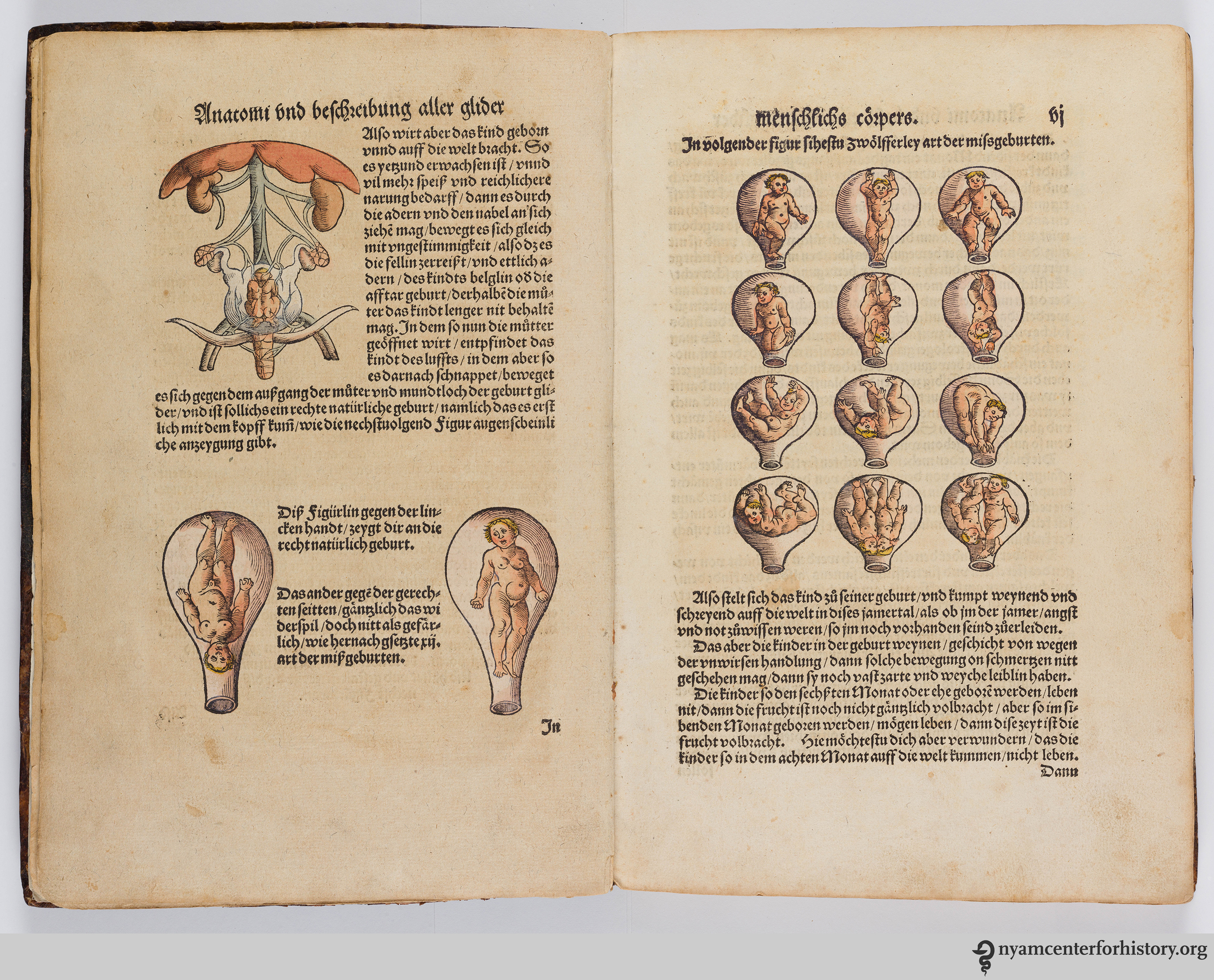
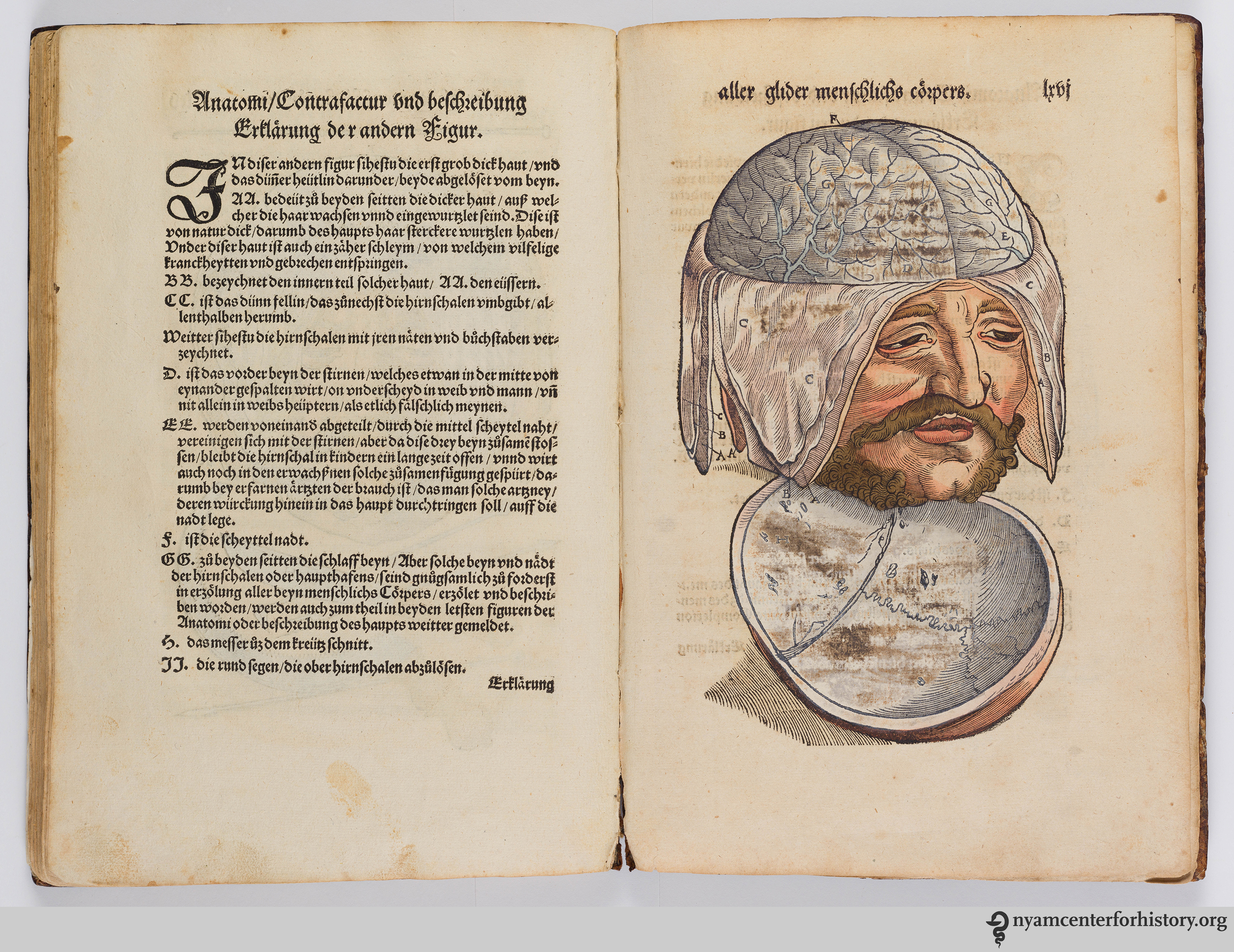
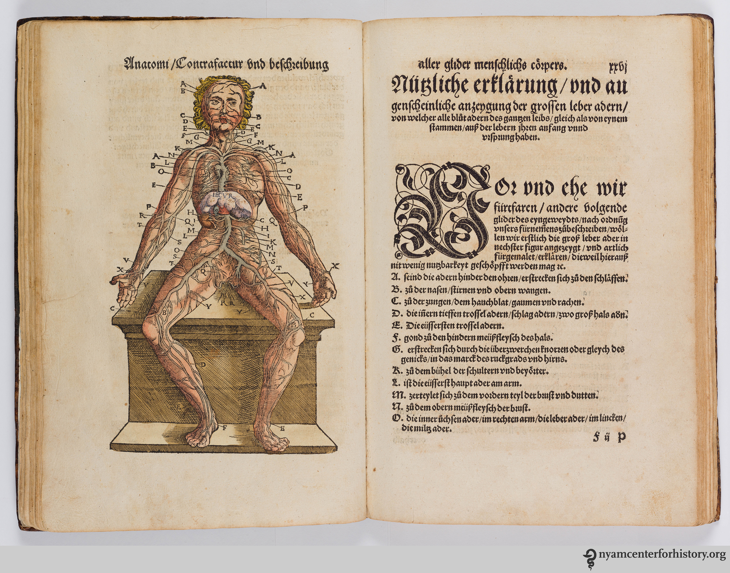
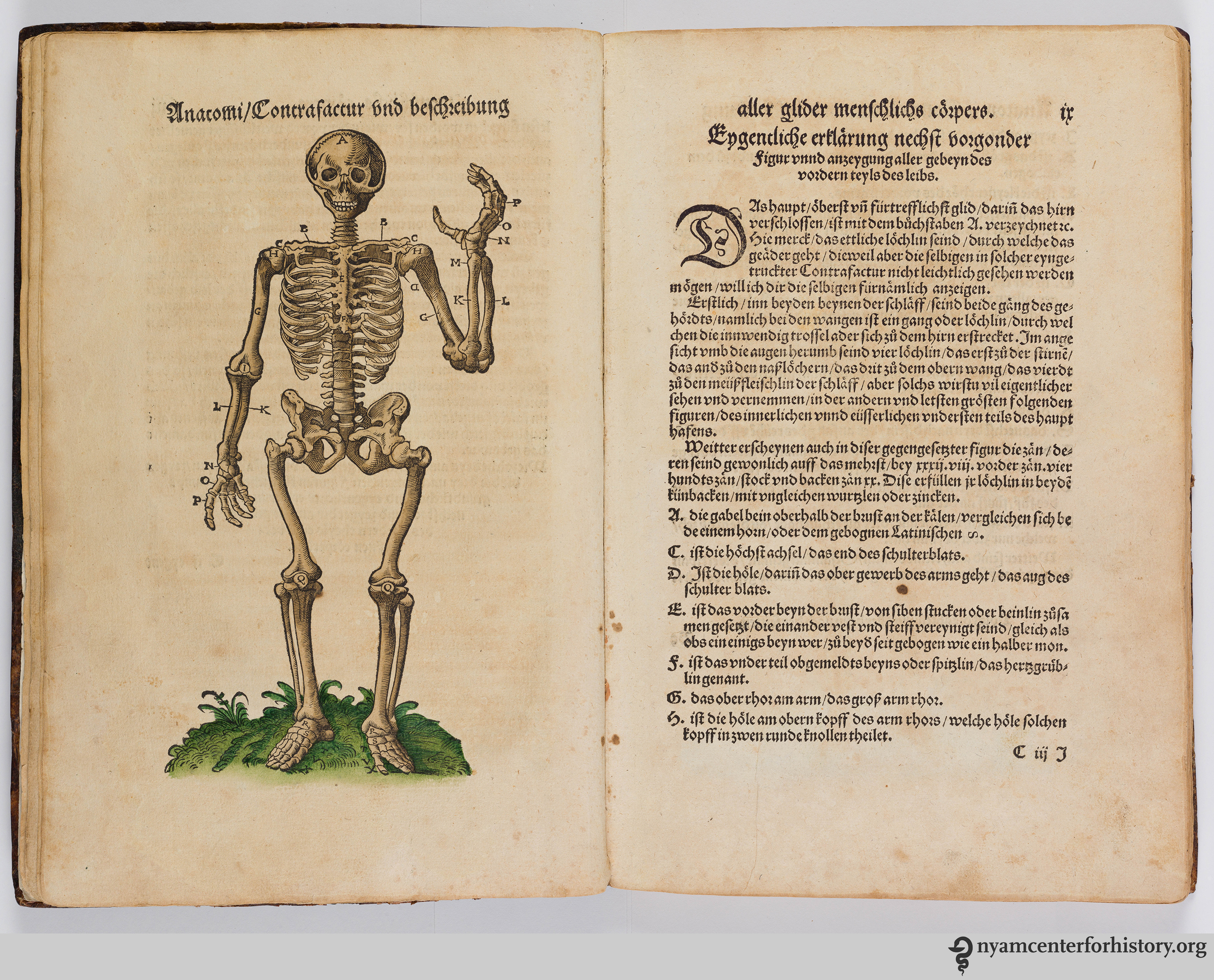
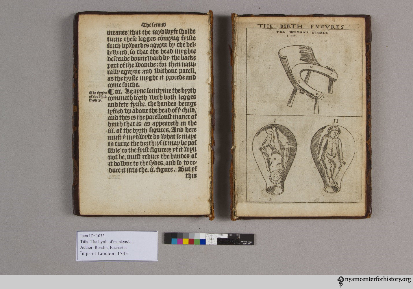
Our extensive collection of anatomical atlases demonstrates the close relationships of physicians and artists, who frequently collaborated to create works both for students of medicine and of art. These atlases show both the successes and failures of collaborations between anatomists and artists who worked together to communicate new medical knowledge. For Vesalius, the collaboration was a great success. In a guest post from 2015, our 2014–2015 Helfand Research Fellow Laura Robson discusses the way Andreas Vesalius’ great milestone work of 1543, De Humani Corporis Fabrica, relies on the synergy between plates and text, and how a later work that uses the Vesalian plates suffers when the anatomist’s text is eliminated. Another guest post by New York physician Jeffrey Levine explores the visual imagery of Vesalius’ famous frontispiece of this same work. Other writers use illustration to signal authority and knowledge. A 2015 post on Walther Ryff explores the ways that Ryff’s use of the counterfeit style in his illustrations implied eye-witness discovery.
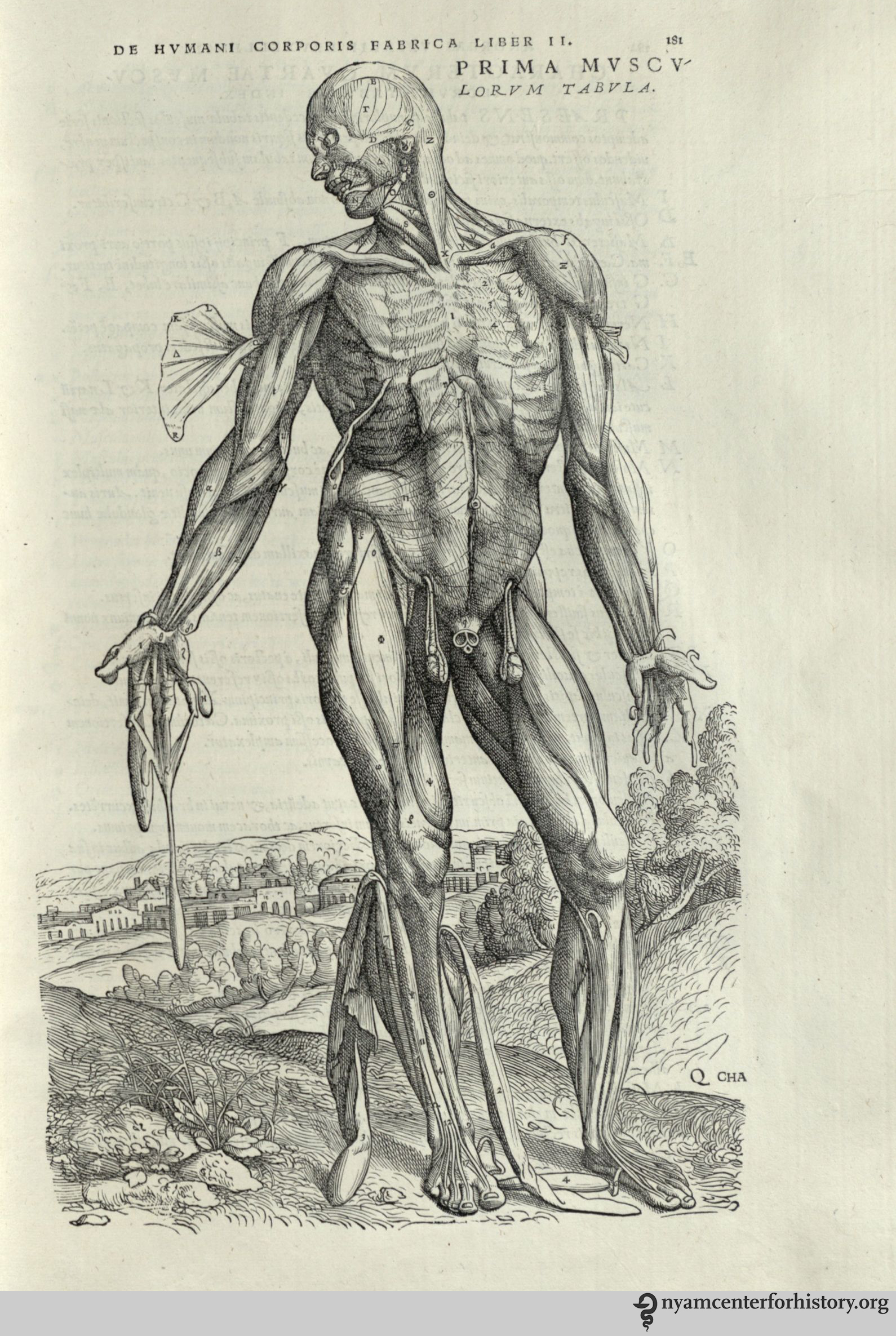
Andreas Vesalius (1514-1564). De humani corporis fabrica libri septum. Basel: Johannes Oporinus, 1543.
Our 2014 festival Art, Anatomy and the Body: Vesalius at 500 offered ample opportunity for critical thinking about the relationship between art and the body. Guest curator and visual artist Riva Lehrer describes her personal experience of the ways the body informs identity, and how that has shaped her own work as an artist in a 2014 post. A selection of images from several of our early anatomical atlases are featured in “Brains, Brawn and Beauty,” an exhibit that accompanied the festival, and are discussed here.
Finally, two posts on skeleton imagery highlight the tradition of danse macabre imagery in anatomical illustrations. Brandy Shillace’s guest post, “Naissance Macabre: Birth, Death, and Female Anatomy” examines depictions of the female body over time. For a look at the evolution of anatomical imagery with special attention to the tradition of portraying the human skeleton in vivo, visit our blog here. You’ll find a slide show hosted by Flavorwire featuring spectacular anatomical images from our collections.

Surgite mortui, et venite ad judicium (Arise, ye dead, and come to the judgment). Table 6. Click to enlarge.
Next month, the New York Academy of Medicine library will be undertaking an artistic project of our own. Capitalizing on the current coloring craze, we are starting a week-long special collections coloring celebration on social media, using the hashtag #ColorOurCollections. We’ll share images from our collections, as will friends at other institutions. We encourage you to color them, and share your colored copies on social media. Read more about how you or your institution can participate.

Coloring a camel from Conrad Gesner’s Historia Animalium, Liber I, 1551.


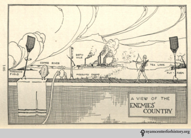


![From: McDermott, S. Metaphysics of Raw Foods. Kansas City, Mo. : Burton Pub. Co.; [c1919]. Click to enlarge.](https://nyamcenterforhistory.org/wp-content/uploads/2015/12/mcdermott_metaphysicsofrawfood_1919_table_watermark.jpg)


