The French Quarter is at the heart of New Orleans. This area is known for its unique architecture. A blend of traditions from its former colonizers, France and Spain, bred a new style. Fires and other calamities since the 18th century may have destroyed most of the original buildings, but a distinct style remains in the neighborhood.
Following the Louisiana Purchase in 1803, New Orleans experienced an influx of people from other states, eager to seize new opportunities. Louisiana and, of course, New Orleans were now part of the United States of America. New rules brought forth new laws. Under the rule of their new country’s leaders, medicine, for one, could only be practiced and administered by licensed professionals.
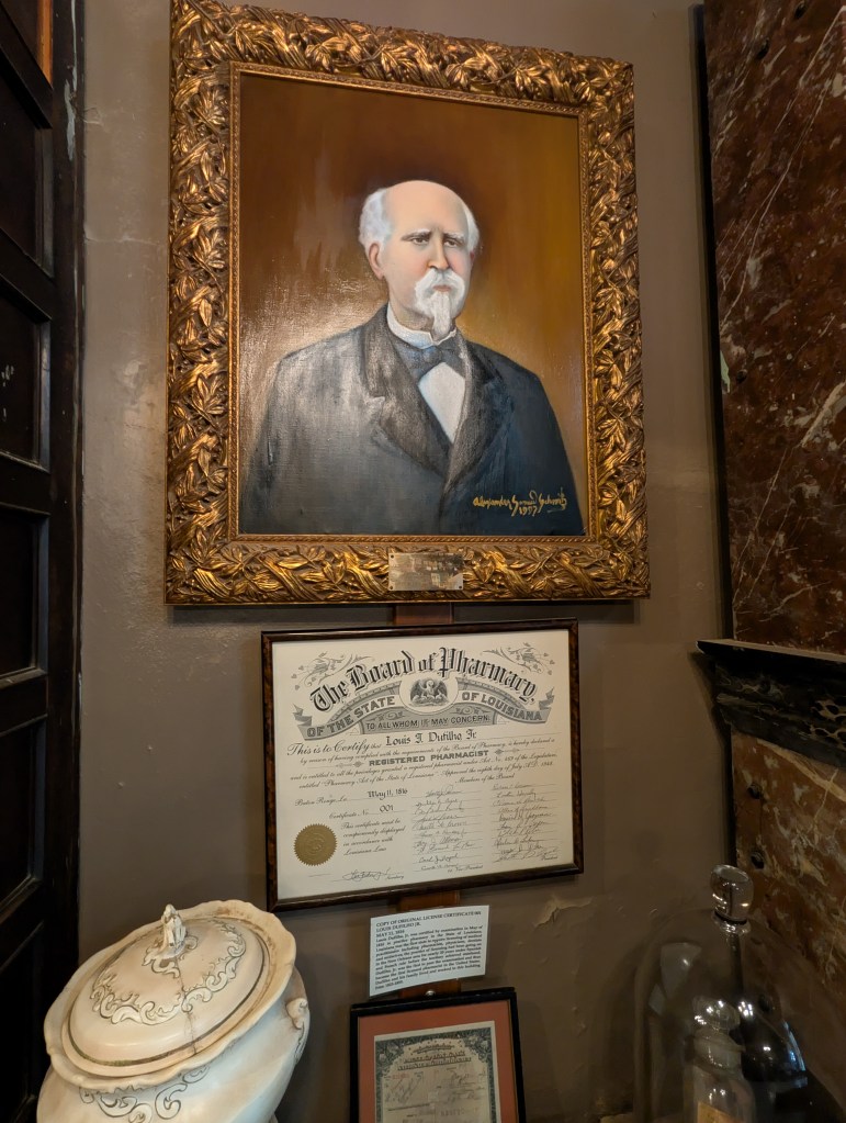
The Board of Pharmacy of the new state of Louisiana granted its first certificate to Louis J. Dufilho Jr. in May of 1816. Dufilho was not only the first Louisianan to do this but also the first licensed pharmacist in the entire United States. In 1823, he and his family came to the French Quarter to establish their pharmacy. From 1823 until 1855, Dufilho served the people of this fast-growing American port.
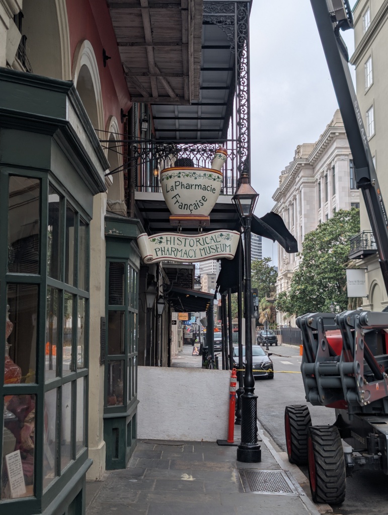
In April of 2025, over 200 years later, a group of us gathered outside a building on a hot New Orleans day. Although the building was going through a brief period of routine touch-ups, it still stood out. A sign with a mortar and pestle declares “Le Pharmacie Française,” with a sign below indicating that this is indeed the “Historical Pharmacy Museum.”
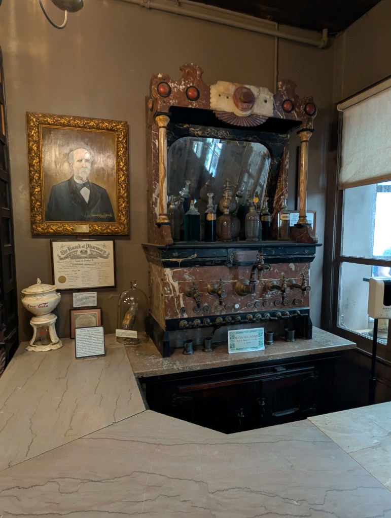
This is the home of the New Orleans Pharmacy Museum at 514 Chartres Street. We were welcomed not only by our tour guide but also by a giant soda fountain from 1855. The decommissioned soda pump still holds the necessary ingredients needed to make proper medicinal concoctions. Before the tour officially started, we were allowed to roam and explore the first floor. New Orleans’ early relationship to the history of a pharmacy was about to be unspooled for us. Our packed crowd consisted of residents from many different states and an entire family from Ireland.
There are two types of tours available here: a self-guided tour, complete with a text guide to help you understand the collection, or a group tour led by a tour guide. The historian we were fortunate enough to have as our tour guide helped bring the sights we were seeing to life. It has been proven that creating an engaging lesson increases the likelihood of retaining that knowledge.
The Pharmacy Museum doesn’t shy away from the realities of the early pharmacy industry. What could easily come across in a sensationalized manner—to engage visitors or promote clicks—is given context. The way they present their material fosters a dialogue with what we know now. In 2018, the Museum even commissioned local artist Kate Lacour to create an illustrated guide to items from their permanent collection. Do No Harm reminds us not to scoff at earlier treatments; the goal has always been healing.
During my visit, something struck me about seeing physical copies of the items I had previously read about. I have seen countless advertisements and trade cards discussing the wonders of Lydia E. Pinkham’s treatments, and now some items were in front of me!
From our digital trade card collection, “Yours for Health, Lydia E. Pinkham.”
This harmony between museums and libraries furthers understanding. The item that I read about is authentic, and that item, which I see before my very eyes, has a history we can explore inside a book.
At the end of the tour, our guide asked us to look at the slate floor we had been on the whole time. She informed us that the floor has not changed since Dr. Dufilho and his family resided there, some 200 years ago! She left us with this thought so that we could think about all those who had also set foot in the building over the years. Knowing what we learned about the evolution from house to historical building, for what purposes had these people come the way that we had?
As we approach summer, with vacation and/or time off approaching, take a moment to think about what you might want to learn more about. Whether you’re visiting an entirely different country, a different state, or even staying local and opening a book, there are plenty of stories waiting to be shared.
Anatomy, Descriptive and Surgical was first published in 1858. The author, Dr. Henry Gray, had intended to create an “accurate view of anatomy” for students and practitioners, for the application of practical surgery. This work was to be an inexpensive textbook from the mind of the celebrated wunderkind, who had already been published, celebrated, and named a Fellow of the Royal Society by the age of only 25.
Illustration From 2nd American Edition of Anatomy…
For the illustrations, Gray re-teamed with Dr. Henry Vandyke Carter, another anatomist/surgeon who was a skilled anatomical artist. Their previous collaboration had been Gray’s 1854 book, On the Structure and Use of Spleen. The new work was to be their biggest collaboration yet. Unfortunately it was also their last: Gray died in 1861 of smallpox, contracted from caring for his nephew. Before his passing, he had completed a second edition of his Anatomy that corrected minor mistakes and added illustrations by a Dr. Westmacott.
Illustration From 2nd American Edition of Anatomy…
Since its original publication in 1858, the book succeeded in both its original purpose and became culturally synonymous with the profession. Descriptive and Surgical Anatomy became a key text, although now the title was shortened to Gray’s Anatomy, both easier to say and in honor of the author. Editions of the book were not just for professionals either. It became part of the Western canon. For example, performance artist Spalding Gray used the title for his one-man piece, later a Steven Soderbergh film, in which he muses on his options after being diagnosed with a rare ocular disease.
The title has never gone out of print. The key textbook even adapted with technology. Back in 2005, as the 39th edition of Gray’s Anatomy was set to be published, one could purchase a virtual edition for an additional $60. At the same time, another Gray—or rather, Grey—would take over the cultural context….
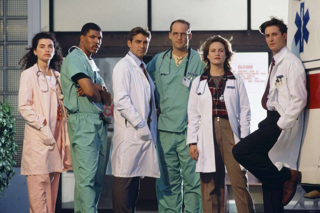
There was no shortage of doctors on television. ER (1994–2009) was created by writer Michael Crichton. Although he never obtained his medical license, Crichton had graduated with his MD from Harvard Medical School. Crichton first scripted ER in the 1970s with the idea of making it a film. It reflected the rotations he took part in. When it was filmed in 1994 as a two-hour television pilot, not much was changed. During his short-lived medical career, he had become disenchanted with how corporate he believed medical care had become. ER is set in the fictional, financially tight, Cook County General Hospital in Chicago. Crichton’s mind-set comes through in some of his other work, including the (also) hospital-set, body-snatching horror film, Coma.
ER set a gold standard for hospital dramas. Crichton believed that for the first time on television they provided “realism” of what things were like an emergency room. Subsequent hospital television would have to do something different. This led to a more stylized approach as it went on for Chicago Hope (1994–2000). Scrubs (2001–2010) was different in that it was a comedy that mixed in the reality of the job. Then there were the countless unsuccessful rip-offs that couldn’t find their own unique voice, lasting one season or less. Plus, for early seasons, ER had up and coming actor George Clooney as part of their cast. He would go on to be one of the biggest names in Hollywood.
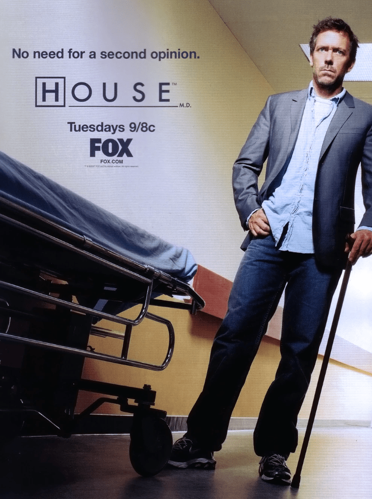
House, MD premiered in 2004 on FOX and starred British actor Hugh Laurie as the titular, misanthropic doctor. Creator David Shore looked to Sherlock Holmes as inspiration for his character. House gave his patients and viewers a methodical approach to his diagnoses, which were, more often than not, some rare disease, disorder, or occurrence. House had managed to give the medical yet another new take.
In 2005, ABC didn’t show much confidence in their latest pilot. It was another medical drama. They couldn’t even agree on a name: Complications, Surgeons, Miss Diagnosis, Grey’s Anatomy, it didn’t matter. They believed nothing they could top ER, which was still a ratings and critical juggernaut ten years in. But they were riding high after successfully rolling out two dramas in the last year, Desperate Housewives and Lost.
First-time creator Shonda Rhimes believed she had a show that would once again break a hospital-set mold. She tended to associate hospitals with “good things.” It was the place where they would “fix” you. Rhimes understood that there had to be a balance between that sentiment and the real lives that those who worked there had. “They’re just people at work.”
With those intentions in mind, executive producer Peter Horton tried to keep it looking “real.” He wanted the characters to look worn out; it’s a tough job! He wanted them to be unglamorous, with little to no make-up. This is most evident in the first episode. They found that even getting real-looking scrubs on the actors wouldn’t make for must-see tv, so they pivoted to it the reality rest on the emotional heft of being in a hospital. The remaining eight episodes of the abbreviated first season reflected that.
Those who worked on the show were skeptical. “It’s doctors with teenage dialogue,” Thomas Burman, a special effects makeup artist recalled thinking. Initial reviews were mixed, including people stating that the show needs “a brain.” Entertainment Weekly had reservations but overall found it enjoyable. ABC could take a loss if this, in their eyes, “generic” mid-season replacement (never a good sign in television terms) fizzled out.
On March 27th, 2005, the first episode of Grey’s Anatomy, “A Hard Day’s Night,” premiered at 10pm, following a season two episode of Desperate Housewives. Audiences were introduced to the main character and narrator, Meredith Grey (Ellen Pompeo), as she and her intern cohort endured their first 48 hours at Seattle Grace. Meredith’s voiceovers give context and comfort. (Coincidentally, a different Gray, Henry Gray, offered a similar “welcoming tone” in the earlier editions of his Anatomy, according to Bill Hayes’s The Anatomist. Unlike Meredith Grey’s, Henry Gray’s voice was taken out of later editions.) Meredith continues to narrate today, 20 seasons in. The episode was the most-watched mid-season premiere in years.
The fresh faces of the season one cast of Grey’s Anatomy
Over the course of the next few weeks, the fervor for Grey’s Anatomy only grew. It was originally only intended to have a four-week run in the coveted post Housewives Sunday slot. ABC kept it for the rest of the season. It would air on Sundays again for the second season, which was even bigger, including a whopping 27 episode order. The stars including Pompeo, Sandra Oh, Katherine Heigl, and Chandra Wilson experienced career highs. There was a career resurgence of 80s teen heartthrob, Patrick Dempsey. Grey’s Anatomy also added to the cultural vernacular! “McDreamy,” Grey’s name for her on-again, off-again lover, Dr. Derek Shepherd (Dempsey), changed the 80’s “Mc-” critique of capitalism to something (or someone) that is craved.
Two years into its run, Andrew Holtz, MPH, wrote a book on the intricate science featured on House, MD. Seeing the success of the other popular doctor show, he wrote The Real Grey’s Anatomy in 2010.As a faculty member, he was given the opportunity for intimate access to the lives of medical students and the patients in care at Oregon Health and State University (OHSU). Grey’s Anatomy was used as the framework to show what reality is like and what a fictional show gets right—or gets wrong. Holtz apologized to fans who were seeking more in-depth analysis of their favorite program.
In the introduction to Holtz’s book, a fourth-year resident laments, “None of them have bags under their eyes…. That is so far away from the reality of interns.” As accurate to “reality” as the show wants to be, especially Peter Horton’s original concerns on their glamour, Grey’s Anatomy is first and foremost a consumer product. Justin Chambers, who played original intern Alexander Karev, notes that “we need to be appointment television every week.” This is why you have event episodes, like the season two, post—Super Bowl bomb scare two-parter, and why difficult surgeries go hand in hand with the complicated interpersonal conflicts the characters go through. It’s art! The show employs medical advisers, and writers work with the objective to only tell stories that have a recorded case. From there, they can tell the story however they want, even if it makes those advisers “roll their eyes” or “pull their hair out”! Real medical terminology has to be learned by the actors, which sometimes is harder for them than a whole monologue.
Current Grey’s Anatomy promotional Image courtesy of ABC.
Grey’s Anatomy continues to be popular as it enters twenty years on air and will conclude its 21st season sometime in May 2025. We know it continues to be one of the most watched shows. Are doctors part of this audience too?
Doctors mention they may get the occasional real-life question that they see stemming from the show. In 2013 Cosmopolitan offered a q+a with a GP, Dr. Emma Wilding, who was asked about some of the more “out there” instances. She was happy to oblige.
Author Eric Berger looked into the television doctor effect for Annals of Emergency Medicine and concluded that while the occasional patient misunderstanding may occur, the show served to open up a dialogue between doctor and patient. If we’re to continue to fight misunderstanding and create personable communication between medicine and people, perhaps having the staff of Seattle Grace—or as it was renamed, Grey Sloan Memorial—as our allies benefits everyone.
By Arlene Shaner, Historical Collections Librarian
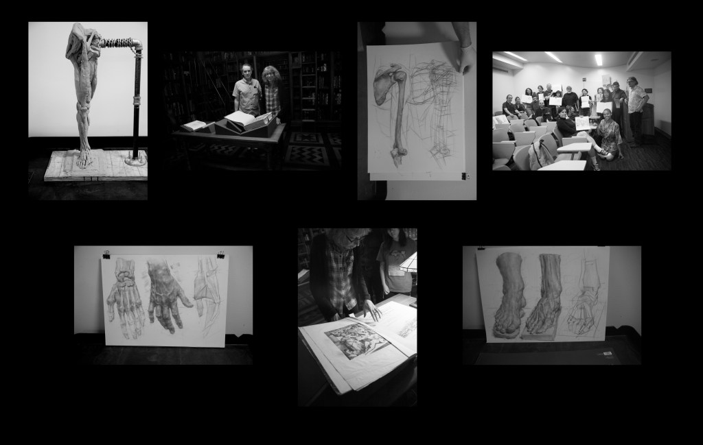
In July 2023, artist and teacher Dan Thompson brought a group of students to the Library’s Drs. Barry and Bobbi Coller Rare Book Reading Room. The students were here in New York for a week-long workshop organized by the Art Students League, “Musculoskeletal Gross Anatomy for the Figurative Artist.” We looked at anatomical atlases dating from the early 16th through the mid-20th centuries. Viewing items from our collection—like the first two images here—and engaging with the students made up the first day of the workshop. The balance took place in the Weill Cornell Medicine anatomy lab, where students worked directly with cadavers.
As the course description explains, “This course presents the study of anatomy as a convergence between anatomical and structural drawing. Motivated students of representational art will have unparalleled opportunities for developing detailed anatomical knowledge through their work in Cornell College of Medicine’s anatomy lab, where they will explore the complexities of the body through the study of prosections and cadavers. Prosections are specially prepared human anatomical specimens, wrapped in a damp preservative, as well as plastinated specimens, which allow for the study of deeper and more isolated anatomical structure. Through laboratory drawing, participating students will become more familiar with the manner of interlocking deeper forms—forms which are not typically clear on anatomical models (due to the haphazard ways that art school skeletons are wired together). Ultimately, students will work towards achieving greater anatomical clarity and validity in their drawing studies, which will be applied to creating higher quality figurative work in the visual arts, from a finer appreciation of human construction.”
Dan teaches at the New York Academy of Art and I have hosted Dan’s New York Academy of Art students here for several years; I first hosted his workshop for the Art Students League in the summer of 2022. This year Dan invited me to visit Weill Cornell’s anatomy lab with the workshop class so that I could gain a deeper understanding of how he teaches with human specimens and watch students make their own drawings and sculptures from cadavers, prosections, and plastinated specimens. Being in the anatomy lab was, for me, a transformative experience, as I had never had the opportunity to see actual cadavers and specimens and think about their relationship to images from historical texts that I share with classes when they visit.
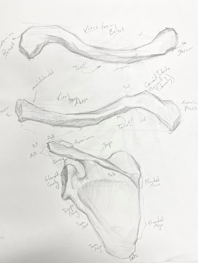
Workshop participant Karina Fuhrman shared images from the visit to the rare book room. The drawings were done by Dan Thompson and the sculpture was done by Karina during her time in the dissection lab.
After the class had ended, I asked if the students would be willing to send their work to me so that we could share it with a broader audience. Many sent images, and it is a privilege to be able to show some of those here.
Artist: Alan LeeArtist: Anna CharuvastraArtist: Chalice MitchellArtist: Eva AvenueArtist: Jae ParkArtist: Kristin duCharmeArtist: Renee Wang
Classes from many local institutions regularly visit the rare book room to engage with materials from our collections. Dr. Evelyn Rynkiewicz, who teaches at FIT, has brought her class “Disease Ecology in a Changing World” more than once. After their 2022 visit, she wrote a blog post about the experience, which you can find here.
If you are interested in bringing your class to the New York Academy of Medicine Library, please reach out to ashaner@nyam.org.
By Dr. Evelyn Rynkiewicz, Assistant Professor of Ecology,. Department of Science and Mathematics at the Fashion Institute of Technology, State University of New York.
My name is Dr. Evelyn Rynkiewicz, I am a professor of ecology at the Fashion Institute of Technology. I teach a course there called “Disease Ecology in a Changing World,” and my background and research is in disease ecology of coinfecting parasites in mice. I wanted to present a course like this for FIT students because diseases are something that affect all of us, everyone has experience being sick, and because emerging infectious diseases are a growing global issue (even before the Covid-19 pandemic, which is of course still impacting us). The challenge in teaching science courses at FIT is that our students mainly have majors in the design and business fields, not in the sciences, so I try to make the course material relate to their backgrounds and experiences as much as possible, to make the content more relevant to them. I also want to increase science literacy in my students, making them comfortable reading, understanding, and talking about science in their personal and professional lives.
I learned about the New York Academy of Medicine Library after seeing the “Germ City” exhibit at the Museum of the City of New York. I got in contact with the Historical Collections Librarian, Arlene Shaner, who set up a visit to show me some of the materials she thought would relate to my course. I was blown away! I knew my students would love to see these historical documents. These materials highlight not only the art and history of how scientists and the public interacted with diseases through time, but also show how intertwined social, economic, and political issues are with how society’s experiences of disease.
Our class took a field trip to the NYAM Library and was shown an array of material; from Hooke’s book on microscopy, Edward Jenner’s work describing his development of the first vaccine, to posters and leaflets used from WWII to the present day to inform people about diseases such as malaria, HIV, or tuberculosis. I am always excited to see what students find interesting from this visit. Many enjoyed seeing the graphic design and illustrations used in the posters, such as those by Dr. Seuss and Keith Haring. Others picked up on how women and marginalized groups were often those who did a lot of the work caring for sick and infected people. Some just liked seeing the historical materials related to New York and being able to see how their home was impacted by diseases in the past.
One of the main assessments for the course is a creative research project where students choose a disease to study and then make a presentation with something creative related to that disease that would help someone learn more about it. I encourage the students to think about how they could use their skills learned from their major and apply it to this topic. The field trip to the NYAM Library provides the initial inspiration for this. I am always so proud and surprised at what they come up with!
Here are some of the things they created:
Arriana Tran, a Fashion Business Management major, created a movie poster. Inspired by the warnings her parents shared with her on the risk of becoming infected with Dengue in her parent’s home country of the Philippines.
Packaging Design major Ethan Wolfsberg designed a malaria testing and monitoring kit that would be able to be used in remote areas that are heavily impacted by this disease. A real-life version would be made in languages appropriate for the area.
To reduce the stigma of taking PreP, Francis Lavery, also a Fashion Business Management major, made an image that emphasizes that this treatment is appropriate for everyone.
Illustration major Leia Garrette wanted to visually show how infection with the agent of Lyme Disease impacts all parts of the body. She created a paper doll where each layer illustrated a different system (e.g. muscles, nervous system) accompanied by an explanation of how each is affected by the infection.
This flyer was created by Sarah Sepulveda from Fashion Business Management. Her plan was for a support group for parents worried about or impacted by Zika virus. There was a focus on Brazil where the outbreak was especially significant in 2016.
Once again, a huge thanks to Arlene and the others at NYAM for their help and insight. I look forward to more collaboration!
Our previous blog posts on this commercial holiday highlighted both cards created for Valentine’s Day as well as trading cards from our collection. At the close of the 19th century, improvements in printing allowed for cheaper goods and paper cards for friends, lovers, and families to send written sentiments.
To celebrate this year, we have created six of our own Valentine’s Day cards featuring images from our collections. One is for the celebration of the popular Galentine’s Day, a celebration of friendship.
Feel free to print out and share with your loved ones!
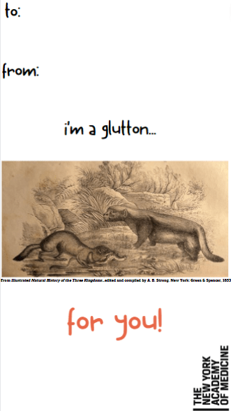
From Sei sparsam!… by Anny Wothe (Leipzig, 1900.)FromHistoriæ animalium... by Conrad Gessner (Zurich, 1551.)From Illustrated Natural History of the Three Kingdoms…edited and compiled by A. B. Strong (New York, 1853.)From Illustrated Natural History of the Three Kingdoms…edited and compiled by A. B. Strong (New York, 1853.)From De motu cordis et aneurysmatibus… by Giovanni Maria Lancisi (Neapoli, 1738.)From Ryzon Baking Book compiled and edited by Marion Harris Neil (New York, 1917.)
By Anne Garner, Curator, Rare Books and Manuscripts, and Robin Naughton, Head of Digital
The Academy Library is thrilled to announce “Facendo Il Libro: The Making of Fasciculus Medicinae, an Early Printed Anatomy.” This online exhibit, focused on an astonishing and influential medical book first published in Italy in 1491, was made possible through the generous support of the Gladys Krieble Delmas Foundation.
Originally collected in manuscript form, the Fasciculus Medicinae (the “little bundle of medicine”) is a richly illustrated collection of medical treatises on uroscopy, phlebotomy, anatomy, surgery, and gynecology. The Fasciculus Medicinae was first published in 1491, but demand for it made it a favorite text for printers. By 1522, it had been issued more than twenty times. Variations in the text and the illustrations through time show the early modern tension between medieval medical ideas and advances in medical understanding forged at the beginning of the 16th century. The exhibit allows visitors to browse full-text scans of all five editions (1495–1522) in The New York Academy of Medicine’s collections; to investigate each edition’s exquisitely illustrated woodcuts and to explore their cultural and medical meanings; and to compare the books’ illustrations in different editions over time. The site includes contributed essays from Dr. Taylor McCall, art historian of material culture and medieval medicine at the Walters Art Gallery, Baltimore, and from Dr. Natalie Lussey Seale of the University of Edinburgh, whose work focuses on early modern Venetian print culture. Dr. McCall’s essay looks at the creation of the text and its accompanying illustrations, while Dr. Seale’s essay offers a window into Venetian printing processes in the 16th century and describes the making of a book in early modern Italy.
Frontispiece, 1495.
The illustrations of the Fasciculus Medicinae offer an intriguing glimpse of medical practice in the 16th century. The book’s woodcuts include narrative scenes depicting the earliest Western depiction of dissection in print, an early illustration of a diagnostic consultation showing a professor analyzing a urine flask, and a physician, holding an aromatic sponge to his nose to avoid infection, attending a sick plague patient confined to his bed. Other woodcuts help us to understand early modern conceptions of health and illness. The Fasciculus Medicinae’s female anatomical figure captures late medieval ideas about women’s bodies, reproduction, and pregnancy. A “Wound Figure” graphically depicts the various threats to the body, from blows to the head down to the prick of a thorn on the feet. Perhaps most surprising of all, the Fasciculus Medicinae’s “Zodiac Figure,” who balances all twelve zodiac signs on his body, conveys the powerful role the stars and planets played in health in the medieval imagination. This figure, who dates to earlier manuscripts from the medieval period, survives well into the twentieth century, appearing alongside horoscopes in a modified form in print in American almanacs produced by pharmaceutical companies.
This slideshow requires JavaScript.
The Facendo Il Libro website has a simple design, but a complex structure. It is both a standalone digital collection and an online exhibit built using Islandora, an open-source digital repository framework. Representing the first full-text internal digitization project for the Academy Library, the five editions of the Fasciculus Medicinae were digitized in the Library’s Digital Lab. The online exhibit was built using an Islandora multi-site to leverage the digital collection repository (Fedora), Drupal Book module, and the current Library branding theme.
The ability to draw from the common repository made it possible to store content once and use it in multiple ways. Thus, the five digitized editions are available in two different places using a single source. The built-in navigational structure for the exhibit makes it easy for users to explore the collection in a linear fashion or by sections.
Replicating the physical experience of touching the text is still a challenge for digital projects. Thus, it was important to create a digital experience that provides the user with some sense of the materiality of the object. For example, the 1500 edition was bound with another text (Savonarola’s Practica medicinae), which is evident from the first digital image of the book. The image shows the thickness of the text and the fact that the 1500 edition begins in middle of the physical object. It shows the user exactly what will be encountered when using the physical item. It also highlights a significant piece of information that could have been lost due to cropping.
Another important aspect of the online exhibit is the illustrations page, where users can see all the illustrations from all editions in one place. When a user clicks on an illustration, the user is immediately taken to a page with descriptions of each illustration as it appears in each edition. To explore the images, users can click on an image and zoom in to see the intricate details.
Facendo Il Libro Illustration Page
Facendo Il Libro Illustration Zoom
“Facendo Il Libro: The Making of Fasciculus Medicinae, an Early Printed Anatomy” offers a great opportunity for users to learn and explore the Library’s five editions of Fasciculus Medicinae in context.
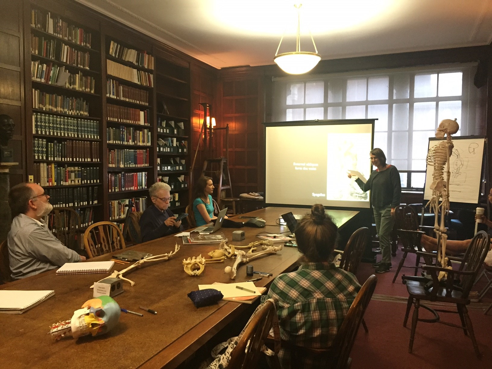
Today’s guest post is written by Kriota Willberg, New York Academy of Medicine’s Artist-in-Residence. Through graphic narratives, teaching, and needlework, Kriota explores the intersection between body sciences and creative practice. This May, Kriota taught a four-week workshop entitled “Visualizing and Drawing Anatomy,” which utilized live models as well as anatomical illustrations from the New York Academy of Medicine’s library. You can read more about Kriota’s work HERE.
The class gets oriented before drawing practice.
The Visualizing and Drawing Anatomy workshop was held at the Academy Tuesday evenings in June. Once again I was impressed by the participants willingness to practice looking underneath our models’ skin to draw the deep anatomical structures that give our bodies form.
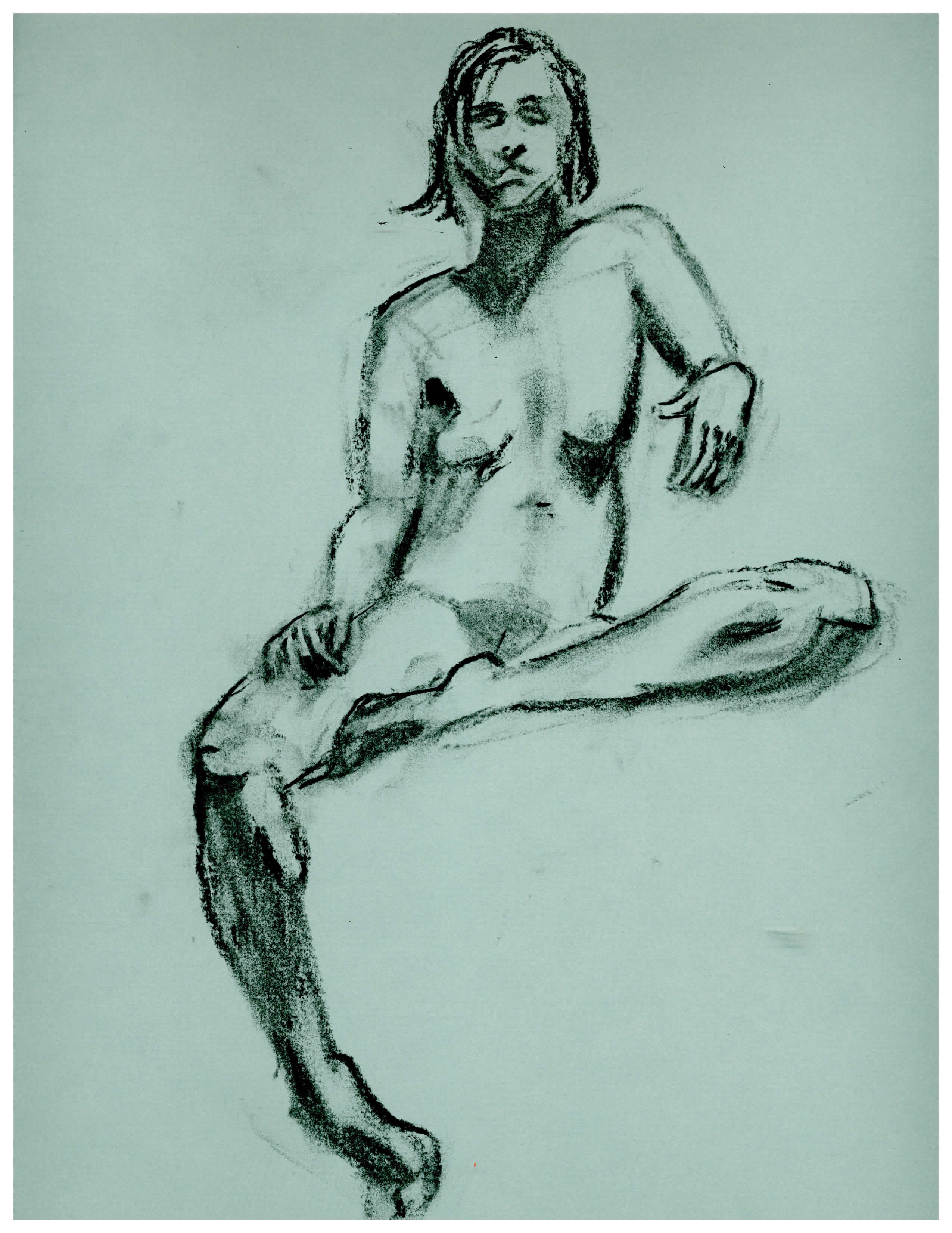
Participants draw using their preferred medium, in this case, paper or an iPad.
Who benefits from this kind of drawing practice? Practically everyone. Trained artists sharpen their skills, and those new to art and drawing learn fundamental principles of anatomy that lay the foundation for drawing the human figure.
Debbie Rabina, who is new to art, took the workshop last year. Since then she has kept a regular drawing practice and she occasionally incorporates anatomy into her work.
Debbie Rabina’s drawing since taking “Visualizing Anatomy” in 2016.
Ellen Zaraoff is a photographer who has just started drawing. Until taking the classes this year she had been focusing on drawing portraits in charcoal. She took the workshop to get an introduction to anatomy, structure, and proportion.
Sarah Wukoson has a BA in art, and works in medical research. She took the workshop this year because she’s interested in the intersection of art and medicine as well as “the interplay of different modes of understanding the body.”
Sarah Wukoson’s 2017 in-class sketches and exercises.
Jim Doolley is a “life-long art lover who decided a couple years ago to take a stab at producing, not just consuming.” His focus is drawing and painting. He took this class to improve his draftsmanship.
Jim Dooley’s 2017 homework.
Susan Shaw is an artist. She says, “I took the class (last year) because I found I was thinking 2 dimensionally when I was drawing and the figures seemed to have no life… I now think about how the body functions when I draw and it makes gesture and weighting much easier.”
Susan Shaw’s figure drawing since taking “Visualizing Anatomy” in 2016.
The variety of participants: artists, illustrators, cartoonists and enthusiastic beginners – all interested in anatomy and the Library’s historical collection make this workshop one of my favorites to teach.
This September 14-October 5, Kriota is offering an “Embroidering Medicine Workshop,” which will take place at the Academy. This four-week workshop explores The New York Academy of Medicine Library’s historical collections, examining relationships between medicine, needlework, and gender. Learn more and register HERE.
Nowadays, “there’s an app for that” for nearly any question or need you might possibly have –not to mention needs you didn’t even know you had. What you might not realize is that apps –in the sense of a handheld device for manipulating data- are hundreds of years old.[1]
Meet the ancestor of your smartphone apps: the volvelle, sometimes called a wheel chart. It’s a (brilliantly) simple paper construction of moving parts; layers of rotating discs with information on them. Externalized, artificial data memory before the printing press, steam power, photography, electricity, ether anesthesia, radar, cars, the internet and wifi. Wow.
Gadgets for working with data are even older than paper volvelles. Think of the astrolabe, which had dials that measured the angle of the sun, allowing you to determine accurate time. Useful as an astrolabe was, it was very fine metalwork and, therefore, expensive. Paper devices were a more economical idea.
Two views of an astronomical volvelle from Federici Chrisogno’s De modo collegiandi pronosticandi et curandi febres (1528). Chrisogno was among the first to posit that the cause of tides was connected to the moon and the sun.[2] Note among the exquisite details the tiny human faces on the sun and moon orbs (in the edges of the top two discs) and the delicate astrological symbols (around the outer disc’s rim).
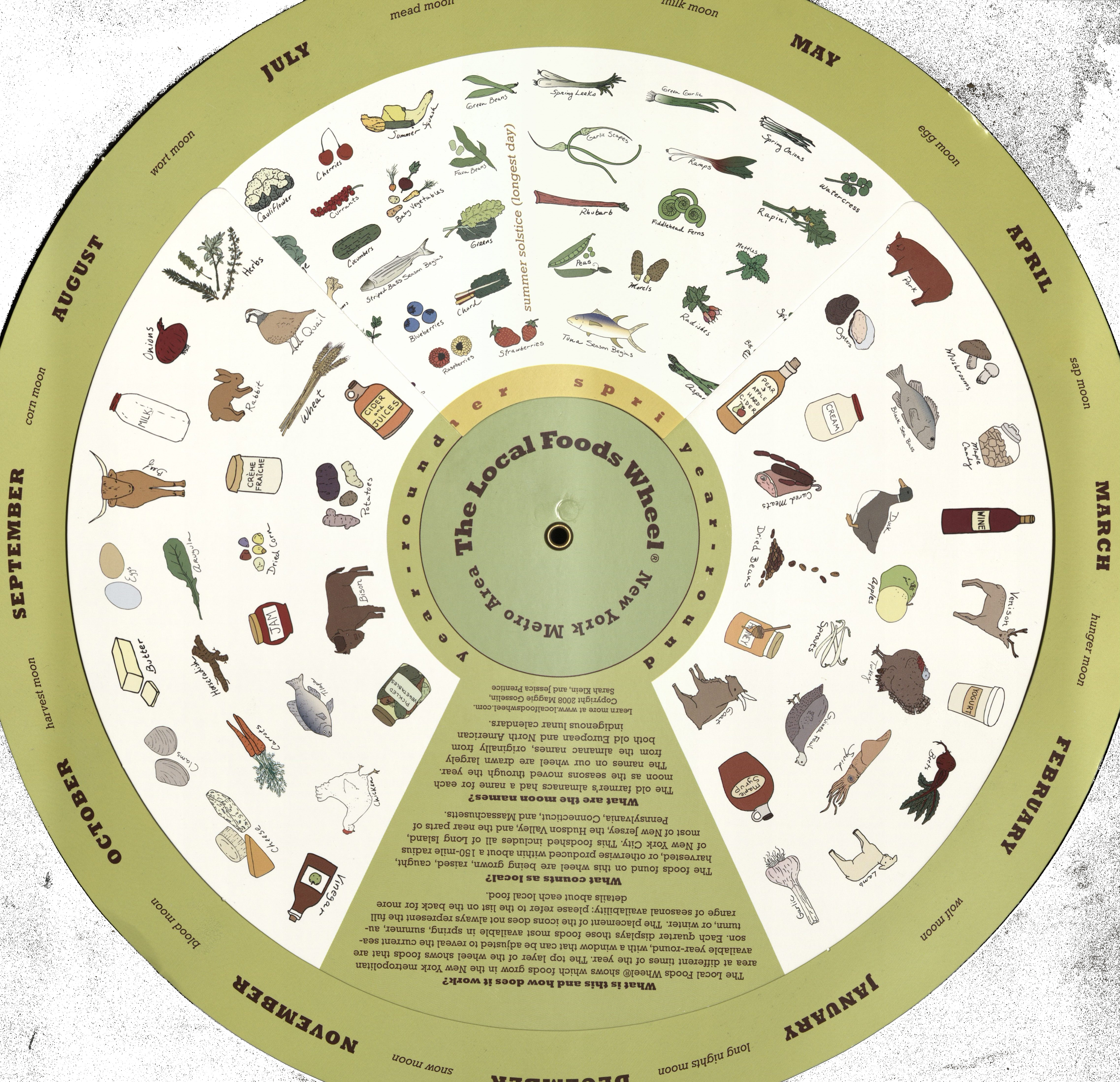
Like many scientific innovations, volvelles came to Europe from the Arabic world during the 11th and 12th centuries in medicinal and astronomical works.[3] One of the earliest extant volvelles was created by Ramon Llull from Majorca (modern day Spain) in his Ars Magna published in 1305. His volvelle, “The Night Sphere,” could be used to calculate the time at night by aligning the device with the pole star –exact times being important to him for knowing the most auspicious times to administer medicine.[4] Incidentally, the European adoption of this useful device is reflected in the name we have for it, volvelle, from the Latin volvere meaning “to turn.” The scope of information that volvelles depict is huge. Besides astronomy, subjects include: verb conjugations, color wheels, metric conversions, fortune-telling, first-aid techniques, and local seasonal foods (such as in the modern example below).
Some volvelle constructions can get very elaborate in form, like this unusual and entertaining piece in our collection, The Bodyscope (1948), by Ralph H. Segal and Theodore I. Segal, with illustrations by William Brown McNett. It is a color-lithographed, interactive anatomical chart designed for the educated lay public. When opened, the chart displays a male figure on the left and a female figure on the right, surrounded by skeletons and muscle men. Each of the large figures houses a volvelle that, when rotated, displays five different views of the internal organs. Additional cut-outs on the front and back of the chart also change as the volvelles move to display additional views of various body parts and systems.
The Bodyscope (1948) by Ralph H. Segal and Theodore I. Segal, with illustrations by William Brown McNett.
Inspired by volvelles in our collections, we’ve gotten creative for the upcoming Museum Mile Festival, Tuesday June 13 from 6-9pm along Fifth Avenue. It’s a delightful cultural block party; seven museums are open for free, and there are special crafts and performances. An evening you won’t want to miss! Especially since we’ve created a DIY volvelle for festival goers to make for themselves.
Our volvelle feature male and female bodies created by the highly influential Dutch physician and anatomist, Andreas Vesalius, for De Humani Corpis Fabrica (1543). The Fabrica was groundbreaking not only for its artistry, but for its promotion of learning about human anatomy through dissection. Understanding of the human body had been dominated in the West since the third century by the work of the Greek anatomist Galen, who performed animal dissection. Vesalius’ work on cadavers revealed anatomical errors in Galen’s work and pushed medical knowledge forward.
Our DIY volvelles let you deepen your own anatomical knowledge by adding in human organs (from the well-known Gray’s Anatomy) and anatomy facts of your choice. See you at the Festival!
Acknowledgments:
Special thanks to Anne Garner for information on The Bodyscope, and the Library extends our gratitude to Harlem Artist & Craftsman for the generous donation of supplies for the Festival.
References: [1] Adam Rothstein. The Original Mobile App was Made of Paper. Retrieved from https://motherboard.vice.com/en_us/article/the-earliest-mobile-apps. [2] Federico Bonelli, Lucio Russo. The Origin of Modern Astronomical Theories of Tides: Chrisogno, de Dominis and Their Sources. The British Journal for the History of Science. 1996; 29 (4): 385-401. [3] David Kahn. On the Origin of Polyalphabetic Substitution. Isis. 1980, 71 (1): 122-127. [4] Rheagan Martin. Decoding the Medieval volvelle. Retrieved from http://blogs.getty.edu/iris/decoding-the-medieval-volvelle/. Accessed March 14, 2017.
By Audrey Sage Lorberfeld, Digital Technical Specialist
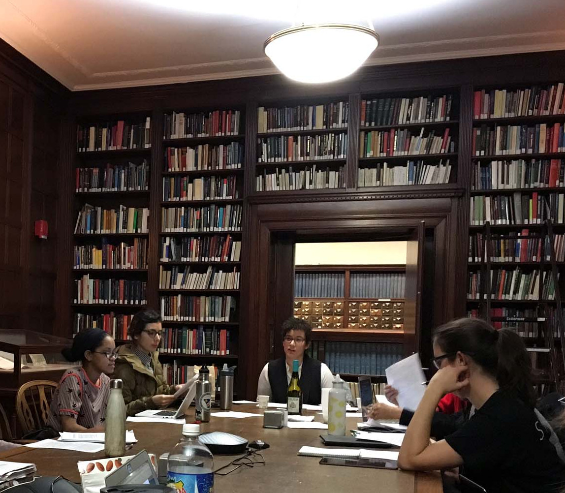
For three hours each Monday evening, January 30 through February 20, the Academy hosted a Brooklyn Institute for Social Research class called Feminist Futures, for which I was lucky enough to be the staff liaison. My classmates ran the gamut from PhD students to artists to professors to web developers to librarians and archivists. Our professor, Danya Glabau, guided us through the intellectual history of the intersection of science studies and feminist theory. Professor Glabau’s syllabus included the writings of such luminaries as Donna Haraway, Bruno Latour, Evelyn Fox Keller, and Emily Martin. To complement these readings, the Academy was able to provide some of its own treasures as well.
One such item was the Traité d’osteologié, published in 1759 with text by the Scottish anatomist Alexander Monro and illustrations supervised by Marie Geneviève Charlotte Thiroux D’Arconville. D’Arconville studied anatomy at the Jardin Du Roi and translated Monro’s earlier text into French for this volume. Although her name does not appear anywhere in the text (her plates were published under the protection of Jean-Jacques Sue, a member of the French Royal Academy), it is generally accepted that d’Arconville is the hand behind the gorgeous images. Among her plates are incredible depictions of male and female skeletons that display features associated with each gender. She renders the male skeleton as large and statuesque and places him in front of a backdrop of Classical architecture. Her female skeleton, on the other hand, is more petite and stands in a less assertive position. Noticeably, her rib cage is extremely narrow while her wide hips and pelvis are very emphasized. There is speculation that the image of a narrow rib cage is meant to associate the skeleton with upper class women who usually wore corsets.
Female skeleton from Traité d’osteologié (1759)
Paired with this item for a unit titled “Feminist Objectivity” were Donna Haraway’s “Situated Knowledges: The Science Question in Feminism and the Privilege of Partial Perspective,” Karen Barad’s “Meeting the Universe Halfway: Realism and Social Constructivism Without Contradiction,” and Michelle Murphy’s “Immodest Witnessing: The Epistemology of Vaginal Self-Examination in the U.S. Feminist Self-Help Movement.” Among other topics, we guessed at what our authors might have thought of today’s quantified-self movement and whether or not data about the self could be categorized as an extension of that self. Further, we asked: what happens to this paradigm when you engage with its exponential commodification? Could self-awareness excuse the self from the ‘wrong type’ of objectification? We also spent a significant part of the class analyzing what Haraway’s idea of “seeing from below” might mean in our current political climate.[1] We queried, is it possible to adopt Haraway’s type of situated knowledge and avoid being ableist?
“Feminist Futures” class taking place at the Academy.Image source: Suzanne Schneider, Director of Operations and Core Faculty at Brooklyn Institute for Social Research.
One of my favorite quotes from this part of the course was “rational knowledge does not pretend to disengagement.”[2] I took this to mean that pushing for a type of feminist objectivity that highlights seeing from below and/or something Barad calls “agential realism” does not mean that you are disengaging from your subject.[3] Rather, it means that you are striving towards a feminist typology of embodiment that focuses its recuperative energies on welcoming emotions and relationships as data, all the while keeping in mind that “no knowledge is innocent.”[4] This was a very powerful idea to me as a woman working at the Academy in a nexus of technology, history, and public service.
We rounded out the class with a viewing of Crania America, a book published in 1889 by Samuel George Morton, a famed phrenologist. Included in his tome are illustrations of different race’s skulls along with commentary on their corresponding mental abilities. He describes his project as demonstrating that “a particular size and form of brain is the invariable concomitant of particular dispositions and talents, and that this fact holds good in the case of nations as well as of individuals.”[5] He goes onto say that:
A knowledge of the size of the brain, and the proportions of its different parts, in the different varieties of the human race, will be the key to a correct appreciation of the differences in their natural mental endowments, on which external circumstances act only as modifying influences….[5]
As you can imagine, this item generated a passionate conversation. Highlights included discovering that the roots of cybernetics (a field which began in WWII) come from the ancient Greek adjective κυβερνητικός, meaning ‘good at steering’ (n.b. the militaristic and authoritative implications); the theory behind Chela Sandoval’s term “US third-world feminist”; and the layered irony within our assigned texts regarding authority and boundaries.
Skull from Crania America (1889)
While this course was challenging, we made sure to keep the conversation approachable and friendly. This litmus test of a Brooklyn Institute for Social Research-The New York Academy of Medicine Library collaboration solidified our belief that:
Together [our two institutions] can make the histories, presents, and futures of science and technology relevant to the lives of work adults, supporting the development of knowledge and interest in these crucial aspects of our complex and ever-changing society. (Professor Glabau)
We hope you join us next time!
References:
[1] Haraway D. Situated Knowledges: The Science Question in Feminism and the Privilege of Partial Perspective. Feminist Studies. 1988; 14(3): 575-599. (Quote on p.583). [2] Haraway D. Simians, cyborgs, and women: the reinvention of nature. New York: Routledge;1991. (Quote on p. 196). [3] Barad K. Meeting the Universe Halfway: Realism and Social Constructivism without Contradiction. Feminism, Science, and the Philosophy of Science. 1996;256: 161-194. (Quote on p.179). [4] Warren K, Cheney J. Ecological Feminism and Ecosystem Ecology. Hypatia. 1991;6(1): 179-197. (Quote on p. 191). [5] Morton S. Crania americana. Philadelphia: London, J. Dobson; Simpkin, Marshall & Co;1839. (Quote on p. 274).
Kriota Willberg, the author of today’s guest post, explores the intersection of body sciences with creative practice through drawing, writing, performance, and needlework. She is offering the workshop “Visualizing and Drawing Anatomy” beginning June 6 at the Academy. Register online.
Cheselden’s Osteographia, 1733, opened to the title page and frontispiece.
Different Disciplines, Same Body
I teach musculoskeletal anatomy to artists, dancers, and massage therapists. In my classes the students study the same raw material, and the set of skills each group acquires can be roughly organized around three distinct areas: representation of the body, kinesiology (the study of movement), and palpation (feeling the body).
As an anatomy teacher I am constantly on the prowl for images of the body that visually reinforce the information my students are learning. The Internet has become my most utilized source for visual teaching tools. It is full of anatomy virtual galleries, e-books, and apps. 3D media make it ever easier to understand muscle layering, attachment sites, fiber direction, and more.
In spite of the overwhelming volume of quality online cutting-edge anatomical imagery, I find myself drawn to historical 2D printed representations of the body and its components, once the cutting-edge educational technology of their respective centuries. Their precision, character, size, and even smell enhance my engagement with anatomical study. Many of these images emphasize the same principles as the apps replacing them centuries later.
The Essential Structure Of The Body
Different artists prefer different methods of rendering bodies in sketches. One method is to organize the body by its masses, outlining its surface to depict its bulk. Another method is to draw a stick figure, organizing body volume around inner scaffolding.
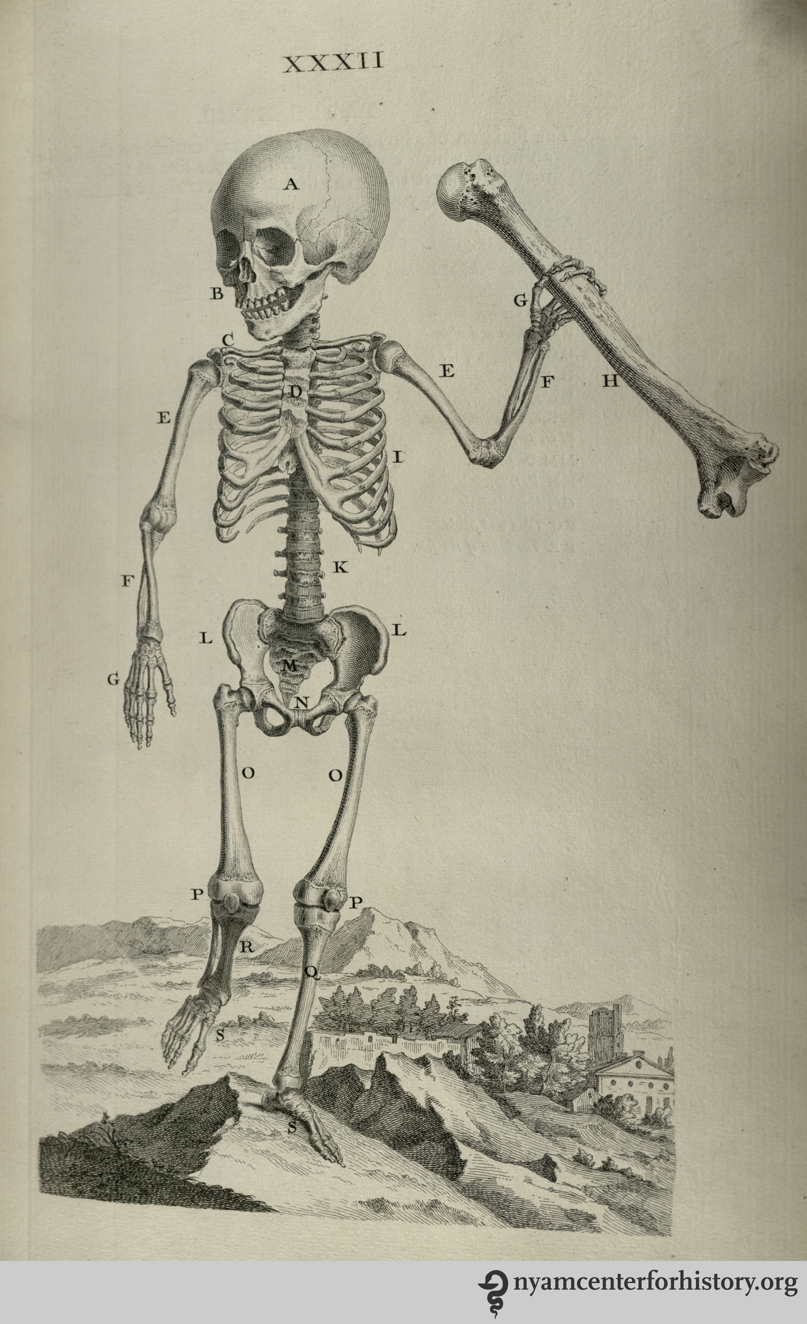
And what is a skeleton but an elaborate stick figure? William Cheselden’s Osteographia (1733) presents elegant representations of human and animal skeletons in action. These images remind us that bones are rigid and their joints are shaped to perform very specific actions. The cumulative position of the bones and joints gives the figure motion. In Cheselden’s world of skeletons, dogs and cats fight, a bird eats a fish, a man kneels in prayer, and a child holds up an adult’s humerus (upper arm bone) to give us a sense of scale while creating a rather creepy theatrical moment.
Muscle Layering
3D apps and other imaging programs facilitate the exploration of the body’s depth. One of the challenges of artists and massage therapists studying anatomy is transitioning information from the 2D image of the page into the 3D body of a sculpture or patient.
Salvage’s Anatomie du gladiateur combattant: applicable aux beaux artes… (1812) is a 2D examination of the 3D Borghese Gladiator. Salvage, an artist and military doctor, dissected cadavers and positioned them to mimic the action depicted in the statue. His highly detailed images depict muscle layering of a body in motion. The viewer can examine many layers of the anatomized body in action from multiple directions, rendered in exquisite detail. Salvage retains the outline of the body in its pose to keep the viewer oriented as he works from superficial to deeper structures.
Bernhard Siegried Albinus worked with artist Jan Wandelaar to publish Tabulae sceleti et musculorum corporis humani (1749). Over their 20-year collaboration, they devised new methods for rendering the dissected body more accurately. The finely detailed illustrations and large size of the book invite the reader to scrutinize the dissected layers of the body in all their detail. Although there is no superficial body outline, the cadaver’s consistent position helps to keep the reader oriented. On the other hand, cherubs and a rhinoceros in the backgrounds are incredibly distracting!
Fiber Direction
Familiarity with a muscle’s fiber direction can make it easier to palpate and can indicate the muscle’s line of pull (direction of action).
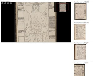
The images of Jacopo Berengario da Carpi’s Anatomia Carpi Isagoge breves, perlucide ac uberime, in anatomiam humani corporis… (1535) powerfully emphasize the fiber direction of the muscles of the waist. This picture in particular radiates the significance of our “core muscles.” Here, the external oblique muscles have been peeled away to show the lines of the internal obliques running from low lateral to high medial attachments. The continuance of this line is indicated in the central area of the abdomen. It perfectly illustrates the muscle’s direction of pull on its flattened tendon inserting at the midline of the trunk.
The Internal Body Interacting with the External World
One of the most important lessons of anatomy is that it is always with us. Gluteus maximus and quadriceps muscles climb the stairs when the elevator is broken. Trapezius burns with the effort of carrying a heavy shoulder bag. Heck, that drumstick you had for lunch was a chicken’s gastrocnemius (calf) muscle.
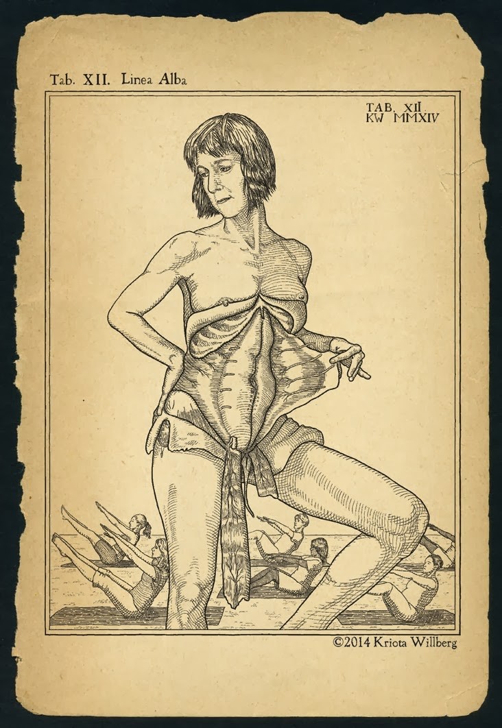
Anatomists from Albinus to Vesalius depict the anatomized body in a non-clinical environment. One of my favorites is Adriaan van de Spiegel and Giulio Casseri’s De humani corporis fabrica libri decem (1627). In this book, dissected cadavers are depicted out of doors and clearly having a good time. They demurely hold their skin or superficial musculature aside to reveal deeper structures. Some of them are downright flirtatious, reminding us that these anatomized bodies are and were people.
Kriota Willberg’s self portrait. Courtesy of the artist.
I am so enamored of van de Spiegel and Casseri that I recreated page 24 of their book as a self-portrait. After my abdominal surgery, the image of this cadaver revealing his trunk musculature resonated with me. In my portrait I assume the same pose, but if you look closely you will see stitch marks tracing up my midline. I situate myself in a “field” of women performing a Pilates exercise that challenges abdominal musculature. And of course, I drew it in Photoshop.


 The Facendo Il Libro website has a simple design, but a complex structure. It is both a