Artist Inspiration: Plant Cure (Part 2)

Todays’ guest post is introduced by Maddy Rosenberg, curator and founder of CENTRAL BOOKING. The New York Academy of Medicine Library and CENTRAL BOOKING collaborated on the exhibition Plant Cure.  For this exhibition, five artists were selected to do research at the Academy Library over six months to produce work with their own unique take on medicinal plants. The project will culminate with an exhibition at CENTRAL BOOKING on the Lower East Side from September 6-October 29, 2017. Part 1 can be read here.

The next two artists featured in the Plant Cure collaboration between CENTRAL BOOKING and the New York Academy of Medicine Library are Susan Rostow and C Bangs. Susan’s sculptural work is extremely textural and beckons to be touched, while with C it’s our eye that takes the journey over the surfaces. Both artists’ works engage us and demand closer scrutiny.

Susan Rostow

I spent many wonderful hours of my childhood reading the encyclopedia. A set of books from A to Z neatly organized on a shelf with the entire world’s information gave me great joy. I may be a romantic, waxing poetic and nostalgic about the past, but that has not stopped me from enjoying the present times of clicking and swiping through Google images and other websites. My ongoing fascination with information, books and images continued to grow through decades and is presently expressed in my sculptural books.

The first time I entered the New York Academy of Medicine Library and was surrounded by rare books dating from the 15th through the 18th centuries, I felt as though I traveled back in time and entered the Middle Ages. I was taken with the smell of the leather covers, amazed by the weight and size of some of the books, marveled at the odd titles on the bindings, and was captured by highly detailed and precise illustrations. Prodigiorum Ostentorum Chronicon (1557) by Konrad Lykosthenes and Osteographia, or, The Anatomy of the Bones (1733) by William Cheselden are a couple of my favorites.

3_Rostow_

Susan Rostow working in studio.

Feeling incredibly inspired, I took my excitement to the studio along with photos of the pictures from the various books I had observed. Armed with a plethora of images and plenty of ideas, I began to work on my vision. Images of medicinal mushrooms and text pertaining to plant cures were put to use by first making carborundum printmaking plates. This is a low tech method used for making plates by hand. This simple, but elegant technique allowed me to connect with some of the similar hand techniques used by the original artists. I printed them with an etching press, a simple press whose basic principle has not changed for centuries. Choosing to use this technique with an old style press made me feel connected to some of the reproductions from the New York Academy of Medicine Library’s rare book collection.

1_Rostow

Susan Rostow, Bone Fungus. 2017, mixed media sculptural book with carborundum prints on paper, dried mushroom, wood, parabolic mirrors, real and plastic bones, sand, glass beads and pigments, 25 x 26 x 26 inches.

After printing hundreds of images of mushrooms and text on paper, the prints were bound together with dried mushrooms, mud, natural glues, and pigments. Paper, tree fungus, roots, soil, and casts from bones merged together creating sculptural books that look, smell and feel like unearthed relics secreted beneath the earth. Hopefully this synthesis captured some of the magic that I felt when I first viewed these incredibly illustrated books.

04_Rostow

Susan Rostow’s sculptural book Bone Fungus (left and center), and detail of Cheselden’s anatomical illustration (1733) (right).

2_Rostow

Prodigioky Ostentory Chronicon (left) William Cheselden’s anatomical illustration (1733) (center), and detail from Susan Rostow’s sculptural book Bone Fungus (right).

C Bangs

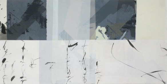
My art investigates frontier science combined with symbolist figuration from an ecological feminist point of view. A decade long collaboration with quantum consciousness physicist Dr. Evan Harris Walker has lead me to incorporate his equations in my paintings in a manner mutually agreed upon, designed to posit questions related to his theories. Functioning as design elements that often speak to the interconnectivity of everything in the cosmos, the equations parallel the sacred writings found in illuminated manuscripts. In recent collaboration with my partner, Dr. Greg Matloff, we investigate consciousness from the point of view of panpsychism philosophically, historically and scientifically.

The books I researched at the New York Academy of Medicine Library included Robert Fludd and Konrad Lykosthenes. What does humankind preserve and what do we eliminate? Fludd had a theory of cosmic harmony and Kepler correctly accused Fludd of being a theosophist. Additionally Fludd is remembered as an astrologer, mathematician, cosmologist, Quabalist and Rosicrucian. His writing centered around sympathies found in nature between man, the earth and the divine.

fpseedsfetus copy

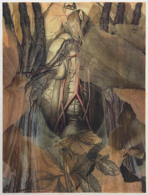
Flowering Pavonis seeds used as an abortifacient with fetus studies. C Bangs (2017).

Maria Sibylla Merian’s Metamorphosis insectorum Surinamensium (1705) at the Brooklyn Botanic Garden ultimately lead me to contact the New York Botanical Garden. Merian wrote that slave women’s use of the peacock flower was deeply political, using it to abort pregnancies forced upon them by their slave owners. The history of abortifacients is nearly as old as the written word and the determination of pregnancy was left to the woman, who was not considered pregnant until she declared herself to be so. When the Catholic Church realized that they could not regulate abortifacients or convict the women who used them, they began persecuting midwives, declaring them witches.[1] The enforcement of religious law and witch burning was an effective tool for breaking a chain of knowledge about abortifacients that had been in circulation for over a thousand years. Despite Merian’s revelation about the peacock flower in her book, widely used by botanists and men of medicine, this knowledge was ignored. Merchants valued the plant’s looks and shipped large amounts of its seeds to their home countries, where the flower decorated many royal gardens.

Cosmic Pavonis Fludd mix copy

Flowering Pavonis and diagrams from Robert Fludd’s Utriusque cosmi majoris scilicet et minoris metaphysica (1617-1621). C Bangs (2017).

Ironically, when I wished to photograph the peacock flower at the Brooklyn Botanic Garden or the New York Botanical Garden, I found that it had been deaccessioned by Brooklyn and is kept in a section not available to the public at the New York Botanical Garden.

Pavoniscosmos1 copy

Flowering Pavonis and images from Konrad Lykosthenes’ Prodigiorum ac ostentorum chronicon (1557). C Bangs (2017).

Reference:
[1] Edwards, Stassa. The History of Abortifacients. Jezebel: 2014, November 18.

Artist Inspiration: Plant Cure (Part 1)

Todays’ guest post is introduced by Maddy Rosenberg, curator and founder of CENTRAL BOOKING. The New York Academy of Medicine Library and CENTRAL BOOKING collaborated on the exhibition Plant Cure.  For this exhibition, five artists were selected to do research at the Academy Library over six months to produce work with their own unique take on medicinal plants. The project will culminate with an exhibition at CENTRAL BOOKING on the Lower East Side from September 6-October 29, 2017.

I approached Lisa O’Sullivan, the Director of the New York Academy of Medicine Library, who I had first met when she participated in one of our panels at the gallery, with an idea for a collaborative project. An important component of CENTRAL BOOKING’s programming has always revolved around art and science as well as artist’s books, therefore a collaboration with the New York Academy of Medicine seemed only natural.

For the project, ultimately named Plant Cure, five artists were selected to do research at the Academy Library over six months to produce work with their own unique take on medicinal plants. The project will culminate with an exhibition at CENTRAL BOOKING on the Lower East Side from September 6-October 29, 2017, featuring the work of the artists in dialog with other artists who have also been intrigued by the theme in their own work. At the Academy, display cases document the research, source material, and working methods employed by each of the five artists in the process of creating their work for Plant Cure.

Over the next few weeks, I am pleased to be able to present here those five artists as they discuss their work and time at the Academy Library. This week we begin with James Martin and Nancy Campbell, both whose final project work is in printmaking, but through very different approaches and results.

James Martin

My questions: how have artists and anatomists from the past chosen to depict what lies beneath the surface of the body? How have botanists and artists portrayed the plants thought to have curative properties? What are the common design elements of these life forms? Have the different printing processes changed the nature of this visual information? And my creative query—how can I re-purpose these incredible pictures from the Academy Library and create something completely new?

I narrowed my focus to anatomical texts that explored arterial and venous networks, attracted to the obvious analogies to plant forms. Historical Collections Librarian Arlene Shaner was able to suggest many fascinating volumes, such as:

The crisp and stylized engravings of John Lizars (1825) use red and blue colors to graphically present the networks of veins and arteries. Antonio Scarpa’s large engravings on the subject of aneurysms are arranged with clarity and artfulness. Closeups of these lethal defects are beautifully abstract. Lithographs of arteries by Richard Quain and Joseph Maclise (1844) have a more poignant quality. The cadavers are not generic bodies, but individuals, often young. Instruments of dissection are part of the still life. Another completely different, but fascinating approach, is Wilhelm Braune’s Topographical Atlas (1888). The color lithographs are accurate renderings from frozen slices of cadavers. Our modern MRI imaging is the closest analogy. Some of these butcher shop portions produce a shiver of revulsion. But, the images are flat and the resulting shapes allow for alternate design opportunities.

Tabulae Arteriarum_watermark

Torso from Frederich Tiedemann’s Explicationes tabularum arteriarum corporis humani (1822).

For my exploration of medical botanicals, I began with the line woodcuts of Fuchs (1542). It could be used as a field guide today such is the clarity and accuracy of its observations. The engravings in William Woodville’s Medical Botany (1793) are even more detailed and nuanced. Structures are clear and complete from root to flower. The addition of color in the Henry Trimen and Robert Bentley’s Medicinal Plants (1880) imparts an even more lifelike quality to the illustrations.

Medical Botany_watermark

Hellebore from William Woodville’s Medical Botany (1793).

As part of my creative process, I took digital photographs of plates contained in the above described books. Back in my studio, I work with these photos with editing software. Beginning with anatomical images, I establish the “bones” of the composition.  These are layered with my photographs of tree bark to provide textures, shapes, and a non-specific context, with the relevant botanicals added to the mix. The finished piece was then printed via an inkjet printer on printmaking paper. I added another element with the application of monotype inks printed from mylar over the digital prints for a slight softening of the sharpness and more richness to the color.

Tree bark photograph used in Torso with Hellebore (Left). Monotype plate for Torso with Hellebore (Right).

My creative mash-ups of these historic images have been inspiring and fun. Thanks to all at the Academy for hosting this project and to Maddy Rosenberg of CENTRAL BOOKING for organizing this residency and the upcoming exhibition Plant Cure.

Torso with Hellebore

Torso with Hellebore by James Martin archival digital print with monotype.

Nancy Campbell

I absolutely adored my time spent in the Drs. Barri and Bobbi Coller Rare Book Reading Room at the New York Academy of Medicine. Handling objects so old, delicate, and precious was a rare treat, indeed.

While I enjoyed studying an array of different volumes in the Academy Library, Okamoto Ippo’s Jūshi kei ryaki wago (1693; 3 vol. book of Moxa-cautery) was a perfect match for me. Medieval Japanese picture scrolls have been a long fascination, and I have studied them in museum exhibitions in Japan and the USA. Of course, I have never held an actual medieval scroll and experienced the sequential unfolding of its story (scrolls being so incredibly fragile). Therefore, handling a 17th century Japanese book during my residence, with its ultra-thin, semi-transparent printed paper, was an amazing first-time experience for me and one that will surely affect my work for years to come.

01.Nancy Campbell.Artemisia

Artemisia by Nancy Campbell.

In my artwork I strive to evoke an Eastern sense of balance between fragility and strength while using a system of highly structured, intricate abstraction. My methods are slow and measured, but I work for a spontaneous result that inhabits an ambiguous realm between the visible and invisible, the logical and the intuitive, the representational and the abstract. Echoed in all of my work is a continuous play of opposites – often found at the heart of Japanese aesthetics.

02.Nancy Campbell, Meridian

Meridian by Nancy Campbell.

My work for the Plant Cure exhibition references text and diagrams that appear to be layered on top of one another. Each page in the Japanese books I viewed has hints of the previous page showing through the thin Japanese paper. I printed and painted on both sides of Japanese papers and used the method of collage (with Japanese glue) to layer multiple sheets together. A large screenprint based on a collage is still in process.

Eyes Turned Skywards

By Anne Garner, Curator, Rare Books and Manuscripts

Ain’t no sunshine when she’s gone….as the song goes, or, on a day like today, when the moon encroaches on the sun. With all eyes turned skywards, we’re taking the long view on star-gazing, looking back to many of our great sixteenth-century astronomy books for inspiration.  Last week, in honor of today’s solar eclipse, we hosted Atlas Obscura in our rare book room for a ticketed event highlighting some of our favorite images of the stars, planets and astronomers– those inquisitive heavenly creatures who made great strides in changing what we know about the physical universe.

1LEFTfleurs_sundetail_doubleimage_watermark

A pocket-sized French book, Les fleurs et secrets de medicine, published around the turn of the 16th-century, offers this partially covered sun, in the image on the left.  On the right, from the same book, our hero, the astronomer.

After Homer’s Iliad and Odyssey, the most popular poem produced by the ancient Greeks was Aratus’ Phaenomena.  Aratus, born in Soli in Cilicia, lived in the late fourth and early third centuries B.C.E.  As a young man, he studied Stoic philosophy in Athens at the school founded by Zeno. Building on a tradition of didactic poetry exemplified by the epic poet Hesiod, the Phaenomena, Aratus’ only complete extant work, explained the constellations and the effects of the planets and stars on human event in verse. A Latin translation of the poem appears in our 1499 Astronomicae Veteres, a compilation of early astronomy texts printed by Aldus Manutius in Venice.  Many of the woodcut images of constellations accompanying the poem date to an earlier Venetian publication of Hyginus’ star atlas, printed by Erhard Ratholdt.

The image of the Pleiades in the illustrated Aratus can likely be attributed to the artist of the famous Hypnerotomachia Poliphili also published by Aldus Manutius in the same year.

The Academy Library has five copies of the Fasciculus Medicinae –a compilation of medical treatises, many from the medieval period first published in 1491 (our earliest edition dates to 1495).  This compiler was probably an Austrian physician named Kircheim, which the Italian publishers corrupted to Ketham.  Kircheim, born in Germany, was professor of medicine in Vienna in about 1460.

The Fasciculus Medicinae contains the earliest realistic anatomical images in print.  The book’s astonishing woodcut illustrations include skilled renderings of medieval prototypes including this one of Zodiac Man, below.  The woodcut offers a visual demonstration of the belief that the planets and stars governed the openings of the body.  The accompanying text advised when bloodletting could be safely done to treat different parts of the body, depending on the dominant sign. A variation of Zodiac Man continues to feature in astrological publications through the early twentieth-century, as a staple feature of the English and American almanac.

6ketham_fasciculomedicina_zodiacman_1522_watermark

Ketham’s Zodiac Man (1522).

The sixteenth-century Spanish physician and surgeon Andrés de León includes this excellent Zodiac Man (below) in his 1590 De Annatomia.

7deLeon_deAnnathomia_1590_ZodiacMan_watermark

de Leon’s Zodiac Man (1590).

The German monk Gregor Reisch is responsible for the astonishing Margarita Philosophica (Philosophical Pearl), first published in 1503. This early general encyclopedia purported to gather together all of the general knowledge considered mandatory for any real Renaissance man. The Margarita was used as a general textbook both for private study and in universities throughout Western Europe.  Our 1517 copy, published in Basil, includes arresting woodcut images, including a scene of Astronomia aiding Ptolemy in his sky-watching ventures, a Ptolemaic armillary sphere, and an image of celestial phenomena.

Images from the Margarita (1517): Astronomia aiding Ptolemy (left), Geocentric World (center), Meteora (right).

It also includes this timely woodcut (below), illustrating the various positions of the Earth, the Sun and the Moon when eclipses occur.

12Reisch_MargaritaPhilosophica_Eclipse_1517_watermark

From Reisch’s Margarita (1517): Eclipse, 1517.

The Dalmatian author Federico Grisogono’s Pronostica offers readers a working volvelle (below) which could be used to predict the critical days of solar and lunar fevers. Attentive and star-savvy caregivers might be able to determine optimal treatment for their patients using Grisogono’s movable diagnostic tool (but don’t ask us to forecast the day your fever will lift, it’s complicated!).

13Grisogono_F_demodoCollegia_1528_volvelle_watermark

Grisogono’s volvelle (1528).

Finally, we’d be remiss if we didn’t include the astronomy publication that causes the big(gest) bang of the century. In 1543, Mikolaj Kopernik (better known to us by his Latin name Nicholas Copernicus) published his watershed De revolutionibus orbium coelestium libri sex, or six books on the revolution of the heavenly spheres, shortly before his death. The book recorded Copernicus’ assertion that the planets revolve around the Sun, and not the Earth.  Copernicus’ ideas are taken by two later Renaissance astronomers who solidify his work. Tycho Brahe uses his heliocentric assertion to collect observations of the sun. Johannes Kepler does the heavy-lifting in terms of calculations, applying Tycho Brahe’s data to Copernicus’ heliocentric assertions and working them out mathematically.

Copernicus’ work created aftershocks for scientific observers attempting to map the physical universe, similar to those produced by Andreas Vesalius when he published his landmark De fabrica humani corporis (thus altering the anatomical map of the body) that same year.  Our edition of De revolutionibus is the second, from 1566.

14Copernicus_DeRevolutionibus_1566_OrderoftheHeavenlySpheres_watermark

Image of concentric circles. Copernicus’ De Revolutionibus (1566).

Incidentally, you can consult another famous astronomer’s work, Cardano’s Libelli quinque, to see this nativity, or astrological chart for Andreas Vesalius’ life (as well as charts for other Renaissance celebrities like Albrecht Durer, Martin Luther, and a Medici or two).

Cardano_libelliquinque_1547_Veaslian_nativity_watermark

Cardano’s Vesalian chart (1547).

You’ll find the two remaining ticketed Atlas Obscura events for 2017 listed here and here.

IMG_3720

Thanks to attendee Jasmine for this great pic!

Just my Optotype

By Emily Miranker, Events & Projects Manager

You’ve probably seen the star of today’s post. Or, rather, peered at it trying to see it clearly (like yours truly). That pyramid of big letters with subsequent lines of more letters getting smaller and smaller: the eye chart.

Snellen Chart_weiss & Sons Catalogue_1898_watermark

The relationship of the distance at which the test is done and the distance as which the smallest figure is (correctly) identifiable defines the patient’s visual acuity. Source: John Weiss & Son (1898).

The German physician Heinrich Kuchler created the first eye chart in 1836 with cuttings from books, papers, and almanacs that he glued to a sheet in ever decreasing size.

Kuchler eye chart_1836_cropped

Kuchler eye chart. Source: SchoolHealth.com

While Kuchler’s example above is not as cleanly designed as this post’s first image, it was a definite improvement over times past. People basically had to self-diagnose themselves or read a piece of text with a doctor and pick the (hopefully) correct lenses. By the nineteenth century, the need for individualized lenses was clear. In 1862 Dr. Franciscus Donders asked his colleague (and eventual successor to the directorship of the Netherlands Hospital for Eye Patients), ophthalmologist Herman Snellen to design a chart.[1] Now called the Snellen chart, it has become one of the most common.

According to Smith-Ketterwell Eye Research Institute scientist and an eye chart design expert, Dr. August Colenbrander, Snellen experimented with dingbats, shapes and even lines of text for the eye chart.[2] But patients could assume the ending of phrases based on context, and symbols were hard to describe. So Snellen stuck to letter forms –but do they look a little odd to you?

Snellen and E Charts_Reynders & Co Surgical Instruments_1889_watermark

To the right of the above Snellen Chart is an E Chart, sometimes called a Tumbling E Chart, which works along the same principles but is used for those who cannot read, like children, or patients unfamiliar with the Latin alphabet. Source: Reynders, John, & Co. (1889)

If your answer is yes, you’re picking up on the fact that Snellen developed a specific kind of letterform called an optotype. Once he concluded that letters were better for vision, he speculated that subjects would identify equally weighted letters of consistent size more easily. So he created a complete typeface in a grid system.

Optotype 5x5 grid

Optotype on 5×5 grid. Source: http://abcdefridays.blogspot.com

Typical typefaces have different line thicknesses and ornamental touches (like the dot on lowercase i’s, the cross-stroke of T’s).  Letter proportionality is usually determined by family groupings (like h, m, n, r, and u). Snellen developed a 5 x 5 grid for his optotypes so the width and height of an optotype is five times the thickness of the line weight.[3] Snellen based his grid on a medical measurement, the arcminute, or one sixtieth of a degree.[4] In optotypes, the weight of a line is equal to the negative space between lines. Typically, C and D would appear wider than Z. The opposite is true of optotypes.

Snellen isn’t the only game in eye chart town. Others include the Jaeger chart, Landolt C, LEA test, LogMAR charts and the Golovin-Sivtsev table. Retired eye surgeon and antique eye glasses expert David Fleishman attributes the Snellen’s widespread popularity even after the advent of other vision assessments to it’s being a “low-tech solution to a complex problem because it was cheap and easy to use.”[5] The 21st century is making its own easy to use -if high-tech solutions– such as the newly released Warby Parker Prescription Check app which utilizes a user’s laptop and iphone to check their vision. The app allows an eye doctor to assess your prescription; though the app stresses it does not replace a comprehensive eye exam.

Warby Parker app

Warby Parker website.

Whatever computer screens hold for the future of vision checks, the Snellen remains one of the top selling posters in the United States.[6]

Special thanks to Avery Trufelman and the 99 Percent Invisible podcast team for inspiration from Episode 242: Mini-Stories: Volume 2.

References:
[1] Kennedy, Pagan. “Who Made that Eye Chart?” The New York Times. New York: May 14, 2013.
[2] Frear, Lori. “What are Optotypes? Eye Charts in Focus,” I Love Typography: July 12, 2015. Accessed 8/1/17.
[3] Frear, Lori. “Examining the Fascinating Typographic History of Eye Charts.” Gizmodo: September 24, 2015. Accessed 8/2/17.
[4] Kalatschinow, Alex. “Optotype: Typography of the Eye Chart,” ABCDEFridays: A Typographic Inspiration Blog: Tyler School of Art of Temple University. Accessed 8/2/17.
[5] Kennedy.
[6] Bordsen, John. “Eye Chart Still the Standard for Vision.” Seattle Times. Seattle: August 9, 1995.
Eye chart blog shop ad

The Other Language of Flowers: The Doctrine of Signatures

By Emily Miranker, Events & Projects Manager

“Is that page winking at me?”

I said this at the office last week, and it’s actually not the weirdest of the sentences I’ve uttered at work here at the library. Some of those include, “That’s the prettiest hairball I’ve ever seen!” and “Yeah, I do wish garlic cured the plague.”

In this case, the sixteenthcentury page in question was winking at me (in a manner of speaking). Page 135 of our 1588 edition of Neapolitan natural scientist and polymath Giambattista della Porta’s Phytognomonica features a woodcut of eyebright. Eyebright is an alpine plant that gets its name for its use treating eye ailments.

della Porta_pytognomonica_eyebright_1588_watermark

As this woodcut aims to make very clear with the frontal and side views on the bottom of the page, the fully open flower resembles a human eye. Image source: Giambattista della Porta, Phytognomonica (1588).

The resemblance of a plant to the body part or malady that it cures is a concept called the Doctrine of Signatures. Along with other early classical scholars, Roman natural philosopher Pliny the Elder and the Greek physician Dioscorides make reference to the Doctrine, but it was best developed by medieval Swiss physician and alchemist Paracelsus (1493-1591).[1] The Doctrine was widely believed in the West, especially in the sixteenth and seventeenth centuries, though it did persist beyond.[2] Nineteenth century American historian and novelist Edward Eggleston observed, “The wild woods were full of creatures (flora and fauna) whose value was written on each of them in the language of signatures … considerately tagged at the creation.”[3] I love this notion, not for its accuracy–it is not accurate, definitely do not eat a plant with heart-shaped leaves if you have heartburn–but because I think it’s a terrific design concept. Simply put, function dictates form and outward appearance reveals therapeutic value.

I’m not alone in affection for the “much-maligned” theory that biologist Bradley C. Bennett called “the Doctrine.” He argues that in many preliterate societies, the association of plant name with its medicinal uses helped people remember useful plants.[4] Similarly, anthropologist G. H. Shepard Jr. suggested such names or signatures are like a mnemonic device for peoples for whom knowledge transmission is oral.[5] Of course, the Doctrine had detractors. Flemish herbalist Rembert Dodoens declared it “absolutely unworthy of acceptance” in 1583.[6] It is inherently subjective (not a good thing for science)–a leaf that looks like a liver to me might look like a kidney to you.

dela Porta_phytognomoinca_hair loss_1588_watermark

Hair loss an issue? Maidenhair fern to the rescue. Image source: Giambattista della Porta, Phytognomonica (1588).

Signatures as a method to remember plants makes sense, particularly with all the scientific advances debunking the medical rationale since della Porta published his book. Bennett conducted an experiment that underscores the memory aid value of the Doctrine “that many valuable herbs were in use before the doctrine and that the organ-plant match was made later to accommodate and validate the doctrine.”[7] Of the over 2,500 plants with heart-shaped leaves, Bennett randomly selected 80. Twenty-one of those were used in medicine, and only three were used in cardiac medicine. So much for every ‘signed’ plant having therapeutic value.

So more accurately, the Doctrine of Signatures is a very human design concept. Indeed, it’s a human-centric design concept; seeing bits of ourselves in bits of plants. This makes sense when you consider that in della Porta’s time it was assumed the universe was created (by God) with mankind at the mortal pinnacle. And remarkably effective, not as a medical truism, but as a memory device.

For what is good design but a simple and powerful solution to a problem, in this case how to remember helpful plants. Not only is 20 percent of our brain devoted to vision, but there is a specific area in the frontal lobe of the brain critical to facial recognition: the fusiform gyrus. “We are hardwired to seek out a round object with two dark bands (one for the eyes, one for the mouth) even before we can see them clearly,” observes neuroscientist Andrew Tate.[8] Is it any wonder that people saw faces (not to mention other body parts) in the plants around them?

Hands and teeth_watermark

Plants resembling the human hand and teeth. Image source: Giambattista della Porta, Phytognomonica (1588).

References:
[1] Bennett, Bradley C. “Doctrine of Signatures Through Two Millennia,” HerbalGram No. 78 (May-July 2008): 34-45.
[2] Simon, Matt. Fantastically Wrong: The Strange History of Using Organ-Shaped Plants to Treat Disease, Wired. Accessed 7/24/17.
[3] Eggleston, E. The Transit of Civilization from England to America in the Seventeenth Century. Appleton and Company: New York, 1901.
[4] Bennett, Bradley C. “Doctrine of Signatures: An Explanation of Medicinal Plant Discovery or Dissemination of Knowledge?” Economic Botany 61 (3). New York: The New York Botanical Garden Press, 2007: 246.
[5] Shepard, G.H. “Nature’s Madison Avenue: Sensory Cues as Mnemonic Devices in the Transmission of Medicinal Plant Knowledge,” Ethnobiology and Biocultural Diversity: Proceedings of the 7th International Congress of Ethnobiology. University of Georgia Press: Athens, GA, 2002: 326-335. Accessed 7/25/17.
[6] Arber, Agnes Robertson. Herbals, their origin and evolution; a chapter in the history of botany, 1470-1670. Cambridge: The University press, 1938
[7] Bennett, p 250.
[8] Tate, Andrew. “10 Scientific Reasons People are Hardwired to Respond to Your Visual Marketing,” Canva. Accessed 7/26/17.

Fantastic Beasts and Where to Find Them: Our Hogwarts Digital Collection

By Anne Garner, Curator, Rare Books and Manuscripts

When Hogwarts librarian Irma Pince first appears in book one of the Harry Potter series, published twenty years ago this week, she is brandishing a feather duster and ordering young Harry out of the library where he’s pursing the noble (and ultimately world-saving) task of looking up the alchemist Nicholas Flamel.

Rare book room

Drs. Barry and Bobbi Coller Rare Book Reading Room.

Pince doesn’t exactly scream poster-child for open access.  And yet, a chance look at our card catalog recently revealed that the Academy Library might have something in common with Hogwarts, aside from its ambiance (The Library’s Drs. Barry and Bobbi Coller Rare Book Reading Room, nestled on a locked mezzanine level of the Academy that visitors sometimes call its “Hogwarts floor,” frequently invites comparisons.)  That something is our collections.

To celebrate the twentieth anniversary of the publication of J.K. Rowling’s Harry Potter and the Philosopher’s Stone, The New York Academy of Medicine Library has launched a special digital collection, “How to Pass Your O.W.L.s at Hogwarts: A Prep Course.” Featuring rare books dating back to the fifteenth century, the collection reveals the history behind many of the creatures, plants and other magical elements that appear in the Harry Potter series.

The digital collection is organized as a fictional study aid for Hogwarts students preparing for their important magical exams, the O.W.L.s. The collection is organized into seven Hogwarts courses, featuring historical content related to each area of magical study. For example, the Transfiguration section focuses on alchemy and the work of Nicholas Flamel—a historical figure who is fictionalized in Rowling’s books.  Both Harry Potter and the Philosopher’s Stone and seventeenth century scientific literature represent Nicholas Flamel as an important alchemist responsible for achieving the philosopher’s stone (the real Flamel was a wealthy manuscript seller, and likely never an alchemist himself).

Nicolas Flamel's Hieroglyphicks_watermark

Salmon, William. Medicina Practica, or the Practical Physician, 1707, featuring Nicholas Flamel’s Hieroglyphics.

The collection’s Care of Magical Creatures section features spectacular centuries-old drawings of dragons, unicorns and basilisks—plenty of prep material here to keep the attention of young wizards during this third year elective course.

The early naturalists Conrad Gessner and Ulisse Aldrovandi both devoted entire volumes of their encyclopedic works to serpents.   Some illustrations depicted snakes as we might see them in the natural world.  Others celebrated more fantastical serpentine creatures, including a seven headed-hydra and a basilisk.  Said to be the ruler of the serpents, the basilisk (from the Greek, basiliskos, for little king) looks a little like a turtle with a crown on his head.

Aldrovandi's Snakes_watermark

Aldrovandi, Ulisse. Serpentum, et draconum historiae libri duo…, 1640, pp. 270-271.

Aldrovandi's Basilisk_watermark

Aldrovandi, Ulisse. Serpentum, et draconum historiae libri duo…, 1640, p. 363.

Off campus proves to be where the wild (er) things are.  In book one of the series, Voldemort gains strength by ingesting the blood of a unicorn.  Rowling’s unicorns have healing properties and can act as antidotes to poison.  The qualities Rowling assigns to these beautiful and rarest of beasts echo their characterization in early modern natural history texts.  Several of these works —illustrated encyclopedias that depict and describe both real and fantastic animals in the sixteenth century—present the unicorn as powerful healers.

We’ve written already about the French apothecary Pierre Pomet’s illustrations of the five types of unicorns, and his assertion in his 1684 history of drugs that unicorn horns sold in most apothecary shops were actually the horns of narwhals.

Pomet's Unicorns_watermark

Pomet, Pierre. Histoire generale de drogues, traitant des plantes, des animaux, & des mineraux…., 1694, p. 9.(Click Here for a coloring sheet of this image!)

Conrad Gessner’s 4500 page encyclopedia of animals, the Historia Animalium, also includes a depiction of a unicorn (below). Gessner writes that unicorn horn and wine together can counteract poisons, and assigns it other efficacious properties.

In Harry Potter and the Philosopher’s Stone, we meet a band of spirited and enigmatic centaurs in the Forbidden Forest.  Centaurs and mer-people fall into a category throughout the series of what Rowling refers to as “half-breeds”:  hybrid creatures who are part man or woman, and part animal. This category of beings is often diminished for being somehow less than fully human.  In the books, half-breeds don’t have the civil rights that other wizarding folk have. Hagrid, Dumbledore, and others are sympathetic to the creatures—In Harry’s fifth year, Dumbledore appoints one as Hogwarts’ Divination Professor.

While the History of Magic taught at Hogwarts is largely fictional, the Academy Library contains books in the real-life history of magic, including the 1658 manual Natural Magick by Giovanni Battista della Porta and a manual for witch-hunters by della Porta’s rival, Jean Bodin—two highlights of the digital collection. Another featured treasure is an actual bezoar (ours comes from the stomach of a cow, ca. 1862), and is used as a key potions ingredient by Hogwarts’ students.

As Hermione Granger says, “When in doubt, go to the library.” We hope you’ll heed her advice and check out our new digital collection, “How to Pass Your O.W.L.s at Hogwarts: A Prep Course.”

Shop Ad for Harry Potter post

The Original ‘App’: Paper Volvelles

By Emily Miranker, Events and Project Manager

Nowadays, “there’s an app for that” for nearly any question or need you might possibly have –not to mention needs you didn’t even know you had. What you might not realize is that apps –in the sense of a handheld device for manipulating data- are hundreds of years old.[1]

Meet the ancestor of your smartphone apps: the volvelle, sometimes called a wheel chart. It’s a (brilliantly) simple paper construction of moving parts; layers of rotating discs with information on them. Externalized, artificial data memory before the printing press, steam power, photography, electricity, ether anesthesia, radar, cars, the internet and wifi. Wow.

Gadgets for working with data are even older than paper volvelles. Think of the astrolabe, which had dials that measured the angle of the sun, allowing you to determine accurate time. Useful as an astrolabe was, it was very fine metalwork and, therefore, expensive. Paper devices were a more economical idea.

Two views of an astronomical volvelle from Federici Chrisogno’s De modo collegiandi pronosticandi et curandi febres (1528). Chrisogno was among the first to posit that the cause of tides was connected to the moon and the sun.[2] Note among the exquisite details the tiny human faces on the sun and moon orbs (in the edges of the top two discs) and the delicate astrological symbols (around the outer disc’s rim).

Like many scientific innovations, volvelles came to Europe from the Arabic world during the 11th and 12th centuries in medicinal and astronomical works.[3] One of the earliest extant volvelles was created by Ramon Llull from Majorca (modern day Spain) in his Ars Magna published in 1305. His volvelle, “The Night Sphere,” could be used to calculate the time at night by aligning the device with the pole star –exact times being important to him for knowing the most auspicious times to administer medicine.[4] Incidentally, the European adoption of this useful device is reflected in the name we have for it, volvelle, from the Latin volvere meaning “to turn.” The scope of information that volvelles depict is huge. Besides astronomy, subjects include: verb conjugations, color wheels, metric conversions, fortune-telling, first-aid techniques, and local seasonal foods (such as in the modern example below).

Local Foods Wheel

The Local Foods Wheel, New York Metro Area; 2015.

Some volvelle constructions can get very elaborate in form, like this unusual and entertaining piece in our collection, The Bodyscope (1948), by Ralph H. Segal and Theodore I. Segal, with illustrations by William Brown McNett. It is a color-lithographed, interactive anatomical chart designed for the educated lay public. When opened, the chart displays a male figure on the left and a female figure on the right, surrounded by skeletons and muscle men. Each of the large figures houses a volvelle that, when rotated, displays five different views of the internal organs. Additional cut-outs on the front and back of the chart also change as the volvelles move to display additional views of various body parts and systems.

bodyscope_1948_front_watermark

The Bodyscope (1948) by Ralph H. Segal and Theodore I. Segal, with illustrations by William Brown McNett.

Inspired by volvelles in our collections, we’ve gotten creative for the upcoming Museum Mile Festival, Tuesday June 13 from 6-9pm along Fifth Avenue. It’s a delightful cultural block party; seven museums are open for free, and there are special crafts and performances. An evening you won’t want to miss! Especially since we’ve created a DIY volvelle for festival goers to make for themselves.

Our volvelle feature male and female bodies created by the highly influential Dutch physician and anatomist, Andreas Vesalius, for De Humani Corpis Fabrica (1543). The Fabrica was groundbreaking not only for its artistry, but for its promotion of learning about human anatomy through dissection. Understanding of the human body had been dominated in the West since the third century by the work of the Greek anatomist Galen, who performed animal dissection. Vesalius’ work on cadavers revealed anatomical errors in Galen’s work and pushed medical knowledge forward.

Our DIY volvelles let you deepen your own anatomical knowledge by adding in human organs (from the well-known Gray’s Anatomy) and anatomy facts of your choice. See you at the Festival!

Acknowledgments:
Special thanks to Anne Garner for information on The Bodyscope, and the Library extends our gratitude to Harlem Artist & Craftsman for the generous donation of supplies for the Festival.

References:
[1] Adam Rothstein. The Original Mobile App was Made of Paper. Retrieved from https://motherboard.vice.com/en_us/article/the-earliest-mobile-apps.
[2] Federico Bonelli, Lucio Russo. The Origin of Modern Astronomical Theories of Tides: Chrisogno, de Dominis and Their Sources. The British Journal for the History of Science. 1996; 29 (4): 385-401.
[3] David Kahn. On the Origin of Polyalphabetic Substitution. Isis. 1980, 71 (1): 122-127.
[4] Rheagan Martin. Decoding the Medieval volvelle. Retrieved from http://blogs.getty.edu/iris/decoding-the-medieval-volvelle/. Accessed March 14, 2017.

Shop Ad for Volvelles post

Launched? Check! Library’s New Digital Collections & Exhibits Website

By Robin Naughton, Head of Digital

VESALIUS_ICONES_SUITE_005_watermark

Content inventory complete? Check.

New and enhanced scans created?  Check.

Content migration complete? Check.

All collections uploaded to repository? Check.

All metadata confirmed? Check.

Backend infrastructure secured? Check.

Design complete?  Check.

Quality assurance complete? Check.

Sign-off? Check.

Then, we’re ready for take-off.

Let’s launch!

We are very excited to announce the launch of our new digital collections and exhibits website.

Starting in 2016, we began working with Islandora, an open-source framework that provides a robust infrastructure for digital collection development.  Our goal was to migrate old collections and develop new digital collections.  Islandora offered a solution that was extensible, easy to use, and built on a foundation that included a preservation-quality repository (Fedora), one of the most extensible content management systems (Drupal), and a fast search (Solr).   With this base, we set about designing the interface, migrating and developing collections, and working to build a digital collection website that would make it easy for the public to explore the amazing collections available at the Library.

You can find us at digitalcollections.nyam.org

The homepage of the website will be your guide to our collections.  There you will find a showcase of our treasures from rare medieval manuscripts to 19th century advertising cards.  From the homepage, you can access a collection by clicking on the image for that collection, search for particular terms using the search box on the right, and browse recently added collections just below the search.  As you explore a collection, you will find that some use the Internet Archive BookReader to provide the experience of turning the pages of a book, while others appear similar to image galleries.  Regardless of the collection design, you can learn more from the descriptive metadata below the object, zoom in on a specific area, and download a copy of the image.

William H. Helfand Collection of Pharmaceutical Trade Cards

The William H. Helfand Collection of Pharmaceutical Trade Cards was donated to the Library between 1986 and 1992 by Mr. Helfand, a leading collector of medical ephemera.  The collection includes approximately 300 colored cards produced in the United States and France in the mid-nineteenth century that advertised a variety of goods. For example, if you’d like a cure for your corns and bunions, then “Ask Your Druggist for Hanson’s Magic Corn Salve.”  Maybe you’d like a solution that will work for multiple ailments such as “Ayer’s Cathartic Pills: the Country Doctor.”   Whatever your ailment, chances are pretty good you will find something in this collection that offers a solution.

As part of the Library’s early digitization efforts and grant funding in the early 2000s, half of the collection was digitized.  This project digitized the rest of the collection.  For the first time, the complete collection, duplicates and all, is available to the public.  Researchers and the general public can explore these trade cards in new and novel ways to gain an understanding of the collection as a whole.

The majority of the metadata on the cards are hyperlinked so that users can easily find information.  For example, if you were interested in a particular manufacturer such as “D. Jayne and Son,” then you can click on that manufacturer’s name to find all the cards associated with that manufacturer.  Also, if you’re curious about all the cards with cats or dogs, then you can search the collection for “cats” to see how many cats appear on trade cards or “dogs” for the number of dogs in our collections.  Let us know how many cats or dogs you find!

Rare and Historical Collections

IslandoraCollections

The website includes a glimpse into our rare and historical collections material.   In one day, high-end photographer, Ardon Bar-Hama, courtesy of George Blumenthal, took photos of a subset of the Library’s treasures.  For example, if you’re interested in cookery, you can page through our Apicius manuscript with 500 Greek and Roman recipes from the 4th and 5th centuries.  Maybe you’re interested in Aristotle’s Masterpiece, or you just want to see the most beautiful anatomical images from Andreas Vesalius’s De Humani corporis Fabrica, or a skunk-cabbage (Symplocarpus Fœtida) hand-colored plate from William P. C. Barton’s Vegetable Materia Medica.  Whatever the interest, this collection offers a broad range of materials from the Library.

Launched? Check!

The Enduring Impact of the X-Ray

Today we have part two of a guest post written by Dr. Daniel S. Goldberg, 2016 recipient of the Audrey and William H. Helfand Fellowship in the History of Medicine and Public Health. Part one can be read here.

X-ray exhibitions were hugely popular all over the country, and the greater NY area was no exception.  At a February 1896 demonstration run by Professor Arthur Wright, director of the Sloan Laboratory at Yale University, a newspaper reported that despite the auditorium being literally jam-packed, students were still crawling through windows 30 minutes into the lecture — and all this despite the fact that none of the audience, save those in the first few rows, could even hear Wright’s discussion.  The deans of multiple Yale schools (Divinity, Law, and Science), the head of the Yale Corporation, and the chief medical examiner were all in attendance.

bostonmedsurgjournal_feb201896_rontgenprocess_watermark

Perhaps the first published X-ray in the United States of a clinical condition. In “Rare Anomalies of the Phalanges Shown by the Röntgen Process,” Boston Medical and Surgical Journal 134(8), February 20, 1896: 198–99.

The pressing question is “why”? Why did X-rays exert such tremendous power across a wide spectrum of social domains? (X-rays were a constant topic of conversation in sermons and religious journals, in women’s journals, in influential satirical periodicals like Punch, and were the subject of a seemingly endless number of political and non-political cartoons, to name but a few).  Although historians of the X-ray have offered a number of plausible answers, I believe there is a key element left unexplored in the historiography: the intellectual frameworks, or ideas, relating to changing ideas of truth, doubt, and objectivity in U.S. society at the time.

Two of these frameworks are most useful in unpacking the stunning impact of the X-ray: the rise of mechanical objectivity, and what can be called “somaticism” within medicine and science.  Historians of science Lorraine Daston and Peter Galison explain that a new model of ‘objectivity’ begins to take hold during the middle decades of the 19th century.  Under this new model, the truth-value of scientific knowledge is a function of the investigator’s ability to remove or eliminate human, subjective influence from the knowledge-making process.  The fact that this is more or less impossible, and that X-rays can be manipulated in all sorts of ways was well-known to contemporaries and remained a source of anxiety for some time.  The important point is the ultimate goal: to let the mechanical processes of nature speak for themselves and reveal their truths.  Ideas of objectivity, as Daston and Galison point out, have for over four hundred years been connected to scientific images, which makes media like photography and X-rays especially significant.

By the end of the 19th century, ideas of mechanical objectivity begin to fundamentally reshape ideas of what is known and what is certain.  This is especially crucial in a century that features so much intense change, including but not limited to governments, family and labor structures, migration patterns, and, of course, industrialization and urbanization.  Late Victorians were beset with anxieties connected to their changing world, and they were especially concerned with artifice and deception — that the world was not what it seemed.  As such, intellectual frameworks that shaped the criteria for truth were hugely influential, and traveled well beyond narrow networks of scientists and medical men.

Somaticism integrates in important ways with constructs of mechanical objectivity.  Historians of medicine have documented the influence of somaticism (or, “empiricism,” as it is also sometimes termed) within medicine over the long 19th century.  The core of the framework is that truths about disease and the body are to be found in pathological anatomical objects.  The existence of these objects can then be clinically correlated with the illness complaints the patient has, or more likely had given that pathological objects are most likely to be located precisely during a postmortem — until the X-ray.  The truths of the sick body are to be found in the natural objects of disease, which makes seeing those objects so essential.  Laennec himself explained that the point of the stethoscope was not to listen; listening was merely a means to an end.  The point, as Jacalyn Duffin explains, was “to see with a better eye.”

Collectively, these frameworks go a significant length in explaining the enormous and enduring social impact of the X-ray.

X Rays for Consideration_watermarked

Article from the New York Record. May 1896.

For example, Morton’s clippings contain a May 1896 article from the New York Record entitled “X Rays for a Consideration: Light in a Human Kidney.”  The article details what may be the first private X-ray laboratory opened in New York City, founded by Mrs. M.F. Martin, and located at 110 East 26th Street.  The lab was intended solely for the use of physicians and hospitals.  One of its first patients was a doctor named George McLane, who traveled from North Dakota to have his kidney X-rayed for evidence of a possible stone.  A surgeon removed McLane’s kidney, and Morton placed it on a plate and subsequently irradiated it with X-rays.  The procedure “revealed the absence of any stone in the organ, demonstrating the entire reliability of doctors to prove the absence of stone in the kidney.”

The X-ray shines its light into the hitherto dark spaces inside the human body, revealing the truth of a disputed question: whether McLane suffered from a kidney stone or not.  The truth resides in the natural object itself, and the mechanism of the X-ray supposedly insulates the production of medical knowledge from the whims and artifices of the investigator (as compared to illustrations and drawings, for example).

Or, as Dr. McLane himself stated at the Post Graduate Hospital (the primary hospital at which Morton cared for inpatients):

“Dr. McLane spoke modestly at the Post Graduate Hospital about the risk he had taken in the name of science . . . ‘Hitherto a great many mistakes have been made owing to the inability of doctors to prove the absence of stone in the kidney . . .’  Now, by a very simple process, the truth can easily be determined.”

It is difficult to imagine how powerful it must have been, in 1896, to witness an X-ray operator remotely anatomize the living body.  Seeing inside the body had been a dream of physicians for centuries prior, and there is every reason to believe that its achievement has not eroded much of its social power.  Americans still perform significantly more medical imaging procedures than virtually any of our comparator societies, and what is most interesting is the evidence that this utilization is driven both by supply and demand.  That is, it is not merely that we have expensive X-ray and medical imaging machines — so we use them.  Across a wide variety of illness paradigms, illness sufferers and patients request medical imaging; they want it to be performed on their bodies.  The history of the X-ray helps us understand the enduring power of these tools, of what it means to delve into the penetralium.

The Early Days of the X-Ray

Today we have part one of a guest post written by Dr. Daniel S. Goldberg, 2016 recipient of the Audrey and William H. Helfand Fellowship in the History of Medicine and Public Health. Dr. Goldberg is trained as an attorney, a historian, and a bioethicist.  He is currently on the faculty at the Center for Bioethics and Humanities at the University of Colorado Anschutz Medical Campus.

After news of Wilhelm Röntgen’s discovery of X-rays was cabled across the Atlantic late in 1895, evidence suggests X-ray experimentation was taken up eagerly all over the U.S. almost immediately.  While scientists and physicians scrambled to build their own X-ray machines, newspapers in major cities throughout the country eagerly reported on their progress, with stories small and large appearing in nearly every significant daily from New York and Philadelphia to Chicago and St. Louis to San Francisco and Los Angeles.  Historians of the X-ray estimate that within only a year of Röntgen’s discovery, literally thousands of articles had been published on the X-ray in both lay and expert periodicals.  Even in the fertile print culture of 1896, this is a significant accounting.

Therein lies the methodological difficulty for the historian of the X-ray.  So often, the craft of history is a tedious search for small scraps of information that may not even exist.  Yet, as to X-rays, the problem is one of feast, not famine.  With so much print material appearing in so many different sources in so many different places all at the same time, sifting through the morass to articulate coherent and important narratives is difficult.

What makes this task far easier is a remarkable collection held at the New York Academy of Medicine Library.  The William J. Morton Collection is a small holding, consisting of only two boxes.  The second box is the true treasure, containing a single folder, approximately six inches thick.  Inside is an unbound series of pages consisting solely of newspaper clippings related primarily to early X-ray use in the U.S.  These are Morton’s clippings, and as far as is known, the order and arrangement of the pages is original to Morton himself.  The collection is astounding, for it represents something of an index or a cipher for the ferment of X-ray use in NYC in the first half of 1896.

Clippings

Newspaper clippings from the William J. Morton Collection, New York Academy of Medicine Library.

There is no question that New York City played an important role in early X-ray use, if for no other reason than the enormous shadow cast by the inventor, Thomas Edison.  There were, however, many other important figures involved in early X-ray use in NYC, including Nikola Tesla[1], Michael Pupin[2], and Morton.  Morton, the son of William T.G. Morton of anesthesia fame, was a prominent physician, a fellow of the New York Academy of Medicine, and a respected neurologist and electro-therapeutic practitioner.

Telegrams_watermark

A telegram dated January 2, 1896 from Dyer & Driscoll, attorneys for none other than Thomas Edison, indicated that Morton visited Edison’s workshop for the purpose of conducting experiments (almost certainly with X-rays) several days earlier.

Because Morton was unquestionably at the forefront of early X-ray experimentation in NYC, his curation is a reasonable index as to important events and moments in the early use of X-rays in NYC.  There are limitations to this approach, of course.  Morton was obviously interested in his own role in early X-ray experimentation, so there is something of a selection bias at work (although it should be noted that there are no shortage of clippings pertaining to Pupin’s important work).

The collection is full of interesting and significant stories in the early history of X-ray use.  For example, in March 1896, strongman Eugene Sandow, considered the father of modern bodybuilding, turned to Morton in an effort to locate the source of a frustrating pain he was experiencing in his foot.  Apparently Sandow had stepped on some broken glass, but even his personal physician could not specify the location of the glass in his foot.  The potential for the X-ray must have seemed obvious, and Sandow reached out specifically to Morton to see if he could be of help.  Morton was eager to oblige.  He turned the X-rays on Sandow’s foot and located the shard of glass disturbing Sandow’s equanimity.  A surgeon subsequently operated and removed the glass, and the story made national news.

How the photograph was made_watermark

The X-Ray of Eugene Sandow’s foot in process.

Interestingly, Sandow was apparently impressed enough with the powerful rays to send an unsolicited telegram to Edison, offering his services as a human subject for any X-ray experiments Edison wished to undertake.

SandowLetter_watermark

Letter to Thomas Edison from Eugene Sandow.

It is difficult to imagine how powerful it must have been, in 1896, to witness an X-ray operator remotely anatomize the living body.  Seeing inside the body had been a dream of physicians for centuries prior, and there is every reason to believe that its achievement has not eroded much of its social power.  Americans still perform significantly more medical imaging procedures than virtually any of our comparator societies, and what is most interesting is the evidence that this utilization is driven both by supply and demand.  That is, it is not merely that we have expensive X-ray and medical imaging machines; so we use them.  Across a wide variety of illness paradigms, illness sufferers and patients request medical imaging; they want it to be performed on their bodies.  The history of the X-ray helps us understand the enduring power of these tools.

Footnotes:
[1] Tesla was heavily involved in early X-ray experiments in his laboratory at 46 East Houston Street; much to Edison’s likely chagrin, given the frostiness of their relationship by the time. The New York newspapers constantly asked Edison about Tesla’s progress.
[2] Pupin, a Columbia University physicist, would in short order — in 1896, in fact —  go on to discover a way of substantially reducing the exposure time needed to produce an X-ray image from hours to minutes.  The basics of Pupin’s method are still used today.