A Show of Hands

By Emily Miranker, Project Coordinator

Welcome to my celebration of one of our collections’ unsung heroes – not a specific book, or author, but a punctuation mark that decorates many of them: the manicule.

berengario_anatomiacarpi_1535_manicule-marginaliap12_watermark

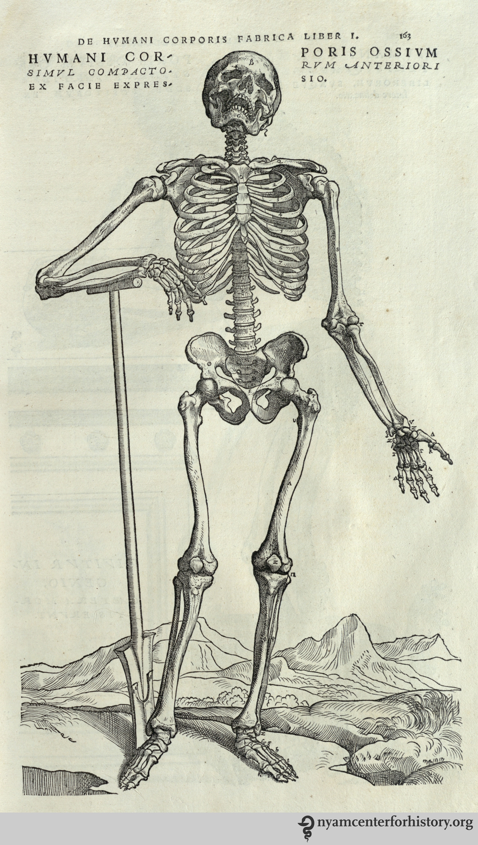
As you can see from the example above, the manicule is a disembodied, pointing hand. In case this, as in many, the manicule also has a little cuff, and a fingernail on the pointing finger that is visible, too. This manicule comes from our Anatomia Carpi by Jacopo Berengario da Carpi, a 16th century Italian physician.

The manicule gets its name from the Latin maniculum for “little hand.” This connection with the actual human body is apparent in some of the other names of the symbol: fist, printer’s first, bishop’s fist, pointer, pointing hand, even mutton-fist.[1] Its usage starts around the 12th century, though there is a record of it appearing in the Domesday Book of 1086 in John Johnson’s Typographia.[2]

Professor William Sherman of York University draws a connection between the classical gesture of “three fingers doubled under the thumb [and index the] finger extended”[3] from formal Roman oration to the strong connection between hands and texts when zeroing in on the manicule in his research on marginalia. Sherman observes the bond between hands and reading is so innate it often goes unnoticed:

“Unless we’re wrestling with an unusually large volume or feeling our way through an unusually delicate book; unless we’re running our fingertips over a text in Braille or our pointers through a Torah scroll; and unless we’re the kind of reader who follows along with our index finger or gives them a good lick before turning the page, it’s probably safe to say that we’re not even conscious of our hands as we make our way through a text.”

It’s certainly compelling to think of the manicule mark as the descendent of an oratory pose and natural human gesticulation.

Manicules are windows into people’s relationships with their books and texts.  Time was taken to read the passage, react to it and record or reference it by hand on the page, which makes sense given the tremendous amount of work, time and expense it once took to get a book into your hands. Keith Houston, author of Shady Characters, observed readers’ investment and attachment to their books was quite unlike the modern day consumer. “It was second nature for a book’s owner to brand it, to annotate and embellish it as they read; to underline pithy phrases and fill the margins with notes.”[4] Indeed, the manicule isn’t really a punctuation mark, something put in by the author for grammatical structural or clarity. Rather, it’s a reader’s mark indicating something to that person – something that could be wholly mysterious, banal, or unimportant to me or you.

doctors-dispensary_page1_watermarked

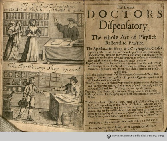
One past owner of our 1657 copy of The Doctor’s Dispensary found many points of interest in the book. Its pages are littered with marginal notes, underlining and manicules. This reader’s manicules are fairly simple, though they do include a cuff –or possibly a sleeve or an arm- while some examples can have long sleeves that drip down to mark the end of the passage of notable text, complete with shading on the clothing or secondary digits pointing things out as well.

As intellectual and cultural knowledge fairly exploded during the Renaissance, ink flowed from readers marking up their books. Then came the world-altering printing press from Johannes Gutenberg in 1440 in Mainz, Germany. Readers continued to annotate their printed books just as they had their handwritten ones; and printed manicules joined the handwritten ones on the page.[5] Printers inserted in-line manicules that indicated authorial notations that were not the readers’ own commentary, subtly (or not) guiding a reader to the ‘correct’ interpretation of the work. Printed manicules became more prominent as authors and publishers took advantage of a means to protect the integrity of their work. The manicule drawn in by hand more or less died out by the 19th century.

berengario_anatomiacarpi_1535_manicule-marginalia1_watermark

The mark lived on in its printed form and got almost annoyingly ubiquitous by the end of the 19th century. Manicules were a bread and butter symbol for advertising typography where they literally pointed the way to headlines, punchlines, venues and notable names.[6] They were even on gravestones, pointing the way to heaven.[7]

manicule-on-a-va-gravestone

This gravestone photo from a Virginia cemetery was snapped by literature professor and medievalist Jonathan Hsy of George Washington University.

The public visually overdosed on the manicule as this point and it fell from such frequent (over)use. The manicule is definitely still around though; on returned envelopes from the US postal service, in design products seeking to impart a vintage flare and in directional signage. And of course in the newest frontier of information and communication; the digital screen where the computer’s cursor was, and sometimes still is, a hand pointing the way.

cursor-hand_freeclipart

References

[1] Sherman, William H. Towards a History of the Manicule, 2005. Accessed November 22, 2016.

[2] McPharlin, Paul. Roman numerals, typographic leaves and pointing hands: some notes on their origin, history and contemporary use. New York: Typophiles, 1942. 47.

[3] Sherman, William H. Towards a History of the Manicule, 2005. Accessed November 22, 2016.

[4] Houston, Keith. Shady Characters: The Secret Life of Punctuation, Symbols & Other Typographic Marks. New York: W.W. Norton & Company, 2013. 170.

[5] Houston, Shady Characters. 178.

[6] Palmer & Rey. Palmer & Rey’s Type Specimen Book. San Francisco: Palmer & Rey, 1887.

[7] McPharlin, Roman numerals, 1942. 65.

Item of the Month: John E. Stillwell’s Prize Notebook

By Becky Filner, Head of Cataloging

In the 19th and early 20th centuries, medical schools offered academic prizes, frequently accompanied by a monetary award, for the best essays, examinations, and student notebooks.[1] The New York Academy of Medicine’s Library holds several examples of prize-winning student medical notebooks, including John E. Stillwell’s Report of Prof. Thomas’ Gynecological Clinics, Session of ’73 and ’74. This notebook is an ornate presentation copy, not the rough notes Stillwell would have taken during the clinics. Written in a neat, legible hand, it also includes a calligraphic title page and twenty-nine watercolor illustrations. The notebook is bound in full leather with blind-stamped fleurs-de-lis and shamrocks on the cover and spine. The notes are from a series of clinics offered during the 1873-1874 school year by Theodore Gaillard Thomas, a professor of gynecology at New York’s College of Physicians and Surgeons and author of A Practical Treatise on the Diseases of Women.

stillwell_title_page_watermarked

Calligraphic title page of John E. Stillwell’s Report of Prof. Thomas’ Gynecological Clinics: autograph manuscript, 1873-1874.

Stillwell’s notes, like Thomas’s lectures, are organized into a series of case studies. In the case study shown below, a woman named Annie Coyle reports that “her friends noticed her abdomen increasing in size” and her “menses … are ‘larger’ and … come twice as often as they ought.” When she had an examination at the dispensary, the examiner “pronounced her pregnant”; she came to Dr. Thomas for an examination because she was “unwilling to rest under such unjust suspicions.”

Stillwell’s carefully transcribed lecture notes and a watercolor showing a woman with an ovarian tumor, from Stillwell’s autograph manuscript, Report of Prof. Thomas’ Gynecological Clinics, 1873-1874.

Dr. Thomas notes that he found an abdominal tumor and gives details on how he determined that it is a “fluid tumor” rather than one “that is filled with air or that is solid.” He rules out pregnancy because he cannot feel any movement when he places his hands on her abdomen and her mammary glands are not enlarged. His conclusion is that she has an ovarian cyst and requires an ovariotomy to remove it. Stillwell’s account of the clinic is accompanied by a watercolor of a female figure with an enlarged abdomen labeled, “Ovarian Tumor.” Other clinics in the notebook cover problems of the uterus and cervix, tumors, peritonitis, fibroids, complications during and after pregnancy, menopause, dementia, and sterility. There is even an account (with an illustration) of a woman who has two vaginas.

stillwell_retroflexed_uterus_watermarked

Watercolor by John E. Stillwell of a retroflexed uterus, from his Report of Prof. Thomas’ Gynecological Clinics, 1873-1874.

Student medical notebooks were usually submitted anonymously to ensure that the judging would not be biased. In this case, we know that Stillwell submitted this prize-winning notebook (even though the notebook does not contain his name) because the Library acquired the notebook along with the prize itself, a wooden case of gynecological instruments with a plaque that reads, “A Prize Awarded for the best Gynecological Report of 1874 in the College of Physicians and Surgeons N.Y. by Prof. T. Gaillard Thomas to J.E. Stillwell.”[2]

stillwell_prize_plaque_watermarked

A plaque on the wooden case indicates that Stillwell received this prize from Prof. Thomas for the “best Gynecological Report of 1874 in the College of Physicians and Surgeons.”

stillwell_prize_instruments_watermarked

Stillwell’s prize consisted of a full set of gynecological instruments stored in a sturdy wooden box lined with purple velvet. The instruments were made by G. Tiemann & Co., Manufacturers of Surgical Instruments, 67 Chatham St., N.Y.

The tools include all the necessary implements for a gynecology practice in the 1870s. Thomas describes many of the tools in his A Practical Treatise on the Diseases of Women (which was a required textbooks for medical students at the College of Physicians and Surgeons around this time). Shown below are drawings of several of the tools, including “Buttles’ spear-pointed scarificator,” a “hard rubber cylinder for dry-cupping the cervix uteri,” cauterizing irons, and tools for sutures, and descriptions of how they were used.[3]

Taken from T. Gaillard Thomas’s A Practical Treatise on the Diseases of Women, 2nd ed., these drawings show obstetrical tools and give brief descriptions of how they were used.

Taken together, John E. Stillwell’s prize notebook and the handsome case of obstetrical tools that he won for his efforts provide an interesting window into both 19th-century medical school competitions and 19th-century obstetrics and gynecology.

References

[1] Contemporary handbooks from medical schools list the types of prizes awarded and the prize money attached to them. See, for example, the section on “Prizes” under “School of Medicine” in Columbia College’s Handbook of Information as to the Several Schools and Courses of Instruction 1886-1887, p. 222-225.

[2] An account of the commencement exercises of the College of Physicians and Surgeons in The Medical Record confirms Stillwell’s receipt of the Thomas prize. “The Thomas prize was awarded to J.E. Stillwell, for a report on ‘Cliniques for Diseases of Women’” (The Medical Record, ed. George F. Shrady, New York: W.M. Wood & Co., v. 9, issue of March 16, 1874, p. 158).

[3] Thomas, Theodore Gaillard.  A Practical Treatise on the Diseases of Women, 2nd ed. (Philadelphia: Henry C. Lea, 1869).

12 Gifts for the Medical History Buff in Your Life

By Emily Miranker, Project Coordinator

Search no further for one-of-a-kind gifts for the medical history buff in your life: the Library’s online shop has you covered with over 3,000 products to choose among. Find a few of our favorites below. And take an extra 15% off as our holiday gift to you: use code ZAZZLETHANKS at check out.

  1. This sturdy tote bag with a vintage advertisement for Tolu’s Rock and Rye cough tonic – good for what ails you. And groceries.

rockandrye-tote

  1. Our skeletal musicians give a whole new meaning to death metal. They might be from 1779; but our headphones are totally 21st century with a 20hz – 20,000hz range, built-in answer button and microphone to seamlessly take calls, and vegan leather padding.

gamelin-skeletal-musician-headphones

  1. This cheery orange fruits and leaves lunchbox includes a large sandwich container, two small containers and an ice pack. Dishwasher safe and BPA-free. Mangia!

culpeper-lunchbox

  1. Speaking of eating, food goes great with wine. These wine charms featuring skulls by 16th century Flemish anatomist Andreas Vesalius go great with glasses of wine.

vesalius-skulls-wine-charms

  1. Since red wine is good for your heart, admire this panel of an exquisite engraving of an anatomical heart by Scottish surgeon Charles Bell on your wall while you sip.

bell-heart-engravingw-with-close-up

  1. Keep your life in order with this desk organizer, the illustration of one of several poems gathered by Hugo Erichsen to “amuse the busy doctor in leisure hours.”

desk-caddy

  1. This flask is from a New York-based surgical supply company’s turn of the century catalogue. Chemistry-lovers, you’re welcome.

gold-flask-locket

  1. Never be too far away from a good book with this rather cellular-looking red marbled book endpaper wallet.

marbled-paper-wallter

  1. Take ‘digital’ back to its roots of actual fingers with this artificial hand by French surgeon Ambroise Paré on your laptop case.

atrifical-hand-laptop-case

  1. Just in case you feel naked without your actual stethoscope around your neck: this tie.

stetho-tie

  1. Once upon a time, your garden was your pharmacy. Peonies were used to treat spasms and cramps, gout, headaches, and fatigue. Caffeine is more popular for fatigue now…

peony-travel-mug

  1. Baby, it’s cold outside! Magnify your warmth by snuggling up with these microscopes.

microscopes-throw-pillow

Lastly, for the person who really and truly has everything already—or more likely just has no space—give the gift of membership to our Friends of the Rare Book Room, the people and programs that explore and support the books where all these remarkable images come from. All proceeds from the shop support the library’s collections’ preservation and public programming, and all Friends memberships are tax-deductible. Happy Holidays!

holiday-rabbit_aldrovandi

Found in the Eyes of Rams: The Bezoar and its Powers

By Emily Miranker, Project Coordinator

This post title is not strictly true. Or remotely true, actually. Bezoars are not found in rams’ eyes (to the relief of sheep everywhere, I’m sure). Maimonides, the 12th century Sephardic Jewish philosopher, reported an Eastern belief that bezoars could be found “in the eyes of rams,” though he then went on to note that “it is found in their [rams] gallbladder and this is true.”[i] Bezoars are in fact found in goats’ stomachs and gastrointestinal tracts, as well as that of other animals such as sheep, cows and us humans.

bezoar_watermark_19xx

Our library’s trichobezoar, ca.1862. Basically the coolest hairball you’ll ever see.

A bezoar is a mass of undigested or inedible material found in the GI tract. Today, they are typically grouped into four categories: phytobezoars (made of vegetable or fruit fibers), lactobezoars (made of milk proteins), trichobezoars (made of hair and food particles) and pharmacobezoars (aggregates of various medications).[ii] Nowadays, if a bezoar doesn’t pass through the digestive system on its own they can be treated through medication to dissolve the mass, lavage therapy, and even surgery.

Once upon a time you may have wanted one in your system. I referenced this in Poisons, Pirates, and Professors in September for National Talk Like a Pirate Day. If you had been poisoned by an attacking pirate, you’d want to swallow a bezoar to cure yourself. Pierre Pomet, 17th century French druggist, wrote of bezoars curing all manner of things from smallpox to epilepsy, ending with its ability to work as an antidote to poison.[iii]

pomet_hist-of-drugs_1725_bezoar-uses-watermarked

pomet-bezoar-goat-_1725_watermarked

The word bezoar comes from the Arabic bazahr or badzehr, meaning counterpoison[iv] and it is also mentioned in ancient Hebrew texts as bel zaard, “master” or “master of poison.”[v] Its power to counteract poison may come from a near eastern goat, the markhor. In Persian, mar is snake and khor means to eat. Snake-eater. So presumably immune to venom. Except that the markhor is an herbivore dining upon grasses and leaves. Misnomer alert! The name may have to do with their corkscrew-like horns (reminiscent of a winding snake) or that they are known to kill snakes on occasion.[vi]

Berlin_Tierpark_Friedrichsfelde_12-2015_img02_Markhor.jpg

This handsome markhor is clearly eating carrots or yam, not a snake.  Source: A. Savin, A Markhor in Berlin Tierpark, Wikimedia Commons.

Whatever the origins of the belief in curing poison, bezoars were popular in the Middle Ages and into the 17th century as antidotes. They were carried as charms, included as decor or attached to drinking and eating vessels to protect the diner, and tests were even designed to detect fakes –the selling of which was a punishable offense.[vii]

bezoar-stone_med-journal-of-aus-1986_watermarked

basin-with-bezoar_med-journal-of-australia-1986_watermarked

Another Frenchman represented in our collections’ holdings, barber surgeon Ambroise Paré, conducted an experiment to test the healing properties of a bezoar stone in the 1500s.[viii] A royal cook caught stealing silver had been sentenced to death. The cook was offered the alternative of being poisoned and then being given a bezoar under Paré’s supervision. If the cook survived the poisoning, he’d be spared. The cook lived only seven hours after the poison was administered, and Paré concluded the bezoar could not cure all poisons.

Still, the bezoar as antidote and mythical token lives on in the popular imagination. In J.K. Rowling’s Harry Potter stories, Professor Snape quizzes Harry on where he’d find a bezoar in his first potions class and later when his friend Ron Weasley is poisoned with mead –intended for Professor Dumbledore- Harry quickly shoves a bezoar down Ron’s throat.

harry-potter-bezoars

Box of bezoars and Half Blood Prince movie still.  Source.

Join us for a free First Mondays lunchtime tour in our Drs. Barry and Bobbi Rare Book Reading Room for a chance to see our bezoar in person. It’s well worth a visit even if poison and goat guts aren’t high on your to-do list; the Rare Book Room is pretty much the real life Hogwarts.

nyam_rbr_106

References:
[i] Rosner, F. (trans.) 1988. Maimonides’ Medical Writings, Treatises on Poisons, Hemorrhoids, Cohabitation. The Maimonides Research Institute, Haifa, 1988, 49-50.
[ii] Eng, Katharine and Marsha Kay. “Gastrointestinal Bezoars: History and Current Treatment Paradigms,” Gastroenterology & Hepatology. Vol. 8, Issue 11, November 2012. 776.
[iii] Pomet, Pierre. A compleat History of drugs. Bonwick, London. 1725.
[iv] Williams, Randolph S. “The fascinating history of bezoars,” The Medical Journal of Australia. Vol. 145. December 1986. 613.
[v] Barroso, Maria Do Sameiro. “The bezoar stone: a princely antidote,” Acta Med Hist Adriat. 2014;12(1):78.
[vi] “Capra falconeri – Markhor.”Brent Huffman. An Ultimate Ungulate Fact Sheet. Accessed November 11, 2016.
[vii] Williams. 613.
[viii] Thompson, C. J. S. (1924) Poison Mysteries in History, Romance and Crime J.B. Lippincott, New York, 61-62.

Today is #GivingTuesday

After Black Friday and Cyber Monday, two whirlwind days for getting deals, #GivingTuesday is a day for giving back.  Through this campaign, millions of people have come together to support and champion the organizations and causes they value. On this day, please consider donating to the New York Academy of Medicine Library.

Open to the public since 1878, the library is home to a collection that spans 12 centuries of learning.  It is a place where world-renowned historians and students alike come to learn, to be inspired, and to form the foundation of knowledge that opens the door to a future discovery.  With your generous contribution, we can foster this discovery for years to come.

donate

As we look to the future, please enjoy this look back at the past year through the eyes of our library staff.

“From Dr. Dorothy Boulding Ferebee, the African American physician leading the Mississippi Health Project during the Great Depression; Mexican physicians marching on the street for reform in the 1960s; to the doctors and nurses at the Lincoln Hospital creating a model for medical activism in the 1970s; this year’s “Changemakers” series was an important reminder that creating social and political change requires energy, engagement, and commitment, at any time in history.”  –Lisa O’Sullivan, Director

archivespanel“On October 26th, the Academy Library convened  ‘Archives, Advocacy and Change:  Tales from Four New York City Collections.’  I moderated a lively conversation with all-star panelists Jenna Freedman (Barnard College), Steven Fullwood (In the Life Archive), Timothy Johnson (Tamiment Library, New York University) and Rich Wandel (The Lesbian, Gay, Bisexual & Transgender Community Center).  The discussion hit on a lot of fascinating issues, including how archivists shape the historical record with the selection and acquisition choices they make, issues of privilege, and access for communities.”  –Anne Garner, Curator

edwardjenner“I was excited to learn in 2016 that the Library holds autograph letters from Edward Jenner in which he discusses the smallpox vaccine that he helped pioneer. These fascinating letters are available to the public for consultation. They demonstrate how strongly Jenner believed that inoculation with cowpox would protect people from the scourge of smallpox.”  –Rebecca Filner, Head of Cataloging

“As a new staff member, I really enjoyed the Rare Book Room tour that Arlene Shaner gave me during my first few weeks at the Academy. I got to see some absolutely incredible items and learned so much about the building’s history. As a little aside, these tours are free and open to the public! They happen from 12-1 on the first Monday of each month.”  –Audrey Lorberfeld, Digital Technical Assistant

pendleton_bugsandnuts_1924_cover_watermark“Most of my works has a lot to do with satisfaction—satisfactions from cleaning dusted books, from placing crumbled health pamphlets into clean, acid-free envelopes, from making fitted enclosures for damaged books, from putting torn pieces together, and so much more. But all of these satisfactions is ultimately coming from that I’m contributing in preservation of the library materials to be more accessible and usable for the future! One of the most memorable item I worked through in Health Pamphlet Rehousing Project this year is this “Bugs and Nuts” pamphlet by Andrew Lenis Pendleton, with so many absurd and eerie illustrations.”  –Yungjin Shin, Collections Care Assistant


color-our-collections“A recent highlight for me would be our first #ColorOurCollections week, held February 1-5, 2016. Over 200 libraries, archives, and cultural institutions around the world participated by creating collections-based coloring sheets and sharing them freely online. It was exciting to connect with other institutions and new followers, and it was especially rewarding to share our collection and see people engage with it in new and creative ways. I can’t wait for the next #ColorOurCollections, coming up on February 6-10, 2017!”  –Rebecca Pou, Archivist


shop1

“One thing I particularly enjoyed this year was developing an online store featuring images from our collections on a variety of products. It allowed me to delve into and share the collections in a new and often very quirky ways. A 1910 health pamphlet on a beer koozie, a 17th century microscopic slice of rock as your party clutch, a poster of vintage stethoscopes to adorn your walls, a refrigerator magnet with a an octopus, or beautifully calligraphic roman numerals from a 9th century Roman cookbook decorating a bookmark – these truly breathe new life into elements of the collection.”  –Emily Miranker, Team Administrator/Project Coordinator

“In sitting down to go through the William J. Morton Papers in connection with my residency as The Helfand Fellow, I was just stunned to find a 7-in thick stack of newspaper cuttings curated by Morton himself and preserved in their original order. The subject of my research is the history of the X-ray, and the difficulty is dealing with the voluminous print matter that appeared almost instantly. Yet Morton essentially curated some of that for me, by clipping articles that reflected his view of what was relevant to the New York metropolitan area and the networks of physicians and scientists in which he traveled. The collection is a gift for the historian, not just for its content, but because of its window into what one prominent NYC physician deemed worth noting about X-ray fever.”  –Daniel Goldberg, Audrey and William H. Helfand Fellow

columbia-dermatology“Dr. Paul Schneiderman brought his Columbia dermatology residents to visit the rare book room on August 26th so that we could explore the history of dermatology. Looking at highlights from the dermatology collection from the 16th through the 20th centuries gave the residents a chance to think about the many ways in which their specialty has changed over time, especially since dermatology relies so heavily on visual representation. We looked at hand colored engravings, chromolithographs, photographs and stereoscopic images and the residents and their mentor engaged in lively debates about whether the descriptions and images matched with current information about some of the diseases that were shown. Not only did the residents have the opportunity to see these wonderful materials, but I had the pleasure of learning more about how to interpret the images from them.”  –Arlene Shaner, Historical Collections Librarian

Shoot That Needle Straight (Item of the Month)

By Allison Piazza, Reference Services and Outreach Librarian

November is National Diabetes Month.  As one would assume, the New York Academy of Medicine Library has a large collection of books about the disease.  As I perused the stacks, however, one title jumped out.

Shoot that Needle Straight by Robert Rantoul was published in 1947, and tells the story of Richard “Dick” Hubbard. The book opens with Dick ill, and home from boarding school.  His symptoms are numerous and puzzling, including dry tongue, a constant craving for sweets, headaches, weakness, and a drastic increase in height.  It’s not long before Dick is told he has Diabetes Mellitus, a diagnosis that elicits a “What the hell is that?” from Dick.

rantoul_shootthatneedlestraight_watermarkWhat follows is an engaging, often-times laugh-out-loud narrative of Dick’s new life with diabetes.  Accompanying each chapter are charming illustrations by W. Joseph Carr.

After he is diagnosed, Dick and his mother travel to Boston, where he will have a two-week stay at a “diabetic home” known as the Carver Home, and see a diabetes specialist by the name of Dr. Anderson.  At the Carver Home, Dick learns about diabetes and how he must control it through a home routine of proper diet and exercise.  He also learns how to give himself insulin injections from his nurse, Miss Carver:

“Disinfect before you begin.
Press the needle firmly in.
Squeeze the plunger way down far.
Withdraw the needle and there you are.”

Dick’s new life as a diabetic is not without its hiccups.  In one chapter, he goes to see a physician who claims he can cure diabetes, which he explains is the result of “a nervous condition brought on by destructive and fearful thinking processes, as a result of strain, over-worry or disasters.”  Of course, the hope for a cure is too good to be true.  The doctor in question turns out to be the head of an international narcotic ring, wanted in Argentina, Mexico and California for peddling an “iron tonic” full of morphine to unsuspecting diabetes patients.

rantoul_shootthatneedlestraight5_watermarkIn another chapter, Dick agrees to be a diabetes research participant at a lab.  In a passage that would make any 21st century Institutional Review Board member cringe, the doctor explains to Dick what it means to be a “guinea-pig”:

“For this period you must be willing to do anything we ask, regardless of your feelings…. At certain times our requests will be difficult and will, no doubt, upset you emotionally, but you must realize the emotions cannot stand in the way of medical science, and content yourself with the thought that we look upon you as a medical specimen rather than a human being.”

rantoul_shootthatneedlestraight6_watermarkAnother highlight of the book is Dick’s (somewhat inexplicable) trip to Munich, Germany with his mother during Adolf Hitler’s reign.  During the trip, Dick is hospitalized with painful sores.  The situation is, understandably, quiet stressful for Dick, but he takes it in stride:

“Nazis. Heil Hitler! The Third Reich! You read ominous stories about them, you shuddered at what people said they intended to do to America and the world, but never in your wildest dreams did you imagine yourself sick and alone among more than one thousand of them in their homeland.  What a story to take home!”

These are just some of the situations Dick encounters as a diabetic.  While highly comical, the book is also meant to educate and inform the diabetic patient.  The American Journal of Digestive Diseases reviewed the book favorably in 1948, saying:

“This is a book that might safely be presented by a physician to a diabetic patient or by anyone to a friend suffering from the disease.  …  The general dietary regimen and the insulin therapy are described in exemplary fashion.”[1]

Another review in Science Education also speaks highly of the book, which they mention “has been checked for accuracy by eminent doctors.”[2]

While Shoot that Needle Straight may no longer be medically (or politically) correct, it is one of the gems of our collection.

References

[1] The American Journal of Digestive Diseases 1948 15(3):103.

[2] Science Education 1950, 34(4): 274-275.

Blood Transfusion: 350 Years

By Paul Theerman, Associate Director

Three hundred and fifty years ago, on December 17, 1666, the Philosophical Transactions published the first account of blood transfusion, in the form of a letter from physician Richard Lower to chemist Robert Boyle.1 Lower’s experiments transfused blood from one dog to another. The article provided his methods, specifying where the arteries and veins were to be cut, how the quill was to be inserted that formed the blood’s conduit between animals, and many other details of the operation.

richard-lower-portrait

Richard Lower (1631-1691), anatomist. Oil painting by Jacob Huysmans. Source: Iconographic Collections, Wellcome Library, London, accessed through Wellcome Images.

In addition to reporting on dog-to-dog transfusions, Lower also mentioned experiments between sheep, and interspecies transfusion between dogs and sheep, alternating donor (“emitter”) and recipient. In this first communication on the subject, Lower also laid out a broad experimental program:

’Tis intended, that these tryals shall be prosecuted to the utmost variety the subject shall beare: As by exchanging the bloud of Old and Young, Sick and Healthy, Hot and Cold, Fierce and Fearful, Lame [i.e., Tame] and Wild Animals, &c. and that not only of the same, but also of differing kinds.

The 1660s were the first heady days of the new Royal Society, whose motto, Nullius in verba (“nothing through words”), was the hallmark of the new “experimental philosophy.” And so these experiments have a bit of the quality of “ringing the changes”: try all kinds of animals, in all kinds of conditions, and see what happens!

Underlying the work, though, was the stronger sense of “blood as medicine.” Seventeenth-century physicians were well aware that too little blood led to death. But more, there was a general notion that blood and health were linked, a notion that came straight out of the humoral tradition. Sanguineous dispositions were healthy ones, in distinction to the melancholic ones that too much black bile created. A balance of humors, of course, was the best, but ruddy blood had an implicit edge.

In the same letter, then, Lower made these observations:

It seems not irrational to guess afore-hand, that the exchange of bloud will not alter the nature or disposition of the Animals, upon which it shall be practiced . . . . The most probable use of this Experiment may be conjectured to be, that one Animal may live with the bloud of another; and consequently that those animals that want bloud, or have corrupt bloud, may be supplied from other with a sufficient quantity, and of such as is good . . . .

Blood transfusion could serve as a type of reverse blood-letting; for those patients with too little blood, more could be supplied, and especially “good” blood rather than “corrupt.” Animals would serve as the blood source without fear that people would take on animal natures.

Human experimentation followed quickly. Similar experiments had been going on in France, and in June 1667, physician Jean-Baptiste Denis undertook the first transfusions into a human. His account, translated and published in the Philosophical Transactions, was called “Touching a Late Cure of an Inveterate Phrensy, by the Transfusion of Blood.”2 Calves’ blood served to restore to his right mind a man under the influence of a “phrensy.” In this case, the animal was chosen for “its mildness and freshness,” in order to temper the blood of the unfortunate—a different understanding from the English!

major_watermark

This image relates more to infusion than to transfusion, that is, placing medicines directly into the body through the bloodstream. The technique lent itself to infusions of blood itself. Source: Johann Daniel Major, Chirurgia infusoria, placidis cl. virorum dubiis impugnata, cum modesta, ad eadem, responsione (Kiloni [Kiel]: Sumptibus Joh. Lüderwald, imprimeb. Joach. Reumannus, 1667), p. 2

On November 23 of that same year, Lower and his colleague, physician Edmund King, transfused sheep’s blood into a Mr. Arthur Coga, also of unsound mind. The transfusion seemed to have a good effect:

The Man after this operation, as well as in it, found himself very well, and hath given in his own Narrative under his own hand, enlarging more upon the benefit, he thinks, he hath received by it, than we think fit to own as yet.3

purmann_watermark

An image attributed as Lower and King’s 1667 transfusion of Arthur Coga. Source: Matthias Goffried Purmann, Grosser und gantz neugewundener Lorbeer-Krantz, oder Wund Artzney (Frankfort; Leipzig: Widow & heirs of M. Rohrlach, Leignitz, 1705), opposite page 292 of part 3.

But transfusion as a therapy soon petered out. One case in France died. Though Coga survived, he did not fully recover, and he was soon drinking up the 20 shilling fee he received for undertaking the procedure. Blood transfusion was mocked and abandoned in England, and outlawed in France.4

Blood transfusion only revived in the middle of the 19th century, chiefly as a way of restoring blood volume. With the investigation of blood types before World War I, and Rhesus factors before World War II, transfusion became mobilized for war with the establishment of blood banks. In the last half of the 20th century, transfusion from banked blood became standard medical practice in surgeries of all kinds, and (using blood products) for hemophilia.

Suggested Reading:

  1. Holly Tucker, Blood Work: A Tale of Murder and Medicine in the Scientific Revolution (New York: W.W. Norton and Company, 2011).
  2. Anita Guerrini, Experimenting with Animals and Humans: From Galen to Animal Rights (Baltimore: the Johns Hopkins University Press, 2003.)

References

  1. “The Method Observed in Transfusing the Bloud out of One Animal into Another,” Phil. Trans. 1665-1666 1, 353-358. Accessed November 7, 2016.
  2. “An Extract of a Letter, Written by J. Denis, Doctor of Physick and Professor of Philosophy and Mathematics at Paris, Touching a Late Cure of an Inveterate Phrensy by the Transfusion of Blood,” Phil. Trans. 1666-1667 2, 617-623. Accessed November 7, 2016.
  3. “An Account of the Experiment of Transfusion, Practised upon a Man in London,” Phil. Trans. 1666-1667 2, 557-559. Accessed November 7, 2016.
  4. Elizabeth Yale, “First Blood Transfusion: A History,” JSTOR Daily: Where News Meets Its Scholarly Match, April 22, 2015. Accessed November 7, 2016.

When Mexican Physicians Take to the Streets and to Villages

Today’s guest post is written by Dr. Gabriela Soto Laveaga, Professor of the History of Science at Harvard University. Her book Jungle Laboratories: Mexican Peasants, National Projects and the Making of the Pill (Duke University Press, 2009) won the 2010 Robert K. Merton Best Book prize in Science, Knowledge, and Technology Studies from the American Sociological Association. On Thursday, November 17th at 6pm, Soto Laveaga will give The Iago Galdston Lecture: “When Mexican Physicians Take to the Streets and to Villages.” There is no charge, but please register in advance here.

In late November of 1964 more than two hundred residents and interns from one of Mexico City’s leading public hospitals threatened to strike because they were denied a Christmas bonus. Their unexpected response revealed the financially precarious situation of junior doctors and the worrisome state of many of the nation’s public hospitals. The subsequent walk-out launched ten months of unprecedented actions in hospitals, clinics, and, surprisingly, Mexico City streets.

demonstration

Physician demonstration demanding “a solution to the medical problem in the country.”

As the movement gained momentum, physicians’ demands for living wages and better working conditions shifted to incorporate a call for social justice for peasants and blue-collar workers. The shift away from hospital-based labor demands alarmed an increasingly repressive regime that set out to discredit physicians through media manipulation, intimidation, and incarceration. By March 1965 many young physicians, once heralded as the future of the nation, were portrayed in the government-controlled media as traitors of the state.

codedmessage

Coded message requesting state governors send information about tension in hospitals. Source: National Archives, Mexico City.

Declassified material offers an extraordinary opportunity to learn —via decoded messages, transcribed wire-tapped conversations, and memos to the president— how the government sought to deal with unruly doctors. It is especially interesting to learn how the government used media – television and newspapers – to distort claims and dismiss doctors’ demands as the actions of a “greedy” profession. Especially revealing is, for example, how secret service agents infiltrated hospitals to gain first-hand knowledge of a movement that quickly became national in scope.

Throughout the multiple walk-outs, hospital emergency rooms remained opened but newspapers created a sense of growing dread among the population. In news stories doctors were often labeled “lazy,” “traitorous,” “murderous,” and, most often, as elites disconnected from the rest of society.

secretservicepicture

Secret Service picture of physicians protesting in May 1965. Source: National Archives, Mexico City.

Days before the 1965 State of the Union address, President Gustavo Diaz Ordaz sent military personnel to oust doctors from key hospitals. In his address the president spent more than thirty minutes speaking about the irresponsible “homicidal actions” of striking physicians. In the aftermath of the movement, more than five hundred physicians lost their license to practice medicine (most were able to practice again in the next presidential administration) and for the following fifty years, until summer 2015, there were no national, doctor-led movements in Mexico.

silentprotest

Doctors taking over downtown streets in silent protest, May 1965. Source: Página 24.

Of note is that after the social movement was unceremoniously truncated a handful of striking doctors joined an urban guerrilla Movimiento Revolucionario del Pueblo (People’s Revolutionary Movement) intent on destroying the government through violence. These doctors were, in turn, captured and together with other members of the guerrilla spent nearly a decade in Lecumberri prison for acts of treason.

The “medical” movement, as it came to be known, was really about two (often at odds) issues: the role of physicians in a rapidly changing society and the country’s need to provide proper healthcare to all working Mexicans, a right established in the 1917 Constitution. In fact young doctors’ reactions may be rooted not in 1960s urban discontent but, curiously, in experiences of city doctors in rural Mexico.

nurses

Nurses forming a  human barrier to protect striking physicians, 1965.

Starting in 1936, all Mexican medical students were required to spend time in remote, poor, and/or indigenous areas provided much-needed primary care. This mandatory social service was later written into the Mexican Constitution. For many city physicians their social service time was a transformative experience. For example, treating patients in extreme poverty while living with them as neighbors and facing similar hardships (such as lack of electricity or running water) sensitized many physicians to the complexity of providing care in Mexico. In addition, these doctors experienced, often for the first time, the deep socio-economic divisions in the country. It was young doctors most moved by their social service year who, oral histories reveal, were more likely to join a social movement.

Currently Mexico’s public healthcare system is going through dramatic shifts, and the 1965 movement is a reminder of the powerful and evolving role that physicians have played in transforming care in the country.

#PageFrights & Pumpkin Carving

skeletoncat

Our library cat is very skittish and only sighted in the weeks leading up to Halloween.

Halloween is just days away and we’re sad to see the end of #PageFrights, a social media celebration of Halloween, library & archives-style. All month long, libraries and other cultural institutions have been showing their scary side on social media, using the hashtag #PageFrights.

With shelves of anatomical atlases, books on bones, and natural histories filled with peculiar creatures, our collection has plenty of strange, frightful content to share, and this month has been spectacularly spooky fun.  The campaign was spearheaded by the Biodiversity Heritage Library, Medical Historical Library at Yale University, Smithsonian Libraries, and us, and many other fantastic institutions are participating!  Be sure to check out the hashtag if you haven’t yet.

In addition to exploring all the #PageFrights action on social media, you can take part by carving pumpkins. Now, we are strongly opposed to the presence of pumpkin pulp around books (a truly terrifying thought to librarians), but #PageFrights participating institutions have created a number of collections-inspired pumpkin carving patterns! These designs are available here. We created two patterns using sources in our natural history collection: Gesner’s Historia Animalium Liber IIII, 1558, and Aldrovandi’s Serpentum, et draconum historiae libri duo, 1640. If you use them, be sure to post your creation to social media with the hashtag #PageFrights!

gesner_historiaanimalium_v4_1558_870_octopus_watermark  pagefrightspumpkinpattern_octopus1

Octopus from Gesner, Historia Animalium Liber IIII, 1558. Click to enlarge.

aldrovandi1640-423_watermark pagefrightspumpkinpattern_aldrovandi_dragon1-revised

Dragon from Aldrovandi, Serpentum, et draconum historiae libri duo, 1640. Click to enlarge.

For even more Halloween content from our collections, click the links below:

Happy Halloween!

bhlpumpkinslitandregularCarved #PageFrights pumpkins, courtesy of the Biodiversity Heritage Library

A Visit to the Drs. Barry and Bobbi Coller Rare Book Reading Room

Dr. Patrick Brunner, the author of today’s guest post, is Instructor in Clinical Investigation at The Rockefeller University.

On July 26 2016, a group of young physician-scientists from The Rockefeller University visited the Drs. Barry and Bobbi Coller Rare Book Reading Room at the New York Academy of Medicine. As part of the Clinical Scholars curriculum, led by Dr. Barry Coller and Dr. Sarah Schlesinger, these researchers regularly meet for educational tutorials, and the excursion to the Rare Book Room has clearly been one of the highlights of this past semester.

Arlene Shaner, the curator of this exceptional collection, presented seminal works to the group, and her deep insight and passion for the history of medicine made the excursion a unique experience. Ms. Shaner started the tour with the presentation of one of the most outstanding works of Western medicine – Andreas Vesalius’ opus magnum “De humani corporis fabrica libri septem” (On the fabric of the human body in seven books) from 1543. Ms. Shaner comprehensively and clearly outlined the historical context in which this book had been published, and fascinated her audience with a display of the book’s iconic woodprints. This artwork, which everyone in the room had seen in numerous reproductions, now laid open in its original form – showing the famous muscle man posing in front of an Italian landscape, and the skeleton, leaning on a spade, gazing towards the sky.

vesaliusgravedigger_watermark

Skeleton from the 1543 Fabrica. Click to enlarge.

A letter, sent from Oxford, dated July 7th, 1909, had been incorporated into the book as an inscription. From this letter one can learn that Sir William Osler himself donated the book to the New York Academy of Medicine. Ms. Shaner clearly knows each and every inch of this version of Vesalius work, one of three copies that the New York Academy of Medicine holds.

osler_detail_watermark Inscription by Sir William Osler found in our 1543 Fabrica. Click to enlarge.

Vesalius’ Fabrica has undoubtedly been one of the most influential books on human anatomy, overthrowing the observations and influences of the Greek physician Galen, which had been uncontested by Western medical science for more than 1300 years. And it was not until 1628 that another seminal work, which had also been put on display for the evening, William Harvey’s treatise “Exercitatio anatomica de motu cordis et sanguinis in animalibus” (On the motion of the heart and blood in animals), established that blood circulates in a closed system, and that the heart acts as a pump – a manuscript considered by many scholars to be the single most important publication in the history of physiology.

The visitors from The Rockefeller University were greatly impressed by the richness of this library – especially as they learned that all the books are available for review through the library’s archives, be it the “Anatomia hepatis” (The anatomy of the liver) by Francis Glisson, or the first atlas of skin diseases by the dermatological founding father Ferdinand von Hebra.

Arlene Shaner also presented Bernhard Siegfried Albinus’ “Tabulae sceleti et musculorum corporis humani” (Tables of the skeleton and muscles of the human body), first published in Leiden in 1747, which not only depicts anatomical studies in a monumental fashion, but presents the models within elaborate and artful surroundings – overall, an impressive testimonial of its time.

The climax of the visit was the display of a very special gift donated to the New York Academy by Sir Alexander Fleming – a capsule containing a colony of Penicillium, taken from the original culture that produced one of the world’s first antibiotics for medical use. And it has only been about 70 years since this medication became available!

fleminggrayobverse_watermarkCapsule containing a colony of Penicillium, donated by Sir Alexander Fleming. Click to enlarge.

Seeing all these treasures that irreversibly changed the world, and learning about the stories behind them in the context of both medical and art history, was a unique, and almost sensual, learning experience for the visitors, and Ms. Shaner’s never-ending expertise helped everyone in the room to deeply dive into history.

Aldous Huxley once said: “The charm of history and its enigmatic lesson consist in the fact that, from age to age, nothing changes and yet everything is completely different.”  Understanding the challenges that these authors face during their life times, which may not have been quite so different from the ones that we face today, while, at the same time witnessing the dramatic changes that have been instigated by their works, was a true inspiration. The afternoon passed quickly, and everyone agreed that they wanted to come back and further explore this treasury in the middle of New York City.