Missing: Very Vicious Red Cow

By Rebecca Pou, Archivist

This is part of an intermittent series of blogs featuring advertisements found in our collection. You can find the entire series here.

I recently cataloged a small volume of clippings and manuscript notes. As is common in books of clippings, the clippings were pasted to the pages, with the article of interest facing up and whatever happened to be on the back facing the page, hidden from the reader’s view. In this case, some loose items gave me the chance to look at the back side of the clippings, which contained classified advertisements. Dated 1802, the ads were all intriguing, but one in particular stood out. Between a tailor advertising his services and a help wanted ad for a dry goods store, it read:

Strayed or Stolen, from the Subscriber lately a Red Cow with lofty horns, a white tail, a spot near her udder and very vicious. Any person giving information where she may be had, shall have two dollars reward with reasonable changes by applying to Robert Sleith.

An advertisement for a different lost cow offered three dollars as a reward. I suspect she was of a gentler disposition.

Pascalis_Clippings_1802_1back_watermark

The back of a clipping in Clippings from “The True American” and manuscript notes, circa 1802. Click to enlarge.

The back of another clipping showed cows were not the only things gone missing:

As amusing as the classified ads are, the clippings and manuscript notes hold the real appeal. The volume was the work of Felix Pascalis Ouviere (1762-1833), a French-born physician. Pascalis, as he is commonly known, studied in Montpellier, lived for a time in St. Domingo, and moved to America. He co-edited the Medical Repository, the earliest American medical journal. He also wrote about yellow fever, commenting on the outbreaks in Philadelphia and New York.1 The clippings in this volume are of his “Advice to the inhabitants of Philadelphia,” a series of nine parts published in the True American and Commercial Advertiser regarding yellow fever.

The volume of clippings will soon be available to readers after a visit to the Gladys Brooks Book & Paper Conservation Laboratory. In addition to this volume, our collection also holds several printed works by Pascalis, as well as considerable other materials (correspondence, manuscripts, diplomas, and more) that are not currently represented in our online catalog. If you are interested in these materials, please email us at history@nyam.org.

Reference

1. Kelly, Howard A. (1928) Pascalis-Ouviere, Felix A. In: Dictionary of American Medical Biography. New York: D. Appleton and Company.

The Bookplates of Medical Men (Item of the Month)

By Erin Albritton, Head of Conservation, Gladys Brooks Book & Paper Conservation Laboratory

Book owners have forever endeavored to find ways to identify volumes as their own. With the invention of the printing press, however, books became more plentiful and an owner’s need to identify his or her own copy even more pressing. The earliest examples of printed bookplates (also known as ex-libris) come from Germany and date to the 15th century, just as printing began to take off in Europe. Over the following centuries, the use of bookplates became widespread throughout Europe and eventually followed colonists to America.1

In the summer of 2014, conservators in the Gladys Brooks Book & Paper Conservation Laboratory began treating three scrapbooks containing 184 bookplates.2 Frank Place, Jr., NYAM’s reference librarian from 1905 to 1945, collected them and donated the books to the library sometime in the mid-20th century. In compiling his collection, Mr. Place mounted the bookplates onto recycled paper pamphlet covers (measuring approximately 5.25” x 8”) and stored them alphabetically in two-ring binders,3 which were actively damaging the plates’ fragile paper supports and making it difficult to use the collection.

Binder showing ring mechanism and resulting damage to paper supports.

Binder showing ring mechanism and resulting damage to paper supports.

Example of pamphlet cover Mr. Place reused as a support for his bookplates.

Example of pamphlet cover Mr. Place reused as a support for his bookplates.

To remedy these issues and minimize the risk of future damage, conservators modified the binders while retaining as much of the original binding structure as possible, replacing the ring mechanisms with fixed posts and hinging the paper supports onto stubs.

Top: Volume 3, before treatment, in its original ring binder. Bottom: Volume 1, after treatment, in a modified post binding with hinged plates.

Top: Volume 3, before treatment, in its original ring binder. Bottom: Volume 1, after treatment, in a modified post binding with hinged plates.

Before and after binder modification.

Before and after binder modification.

Because the original binder spine pieces were too big for the modified structures, conservators encapsulated the pieces in Mylar and affixed them to protective four-flap enclosures.

Mylar-spine wrappers with encapsulated original spine pieces.

Mylar-spine wrappers with encapsulated original spine pieces.

Mr. Place’s charming collection in NYAM’s library offers a window into what some scholars have referred to as the “golden age of bookplate enthusiasm,”4 which spanned from the end of the 19th century through the first decades of the 20th century. During this time, societies for the collection and exchange of ex-libris sprang up across Europe and the United States. Collectors prized plates for their aesthetic value as miniature pieces of art and often acquired them, not for use in identifying their books, but for the sole purpose of organizing, exhibiting, and exchanging them with others. Many collectors limited their acquisitions to bookplates representing a particular theme and, here, it is no surprise that Mr. Place specialized in the plates of medical practitioners and institutions. Correspondence (included in the scrapbooks) between Mr. Place and other collectors (specifically H. M. Barlow, secretary at the Royal College of Physicians, and Dr. Henry de Forest, a prominent New York physician5) indicates that he was not only interested in growing NYAM’s collection but, in the spirit of the times, was also an active contributor to the collections of others.

While small by comparison to other collections, Mr. Place’s scrapbooks offer some wonderful examples of the broad ranging sizes and styles of bookplates—from modest ornamental name labels:

Ornamental name labels of (left) Samuel Smith Purple, MD and (right) Robert Latou Dickinson, MD.

Ornamental name labels of (left) Samuel Smith Purple, MD and (right) Robert Latou Dickinson, MD.

to elaborately illustrated panels, depicting anything from coats of arms to the owner’s occupation and hobbies. Not surprisingly, in the case of medical bookplates, images such as Hippocrates, microscopes, and the caduceus, along with skeletons, skulls, and beakers tend to figure prominently.

This slideshow requires JavaScript.

NewYorkAcademyOfMedicine_watermarkIt is a pity we do not know whether, in addition to collecting, Mr. Place ever commissioned his own miniature work of art to grace the inside covers of his personal volumes. Thanks to a bookplate, however, we are forever reminded of his contribution of this delightful little collection to NYAM’s library.

Treatment of the third and final scrapbook is in process and the entire collection will be available for use in early 2015. In the meantime, a list of all 184 bookplates can be obtained by contacting history@nyam.org or calling 212-822-7313.


Notes

1. The oldest known American bookplate dates to 1679 and takes the form of a simple label indicating the owner’s name. Curtin, R. G. (1910). “The Book-Plates of Physicians, with Remarks on the Physician’s Leisure-Hour ‘Hobbies’.” Philadelphia: J.B. Lippincott Co. (Reprinted from International Clinics, Vol. II, 20th Series), p. 239.

2. Thanks to Saira Haqqi, 2014 Gladys Brooks Book Conservation Intern, for her work conserving volume 2 of this collection.

3. In an address to the Historical Section of the Philadelphia College of Physicians on November 22, 1907, physician and bookplate collector Roland G. Curtin advises collectors to hinge their plates onto paper cards (measuring 7.5” x 9.5”) and to keep the cards loose, thus enabling the inspection of the backside of plates as well as the display of single plates without endangering the entire collection. Ibid. 253. It seems likely that Mr. Place read Dr. Curtin’s address (a reprint, cited above, was accessioned into NYAM’s collection in 1910) and was endeavoring to follow this advice when arranging his own collection.

4. Pincott, A. “American Bookplates.” Rev. of American Bookplates by W.E. Butler. Print Quarterly, Vol. 18, No. 3 (Sept. 2001), p. 351. http://www.jstor.org/stable/41826267.

5. See blog post by Arlene Shaner, Reference Librarian for NYAM’s Historical Collections, discussing a bookplate and correspondence from Dr. Henry de Forest: https://nyamcenterforhistory.org/2013/03/06/biblioclasts-bibliosnitches-beware/.

What Soldiers Read

By Johanna Goldberg, Information Services Librarian

During World War I, the American Library Association (ALA) undertook a million-dollar campaign to bring libraries to soldiers in United States training camps and cantonments. The ALA detailed these efforts in its regularly published War Library Bulletin and War Libraries, distributed by its Library War Service.

"Action in the New York City campaign." From War Library Bulletin, volume 1, number 6, April 1918.

“Action in the New York City campaign.” From War Library Bulletin, volume 1, number 6, April 1918. Click to enlarge

The ALA asked each United States city to contribute monetarily in “an amount equivalent to 5% of its population” and collected books and magazines at local libraries. These materials went to ALA-established collection centers throughout the country before being forwarded to camps. (The Red Cross and Y.M.C.A. took on the work of distributing reading materials to troops abroad.) By January 1918, the ALA had raised more than $1.5 million dollars to build and staff libraries, buy additional titles, and transport materials.

What kind of reading material did soldiers want?

"What books do the men read?" From War Library Bulletin, volume 1, number 4, January 1918.

“What books do the men read?” From War Library Bulletin, volume 1, number 4, January 1918.

Soldiers also read for pleasure. As Burton E. Stevenson wrote in the January 1918 War Library Bulletin article “What Soldiers Read”:

"Men now have time to read." From War Library Bulletin, volume 1, number 4, January 1918.

“Men now have time to read.” From War Library Bulletin, volume 1, number 4, January 1918.

There is an impression in some quarters that our soldiers have no time to read. Nothing could be further from the truth. Most of them have more real leisure than they ever had before. They are free practically every evening, and not only free, but without the distractions most of them had in civil life. There are no parties, no dances, no social engagements, and many of them find that the most pleasant way to spend an evening in camp is with a book. So, in one camp, one man has started to read Boswell’s “Life of Johnson.” Another is wrestling with Bergson’s “Creative Evolution.” Another has started Gibbon, and is working hard to finish it before he is sent to France. Still others are beginning courses of reading in various branches of English literature, under the direction and guidance of the librarian.

The cover of the June 1918 War Library Bulletin trumpets the campaign’s successes:

The front cover of War Library Bulletin, volume 1, number 7, June 1918.

The front cover of War Library Bulletin, volume 1, number 7, June 1918.

"Delivery counter at Camp Lewis A. L. A. library." From War Library Bulletin, volume 1, number 4, January 1918.

“Delivery counter at Camp Lewis A. L. A. library.” From War Library Bulletin, volume 1, number 4, January 1918. Click to enlarge.

But there was still more to be done: the August 22, 1918 War Libraries issued a new challenge: “We are going to ask the American people, in the week beginning November 11, 1918, for $3,500,000 with which to carry on the Library War Service for another year.”

On the very date selected by the ALA, of course, World War I ended. The next year, President Wilson proclaimed the date Armistice Day; in 1954, November 11 became Veterans Day to honor all American veterans, not just those from World War I.1

By the end of the war, ALA’s Library War Service had raised more than $5 million and distributed more than 10 million books and magazines. There were also long-term results: the Library War Service’s work led to the founding of the American Library in Paris and American Merchant Marine Library Association.2

The back cover of War Library Bulletin, volume 1, number 7, June 1918. Click to enlarge.

The back cover of War Library Bulletin, volume 1, number 7, June 1918. Click to enlarge.

References

1. United States Department of Veterans Affairs Office of Public and Intergovernmental Affairs. History of Veterans Day. Available at: http://www.va.gov/opa/vetsday/vetdayhistory.asp. Accessed November 6, 2014.

2. Online Archive of California. Preliminary Inventory to the American Library Association War Service Records, 1917-1923. Available at: http://www.oac.cdlib.org/findaid/ark:/13030/tf8n39n9nm/admin/#did-1.7.1. Accessed November 6, 2014.

See-Through Science: The Rise of the X-Ray

By Paul Theerman, Associate Director, Center for the History of Medicine and Public Health

One X-ray-related movie scene stands indelibly in my mind: Henri Charrière (Steve McQueen) getting X-rayed in the escape-thriller Papillon. While purchasing his escape from a French penal colony in the 1930s, Charrière dutifully submits to being X-rayed. He lies down, a photographic plate is put under his head, and a big glowing tube is brought near, which then glows even more brightly and loudly crackles for many, long seconds. The scene is repeated with Charrière on his stomach, a new plate next to his chest, and the tube right over his back. When I first saw this film, this X-ray scene brought out loud gasps in the audience. To me, the scene still captures some of the wildness, improvisation, and, viewed through today’s eyes, danger of early X-rays.

Wilhelm Röntgen. From the frontispiece to Charles E. Phillips, Bibliography of X-ray literature and research, 1896–1897; being a ready reference index to the literature on the subject of röntgen or X-rays (London: The Electrician Printing and Publishing Company, Ltd., [1897]).

Wilhelm Röntgen. From the frontispiece to Charles E. Phillips, Bibliography of X-ray literature and research, 1896–1897.

X-rays were discovered on November 8, 1895, by Wilhelm Röntgen (1845–1923) of the University of Würzburg. Quite serendipitously, he found that an active vacuum tube, shielded with a cardboard sleeve so that no visible or ultraviolet light could come out, caused fluorescence on a light- and UV-sensitive screen some distance away. When he inadvertently passed between the tube and the fluorescing screen, he saw a projection of his own skeleton. As X-rays (as Röntgen named the effect; others later called them Röntgen-rays) also affected photographic plates, these ghostly images could be fixed. The first published X-ray was of the human hand, that of his wife Anna Bertha Röntgen. Röntgen presented it in a formal paper at the end of December 1895. A Viennese newspaper picked up the story on January 5, with international papers following—The New York Sun covered it on its front page on January 7—and Röntgen and his rays became a world-wide phenomenon. For his work, he received the first Nobel prize in physics in 1901.1

X-ray of the hand of Anna Bertha Röntgen, submitted by Wilhelm Röntgen, to the Physico-Medical Society of Würzburg, December 28, 1895. Image from Bettyann Holtzmann Kevles, Naked to the Bone: Medical Imaging in the Twentieth Century (New Brunswick, N.J.: Rutgers University Press, 1997), pp. 21.

X-ray of the hand of Anna Bertha Röntgen. Image from Bettyann Holtzmann Kevles, Naked to the Bone: Medical Imaging in the Twentieth Century.

The X-ray’s medical potential was glaringly obvious. By early February the first medical uses of X-rays began to make their way into the published literature. It wasn’t hard to work with X-rays: vacuum tubes and photographic plates, the basic tools, were readily available. In the United States, three different institutions made claim to the first documented use of X-rays for medical purposes. Likely the very first was at Dartmouth (like many claims of priority, this has its competitors) where two faculty brothers, Gilman and Edwin Frost, a physician and a physicist, respectively, took an X-ray of a local boy with a broken arm. Edwin mentioned this case in his February 14 article in the premier journal Science—the first known published American reference to medical X-rays.2

An 1897 set-up for taking an X-ray of the hand. David Walsh, The Röntgen Rays in Medical Work (reprint ed.: New York: William Wood and Company, 1898), opposite page 36.

An 1897 set-up for taking an X-ray of the hand. David Walsh, The Röntgen Rays in Medical Work, opposite page 36.

Perhaps the first published X-ray in the United States of a clinical condition. In “Rare Anomalies of the Phalanges Shown by the Röntgen Process,” Boston Medical and Surgical Journal 134 (no. 8, February 20, 1896): 198–99.

Perhaps the first published X-ray in the United States of a clinical condition. In “Rare Anomalies of the Phalanges Shown by the Röntgen Process,” Boston Medical and Surgical Journal 134(8), February 20, 1896: 198–99.

Two other institutions also took early medical X-rays, in each case resulting from a doctor- physicist pairing. New York surgeon William Tillinghast Bull brought in a case of a person shot in the hand to Columbia scientist Michael Pupin.3 And in Boston, physician Francis Henry Williams, who was both an MIT grad in chemistry and a Harvard Medical School–trained physician, asked MIT physics professors Ralph R. Lawrence and Charles L. Norton to take X-rays of patients. Though unsigned, some attribute a February 20 article in the Boston Medical and Surgical Journal to him, which provides the first published X-ray in the United States of a pathological condition.4 Yale physicist Arthur Williams Wright also did early X-ray work, but not in a medical context.5

The speed with which this technology developed medically is breathtaking. In 1897, British physician Dr. David Walsh—who already identified himself as honorary secretary of the Röntgen Society of London—published The Röntgen Rays in Medical Work (reprint ed.: New York: William Wood and Company, 1898), almost 150 pages of instructional anatomy, physiology, and legal medicine. The American Roentgen Ray Society was founded in 1900, and became a forum for working out some of the safety concerns about X-rays. By 1901, Boston physician Francis Henry Williams (who had done that early work with MIT) oriented his practice completely to the topic, as shown by his magisterial 650-page book, The Roentgen Rays in Medicine and Surgery: As an Aid in Diagnosis and as a Therapeutic Agent, Designed for the Use of Practitioners and Students (New York: The Macmillan Company, 1901)—the first of three editions and, in five years, his eighth book on the subject.

From the sales pamphlet, Roentgen Ray Apparatus, published in June 1897 by the Edison Decorative and Miniature Lamp Department of the General Electric Company, Harrison, NJ.

X-rays are now routine, but scientists continue to develop new modes of imaging. In 2000, the National Institutes of Health founded the National Institute of Biomedical Imaging and Bioengineering. A lot of progress in just over 100 years!

References

1. Bettyann Holtzmann Kevles, Naked to the Bone: Medical Imaging in the Twentieth Century (New Brunswick, N.J.: Rutgers University Press, 1997), pp. 17–23.

2. Edwin B. Frost, “Experiments on the X-rays,” Science New Series 3 (February 14, 1896): 235–36. See also Peter K. Spiegel, “The First Clinical X-Ray Made in America—100 Years,” AJR 164 (1995): 241–43, from http://www.ajronline.org/doi/pdf/10.2214/ajr.164.1.7998549, accessed November 6, 2014, for photographs of what is believed the first clinical X-ray session, at Dartmouth, and for a copy of that X-ray.

3. Kevles, pp. 35–36.

4. “Rare Anomalies of the Phalanges Shown by the Röntgen Process,” Boston Medical and Surgical Journal 134 (8), February 20, 1896: 198–99. See also Abdul-Kareem Ahmed, “The Race to Give X-ray Vision to Medicine,” posted December 18, 2012, http://scopeweb.mit.edu/articles/the-race-to-give-x-ray-vision-to-medicine/, accessed November 6, 2014.

5. National Academy of Sciences, Biographical memoir of Arthur Williams Wright, 15 (1932):252.

The Talking Book Reads Itself to the Blind

By Johanna Goldberg, Information Services Librarian

“Suppose some malevolent power suddenly turned all the books in your library against you. Suppose every book within your reach slammed itself shut and lock its covers. Suppose every printed page were blank. What would it do to your life?”1

So begins “The Talking Book Reads Itself to the Blind,” a pamphlet in our collection likely printed in the early-to-mid 1940s.

In the late 1920s, the American Foundation for the Blind (AFB) began researching ways to provide audio books, or Talking Books, for people with vision loss. Through a partnership with RCA Victor and with government, Carnegie Corporation, and donor support, the Talking Book and Talking Book machine became viable in the 1930s.

In 1931, an act of Congress funded the establishment of the National Library Service for the Blind and Physically Handicapped, part of the Library of Congress. Two years later the government allocated $10,000 for the distribution of Talking Books. By 1934, 17 states had ordered 5,000 Talking Book machines from the AFB; assured that Talking Book machines were sufficiently available, the National Library Service began producing and distributing Talking Books.2

This service was life-changing for many people with vision loss. In the pamphlet, the AFB estimated that three-quarters of the more than 9,200 blind people in the greater New York area did not know Braille. But the Talking Book machines were expensive—the AFB sold them at cost for $42. The AFB’s goal was to amass enough donations to bring 2,500 Talking Book machines to those who lacked the funds to buy one themselves.1

"Sensitive fingers quickly master the simple dials and controls which make the TALKING BOOK read more slowly or a little louder at their touch." From “The Talking Book Reads Itself to the Blind.”

“Sensitive fingers quickly master the simple dials and controls which make the TALKING BOOK read more slowly or a little louder at their touch.” From “The Talking Book Reads Itself to the Blind.”

By the time the AFB printed this booklet, a person with vision loss could apply to the Library of Congress or a local library for the blind to receive Talking Books by mail, free of charge. “Uncle Sam even pays the postage.”1 The AFB offered more than 20 books, and planned to add two to three new ones every month.1

The list of available books is a fun look at what titles were popular in the 1930s and 1940s. Some items remain classics, while others have lost their luster over the years. Maybe it’s time to revive interest in Booth Tarkington’s Presenting Lily Mars or John Masefield’s The Bird of Dawning.

A list of available talking books advertised in "The Talking Book Reads Itself to the Blind." Click to enlarge.

A list of available Talking Books advertised in “The Talking Book Reads Itself to the Blind.” Click to enlarge.

For more on the history of Talking Books, visit the American Foundation for the Blind’s online exhibit.

References
1. The Talking book reads itself to the blind. New York: American Foundation for the Blind.

2. American Foundation for the Blind. AFB Talking Book Exhibit. 2009. Available at: http://www.afb.org/talkingbook/home.asp. Accessed April 18, 2014.

Brains, Brawn, & Beauty: Andreas Vesalius and the Art of Anatomy

By Rebecca Pou, Archivist, and Johanna Goldberg, Information Services Librarian

For our October 18 festival, Art, Anatomy, and the Body: Vesalius 500, we exhibited items from the library’s collections showing the history of anatomical illustration. You can still visit the New York Academy of Medicine to view the exhibit in person on the ground floor. If you can’t make it, we offer a digital version below.

The exhibit on display at the new York Academy of Medicine.

The exhibit on display at the New York Academy of Medicine.

In 1543, Andreas Vesalius was a 28-year-old professor of surgery and anatomy at the University of Padua, one of Europe’s best known medical schools. That year, he published his most famous work, De humani corporis fabrica, translated as On the Fabric of the Human Body. Vesalius dedicated the work to Charles V; he subsequently received the appointment of physician to the imperial family.

Working from three images from the Fabrica—a skeleton, a figure of muscles, and an illustration of the brain—this exhibit shows the many ways Vesalius’ work built on past anatomists, and exerted its influence well into the future.

Images from great works in our collection, from Magnus Hundt’s 1501 Antropologium to Dominici Santorini’s 1775 Anatomici summi septemdecim tabulae, show the evolution of artistic style and scientific understanding. Some show examples of “borrowing” Vesalius’ images and placing them in new contexts.

Click an image to view the gallery.

The NYAM Lectures: Medical Talks by Eminent Speakers (Items of the Month)

By Latrina Keith, Head of Cataloging

WNYC-LogoThe New York Academy of Medicine and New York Public Radio (NYPR) have digitized and cataloged some 40 radio broadcasts produced by NYAM and originally broadcast over WNYC radio in the 1950s. These lectures are drawn from the more than 1,500 original lacquer discs transferred from NYAM to the NYPR Archives in 2008. The digitization and cataloging resulted from a joint project between NYAM’s Center for the History of Medicine and Public Health and the New York Public Radio (NYPR) Archives, and with a grant from METRO, the New York Metropolitan Library Council.

The New York Academy of Medicine and WNYC-FM began their radio relationship in 1946 with the launch of The Laity Lectures—later to become Lectures to the Laity—a popular series of Academy lectures and talks on culture and medicine that had started in 1935. By mid-1950, this series was joined by For Doctors Only, which aimed to bring “the best of the meetings, conferences, and roundtable discussions held at the academy” to the medical profession and also addressed critical analysis of issues of society and medicine, as well as the application of the social sciences to medicine, and provided academic presentations in the history of medicine.

The current periodicals room of the New York Academy of Medicine Library, circa ____.

The current periodicals room of the New York Academy of Medicine Library, photographed by Irving Underhill, 1872-1960.

Lecture topics include gerontology, aging, nutrition, cancer, public health, heart disease, dermatology, psychiatry, and the role of the physician and the law, among others. Along with such informative topics, there were notable guest lecturers such as American anthropologists Margaret Mead and Ralph Linton; Dr. Sidney Farber, the father of modern chemotherapy and pediatric pathology; noted gerontologist Dr. John Steele Murray; and Dr. Leona Baumgartner, the first female commissioner of New York City’s Department of Health from 1954-1962. Baumgartner will be the focus of the upcoming Iago Galdston Lecture, “Making Public Health Contagious: The Life & Career of Leona Baumgartner, MD, PhD” to be presented by Dr. Hilary Aquino on December 4, 2014 at NYAM.

Making these historical hidden treasures available to all is a great achievement for both NYAM and NYPR. We hope that one day the entire radio broadcasts will be restored for the educational and cultural benefit of all.

Creepy Historical Drawings of Skeletons Contemplating Mortality

By Anne Garner, Curator, Rare Books and Manuscripts

Surgite mortui, et venite ad judicium (Arise, ye dead, and come to the judgment). Table 6. Click to enlarge.

Jacques Gamelin. Nouveau recueil d’osteologie. “Surgite mortui, et venite ad judicium (Arise, ye dead, and come to the judgment).” Table 6. Click to enlarge.

One of the greatest pleasures of the vast library collections of the New York Academy of Medicine lies in browsing our fascinating treasury of anatomical atlases and smaller format illustrations of the human body. From early attempts by anatomists like Dryander and Hundt, who depicted the body diagrammatically, to the Baroque and fantastic skeletons of the French anatomist Jacques Gamelin, almost two hundred fifty years later, these illustrations are not only visually transfixing, but offer tantalizing visual evidence of the progress made in understanding and depicting the way the body works. Chief among these milestone illustrations stands the monumental work of Vesalius, whose skeletons and muscle men changed the way the human body was drawn forever in 1543.

In this slide show hosted by Flavorwire, we’ve assembled some of our favorite images by pioneering anatomical illustrators. In honor of Halloween, we’re highlighting skeletons with a gleam in their eyes, a scowl on their faces, and a spring in their step, for optimal thrills and chills.

Reflections on “Art, Anatomy and the Body: Vesalius 500”

Our “Art, Anatomy, and the Body: Vesalius 500″ festival guest curator, artist and anatomist Riva Lehrer, reflects on the event.

Riva Lehrer, left, with Lisa O'Sullivan, director of the Center for the History of Medicine and Public Health. Photo by Charles Manley.

Riva Lehrer, left, with Lisa O’Sullivan, director of the Center for the History of Medicine and Public Health. Photo by Charles Manley.

My approach as co-curator of “Art, Anatomy and the Body: Vesalius 500” was to ask how we use anatomy today to understand what it means to be human. Throughout history, we’ve used metaphor to organize our concepts of the body. We’ve imagined it as a vessel full of roiling humors, as an elaborate clock, as a regulated factory, as a robot and a computer, to name just a few. Even anatomical study is affected by metaphor and symbolism, and often guides what we see.

As science creates new perspectives on human (and non-human) anatomy, society responds by re-imagining new possibilities. When we internalize these visions we live differently in our bodies. “Art, Anatomy and the Body: Vesalius 500” brought together artists, writers, and scholars to discuss about how we see ourselves now and how we construct ourselves as public bodies.

As emcee, I had the pleasure of introducing Ann Fox, Sandie Yi, Dan Garrison, Sander Gilman, Nuha Nazy, Dima Elissa, Bill Hayes, Steven Assael and Alice Dreger, who were among the more than 25 festival participants.

Chun-Shan “Sandie” Yi. Photo by Charles Manley

Chun-Shan “Sandie” Yi. Photo by Charles Manley

Curator Ann Fox and artist Chun-Shan “Sandie” Yi started the day with an overview of contemporary artists who explore identity through medical and anatomical imagery, including artists who tackle our continuing discomfort with HIV/AIDS, a disease that from the very first raised issues of identity and ostracism. Sander Gilman furthered our thoughts about unacceptable bodies by discussing how teaching posture in schools has been used to control and regulate bodies, and make them socially predictable.

Dan Garrison brought us back to the core of the festival via the origin of Vesalius’ Fabrica. He posited some very intriguing ideas around who Vesalius may have been; he may have had a variant body (possibly dwarfism), and this may have contributed to how he created his great work.

Dan Garrison. Photo by Charles Manley.

Dan Garrison. Photo by Charles Manley.

Identity is always a struggle between the specifics of individuality and alliance with group affiliation. This echoes the direction of modern medicine; treatments are becoming increasingly targeted to individual bodies, whether through genetics, prosthetics, or adapted drug regimens. We glimpsed this future during the ProofX presentation, with Nuha Nazy and Dima Elissa. ProofX uses 3D printing to produce extremely precise implants, surgical models, and adaptive devices for a wide range of conditions. It’s anatomical interface at an unprecedented level.

ProofX's 3D-printing demonstration.

ProofX’s 3D-printing demonstration. Photo by Charles Manley.

Bill Hayes engages medical history in order to understand his own biography. His research blends with memoir and hands-on experience, as witnessed by The Anatomist, the remarkable story of Henry Gray, author of Gray’s Anatomy. Hayes steps across the boundary between modern identity and historical precedent, here discussing the history of exercise, in order to show us how we arrived at our present state.

Anatomy and poetics also wove together in the work of Steven Assael. His paintings and drawings are highly (and gracefully) accurate, yet manage to be astute and nuanced examinations of his subjects’ personalities. He transfixed the audience by the lushness of his technique and the drama of his compositions.

Alice Dreger. Photo by Charles Manley.

Alice Dreger. Photo by Charles Manley.

Our excellent final event was a talk by bioethicist Alice Dreger. She traced the origins of contemporary medical photography as well as taking a fresh look at traditional anatomical illustration. Dreger has thought deeply about how we signal our identity through bodily choice. She raised questions about what caused doctors to lose touch with the vulnerability of people in medical settings, and to describe variant bodies in dehumanizing ways. She also pointed out that doctors often can’t admit the taboo pleasure of viewing physical anomalies, and how that covert pleasure affects their relationships with patients.

The human body has its secret, unspoken existence and its public presentation, meant to be decoded by other human beings. Anatomy would seem to be an objective bridge between the two, yet can be just as complicated and interpretive as any form of art. Our festival let us perceive the dialogue between poetics and science, and between inner and outer realities of the body.

For more images of the festival, visit our Facebook page.
Read a summary of the festival by presenter Kriota Willberg: Part 1 and Part 2
Click here for a blog from Hyperallergic.
Read “Seeing is Believing: New York Academy of Medicine’s Vesalius 500th Year Celebrations,” an article by presenter Brandy Schillace, PhD.

Polio: A Fearful Disease Nears Its End

By Paul Theerman, Associate Director, Center for the History of Medicine and Public Health

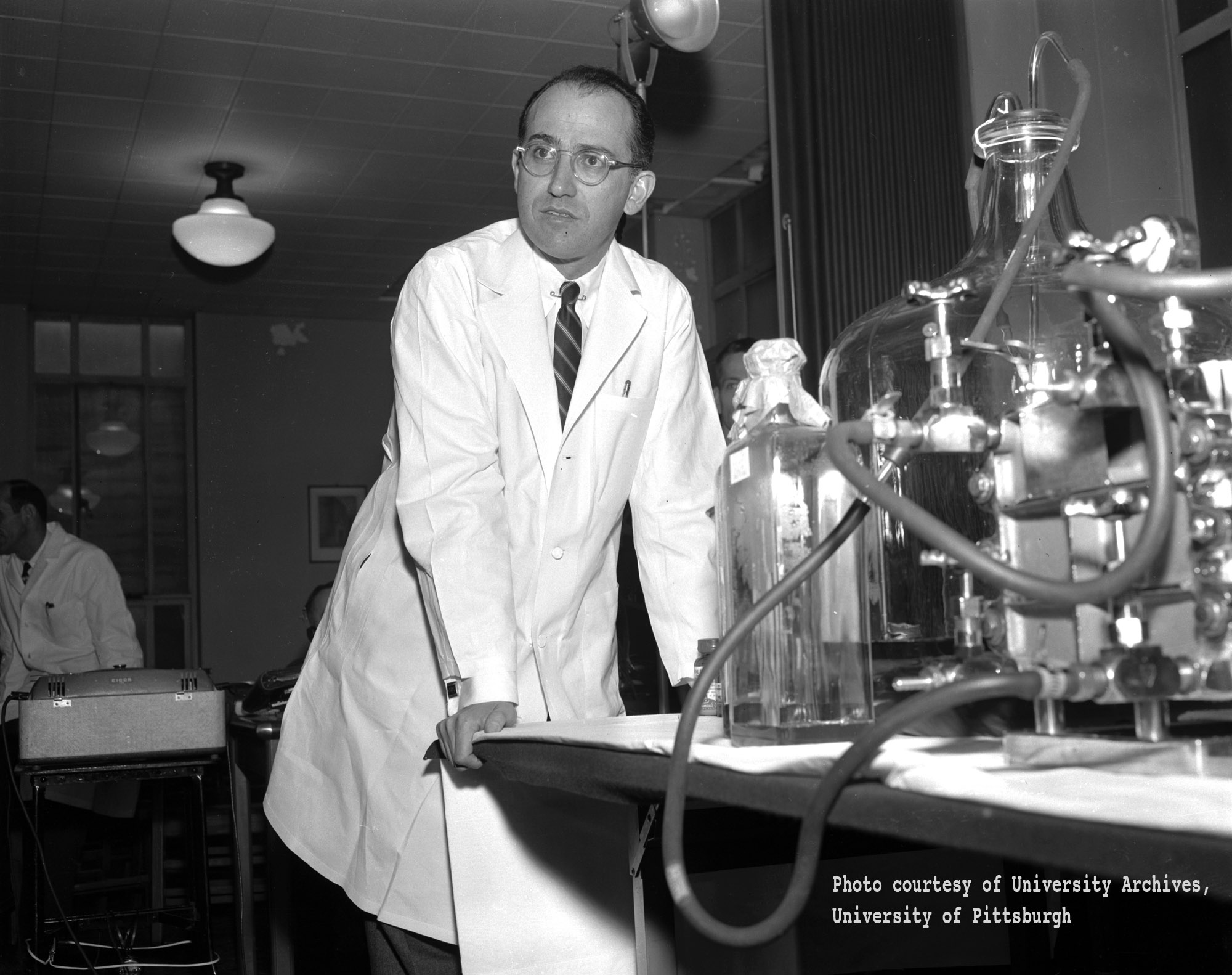
Friday, October 24, is World Polio Day. Inaugurated a decade ago, the day is promoted by the World Health Organization, UNICEF, and Rotary International to mark the coordinated battle to eradicate polio worldwide. The date for World Polio Day honors Jonas Salk, whose 1950s polio vaccine effectively ended the epidemic in the United States. World Polio Day comes just before Salk’s birthday on October 28.

Jonas Salk. Courtesy of  the Steeltown Entertainment Project. Click to enlarge.

Jonas Salk. Courtesy of the University of Pittsburgh, via the Steeltown Entertainment Project. Click to enlarge.

Jonas Salk was born in 1914, and on the centenary of his birth, many celebrations mark his achievement. Here at the New York Academy of Medicine, we are screening a documentary about Jonas Salk on November 18, The Shot Felt ’Round the World, with commentary from his son Dr. Peter Salk, Time magazine writer Jeffrey Kluger, and historian of medicine Dr. Bert Hansen. Elsewhere in New York both City College of New York and NYU Langone Medical School are hosting celebratory symposia, and the Jonas Salk Legacy Foundation maintains a list of events and exhibitions in many different venues.

Though every analogy is partial, the American polio epidemics of the 20th century bear resemblance to the current outbreak of Ebola in West Africa. Both diseases were around and known before their largest epidemics. In 1916 polio broke out in the United States, with New York City having more than 9,000 cases, a quarter of which resulted in death. Another major New York City outbreak occurred in 1931. Even by then, little was known about the disease: it fell under a category now known as “emerging infectious diseases.”1

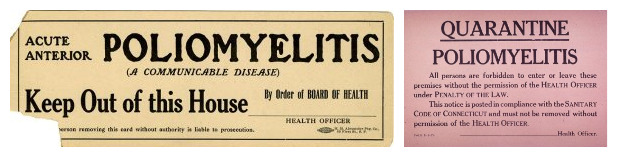
In their 1934 book, Poliomyelitis: A Handbook for Physicians and Medical Students, NYAM Fellow Dr. John F. Landon and his co-author, Lawrence W. Smith, called it a “still obscure disease” (p. vii) with a “particularly baffling” origin and means of transmission (p. 1). There were no effective treatments; the most one could do was to relieve symptoms, which included fever and strong pain, especially in the head and neck. Prevention was difficult if not impossible. Like Ebola, the disease’s spread, write Landon and Smith, could be curtailed chiefly by taking extreme care in physical contact and by quarantining active patients. The Handbook provided several practical appendices on nursing care and aseptic techniques, so caregivers could protect themselves and others from contagion. One appendix reproduced the New York City Health regulations on polio, which specified a three-week quarantine for all patients and a two-week quarantine for those in contact with them, with placarding of premises with quarantine signs.1

Two polio quarantine cards, courtesy of the National Library of Medicine.

Two polio quarantine cards, courtesy of the National Library of Medicine. Click to enlarge.

Chart from Poliomyelitis: A Handbook for Physicians and Medical Students. Click to enlarge.

Chart of the 1931 New York polio epidemic, compiled by the New York Department of Health. In Poliomyelitis: A handbook for physicians and medical students. Click to enlarge.

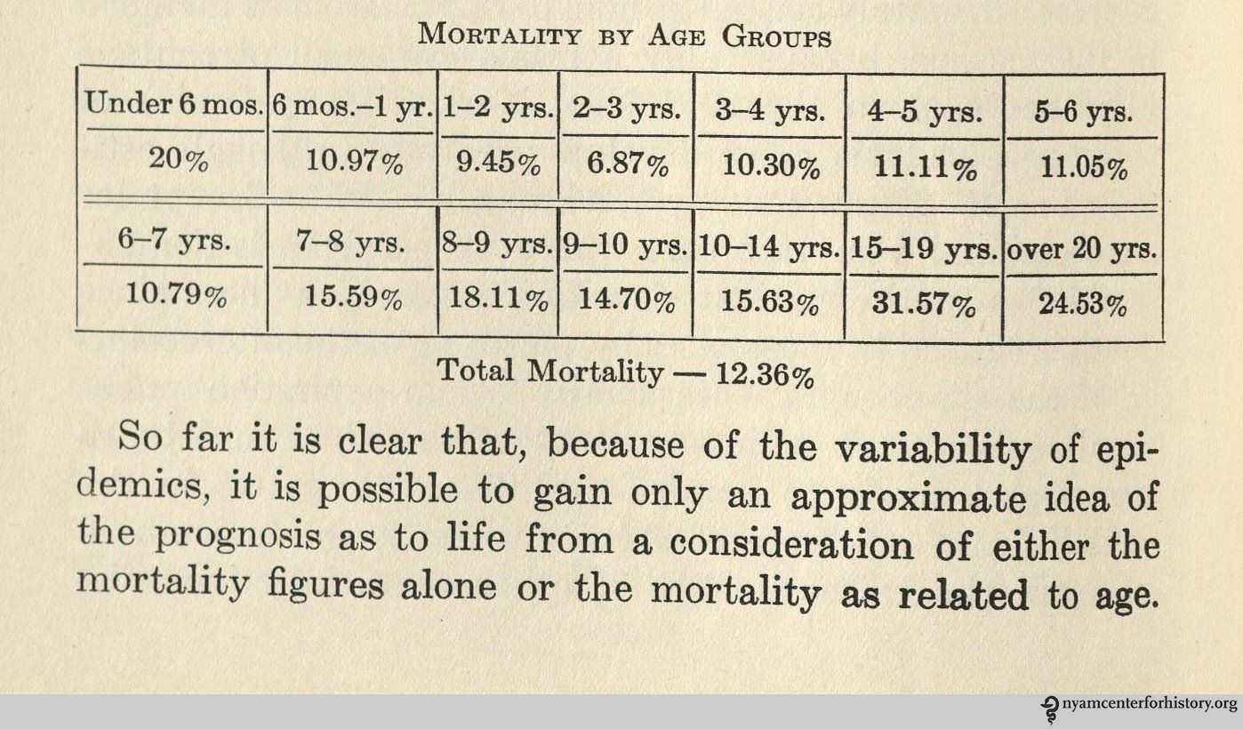
And like Ebola, the disease had terrible effects. The virus can enter the central nervous system, causing both temporary and at times permanent paralysis long after the disease runs its course. And even if the paralysis is temporary, post-polio syndrome can debilitate people years later. But in the early 20th century polio was often fatal, at rates that in 1931 averaged about 10% to 15% overall, but rose to over 20% for those under six months of age, and over 30% for those 15 to 19 years old (p. 158).1 By the time of the post–World War II epidemics, the death rate had dropped, but with increasing numbers of paralyzed survivors.

In 1952, polio struck the United States hard, with 58,000 affected, of which more than 3,000 died and more than 21,000 were left paralyzed to some degree or other.2 This was a huge number, even given the size of the country. Polio was four times as prevalent in the United States then as Ebola is in Liberia today. And while death rates from Ebola are higher, overall death and disability rates are comparable.

With this as a backdrop, the possibility of an effective polio vaccine was electrifying. In 1954, Jonas Salk’s promising new vaccine started widespread field testing, with over a million children taking part. On April 12, 1955, Dr. Thomas Francis Jr., director of the Poliomyelitis Vaccine Evaluation Center at the University of Michigan School of Public Health, pronounced the vaccine safe and effective. Large-scale immunization campaigns quickly started up.3–5 Polio was under control in the United States by the 1960s.

"The 1954 Poliomyelitis Vaccine Field Trial Areas." In Evaluation of the 1954 field trial of poliomyelitis vaccine: Final report. Click to enlarge.

“The 1954 Poliomyelitis Vaccine Field Trial Areas.” In Evaluation of the 1954 field trial of poliomyelitis vaccine: Final report. Click to enlarge.

The disease is one of the few for which eradication rather than control is considered feasible, a goal announced in 1988 by WHO, UNICEF, and Rotary. As of 2013, only three countries worldwide still had polio endemic in their populations—Pakistan, Nigeria, and Afghanistan—and the number of cases stood at fewer than 500, in less than a dozen countries in all.6 Yet polio is in the news again, as war has hindered vaccination programs, health workers have been put under attack, and cases have spread.7 At the eve of eradication, polio is proving difficult, even if it no longer inspires the wholesale fear that it did 60 years ago.

References

1. Landon JF, Smith LW. Poliomyelitis: A handbook for physicians and medical students, based on a study of the 1931 epidemic in New York City. New York: Macmillan; 1934. All in-text page numbers come from this handbook.

2. Salk Institute for Biological Studies. History: Polio today. Available at: http://poliotoday.org/?page_id=13. Accessed October 22, 2014.

3. Francis T. Evaluation of the 1954 field trial of poliomyelitis vaccine: Final report. Ann Arbor: University of Michigan; 1957.

4. March of Dimes. April 12 1955: Polio Announcement. 1955. Available at: https://www.youtube.com/watch?v=2LlDn_MQDkc. Accessed October 22, 2014. The March of Dimes was known earlier as the National Foundation for Infantile Paralysis, the group that underwrote much of the research and testing on polio.

5. Progress report to physicians on immunization against poliomyelitis, advance briefing. Indianapolis: Eli Lilly and Company; 1955. This report was part of the campaign and excitement around the Salk vaccine.

6. World Health Organization. Polio Case Counts. Accessed October 22, 2014.

7. For example: Gladstone R. Amid Iraq’s Political Chaos, a New Polio Vaccination Campaign Faces Challenges – NYTimes.com. New York Times. http://www.nytimes.com/2014/08/12/world/middleeast/amid-iraqs-chaos-a-new-polio-vaccination-campaign.html?_r=3. Published August 11, 2014. Accessed October 22, 2014.