Deafness as a Public Health Issue in the 1920s & 1930s (Part 1 of 2)

Today we have part one of a guest post written by Dr. Jaipreet Virdi-Dhesi, the 2016 Klemperer Fellow in the History of Medicine at the New York Academy of Medicine and a SSHRC Postdoctoral Fellow in the Department of History at Brock University in St. Catharines, Ontario. She is working on her first book, Hearing Happiness: Fakes, Fads, and Frauds in Deafness Cures, which examines the medical history of hearing loss and “quack cures” for deafness. Some of these cures are explored on her blog, From the Hands of Quacks. You can find her on twitter as @jaivirdi.

In 1935, physician Francis L. Rogers of Long Beach read a paper addressing the worrisome statistics of deafness. One study discovered nearly thirty-five thousand Americans were deaf. Another found that out of a million people tested for their hearing, 6% had significant hearing impairment. Yet another study reported three million people had some kind of hearing impairment. This “problem of deafness,” Rogers emphasized, “is primarily of public health and public welfare.” Not only were there too many people failing to adequately care for their hearing, but many cities, schools, and governments lacked the proper infrastructure to educate the public on the importance of hearing preservation. Indeed, as Rogers stressed: “Today the three great public health problems confronting the world are heart disease, cancer, and deafness.”[1]

Image 1

A window display in Detroit (Hearing News, June 1942)

The notion of deafness being statistically worrying as a public health issue actually dates to the late nineteenth century, especially to the work of otologist James Kerr Love of Glasgow. Love conducted several statistical studies of the ears of deaf schoolchildren, discovering that the majority of them were not completely deaf, but had some level of “residual” hearing. With proper medical treatment, the hearing could be intensified enough to warrant a “cure.” For other cases, children could be taught to make use of that residual hearing through invasive training using acoustic aids and other kinds of hearing technologies.

Love’s research concluded that many deafness cases could actually be relieved if the ears of children were examined early and frequently—that is, deafness could be prevented. His “prevention of deafness” concept was influential for the new generation of otologists in America, especially those who were members of the New York Academy of Medicine’s Section of Otology during the first three decades of the twentieth century.

To raise awareness on the necessity of proper medical examinations and frequent hearing tests, these otologists collaborated with social organizations such as the New York League for the Hard of Hearing, which was established in 1910. The League was a progressive group catering to the needs of hard of hearing or deafened persons who were raised in a hearing society rather than in a D/deaf community and communicated primarily with speech and lip-reading rather than sign language. Composed mostly of white, middle-class, and educated members who lost their hearing from illness, injury, or progressive deafness, the League strove to construct hearing impairment as a medical issue. They argued hearing impairment was not an issue of education or communication, but rather a handicap.

Image 2

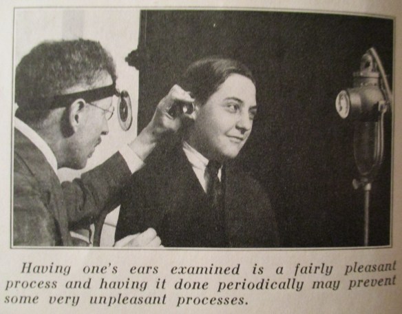
An otologist examining a young patient’s ear (Hygeia, June 1923)

The collaboration between New York otologists and the League eventually created a national network of experts, social services, teachers, physicians, and volunteers who banded together to address the so-called “problem of deafness.” That is, the problem of how to best integrate the hard of hearing, the deafened, and to some extent, even the deaf-mutes, into society. One key achievement of the League was the establishment of hearing clinics to properly assess hearing impairment, especially in children, to ensure medical care could be provided before it was too late. This project was primarily spearheaded by Harold M. Hays (1880-1940), who was recruited as president of the League in 1913, becoming the first active otologist collaborating with the League. After the First World War, Hays set up a clinic for treating hearing loss in children at the Manhattan Eye, Ear, and Throat Hospital.

Image 3

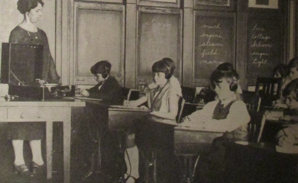
Group hearing tests of schoolchildren, using an audiometer. Headphones are used first on the right ear, then the left. (Hygeia, February 1928)

Hays claimed that hearing impairment might be a handicap, but “the sad part of it is that 90 percent of all hearing troubles could be corrected if they were treated at the proper time.” With regular hearing tests, this was possible. Yet, as Hays argued, regular hearing tests were not considered on par with other hygienic measures under public health services:

We are saving the child’s eyes! We are saving the child’s teeth! Is it not worth while to save the child’s ears?”[2]

During the 1920s, Hays’ activism for regular hearing tests was so instrumental that in 1922, the League’s newsletter, The Chronicle, told its readers “we believe that the League would justify its existence if it did no other work than to prevent as much deafness as possible.”  To achieve this mandate, the League launched a large public campaign to raise awareness on the importance of medical care. Indeed, in one report for the League, Hays remarked that with the increased publicity, there were 10,000 calls to the League in 1918 alone inquiring about aural examinations. A steady increase in patients would follow: 17 clinic patients in 1924, 326 in 1926, and then 1,531 in 1934.

Another publicity campaign spearheaded by the League was the establishment of “Better Hearing Week” in 1926, a week-long awareness program (later renamed “National Hearing Week”). Held in October, the campaign included symposium discussions on the “Problems of the Hard of Hearing,” including topics on the relationship between the physician and his deafened patient, how the deafened could build their lives, and even on newest technological developments in hearing aids. October issues of The Bulletin (the renamed League newsletter) and the Hearing News, the newsletter of the American Society for the Hard of Hearing (ASHH) included reprints of letters from prominent leaders supporting the mandates of “Better Hearing Week,” including letters from President Roosevelt and New York Mayor LaGuardia.

Image 4

Advertisement for Western Electric Hearing Aid, the “Audiophone.” These before-and-after shots were powerful for demonstrating the effects of “normal” hearing, sending the message that outward signs of deafness, such as the “confused face,” could easily disappear once being fitted properly with a hearing aid. (Hearing News, December 1936)

The 1920s publicity campaigns were primarily focused on fostering ties between otologists and the League, in cooperation with hospitals and schools. In 1927, the League purchased audiometers and offered invitations to conduct hearing tests in schools across New York, so children with hearing impairment could be assessed accordingly. Two years later, the League worked with Bell Laboratories to further substantiate the conviction that deafness was a serious problem amongst schoolchildren and that something needed to be done.

At the same time otologists across America established joint ventures between organizations like the America Medical Association and the American Otological Society. They formed committees to write reports to the White House on the national importance of addressing the “prevention of deafness.” Wendell C. Phillips (1857-1934), another president of the League and the founder of ASHH, particularly emphasized the need to address the “psychologic conditions and mental reactions” of the deafened patient, for the tragedy of acquired deafness meant it is a “disability without outward signs, for the deafened person uses no crutch, no black goggles, no tapping staff.”[3] It was an invisible handicap that needed to be made visible if it was to be prevented, if not cured.

References

[1] The Federation News, August 1935.

[2] Harold M. Hays, “Do Your Ears Hear?” Hygeia (April 1925).

[3] Wendell C. Phillips, “Reminiscences of an Otologist,” Hygeia (October 1930).

Many Anatomy Lessons at the New York Academy of Medicine

Kriota Willberg, the author of today’s guest post, explores the intersection of body sciences with creative practice through drawing, writing, performance, and needlework. She is offering the workshop “Visualizing and Drawing Anatomy” beginning June 6 at the Academy. Register online.

Cheselden's Osteographia, 1733, opened to the title page and frontispiece.

Cheselden’s Osteographia, 1733, opened to the title page and frontispiece.

Different Disciplines, Same Body

I teach musculoskeletal anatomy to artists, dancers, and massage therapists. In my classes the students study the same raw material, and the set of skills each group acquires can be roughly organized around three distinct areas: representation of the body, kinesiology (the study of movement), and palpation (feeling the body).

As an anatomy teacher I am constantly on the prowl for images of the body that visually reinforce the information my students are learning. The Internet has become my most utilized source for visual teaching tools. It is full of anatomy virtual galleries, e-books, and apps. 3D media make it ever easier to understand muscle layering, attachment sites, fiber direction, and more.

In spite of the overwhelming volume of quality online cutting-edge anatomical imagery, I find myself drawn to historical 2D printed representations of the body and its components, once the cutting-edge educational technology of their respective centuries. Their precision, character, size, and even smell enhance my engagement with anatomical study. Many of these images emphasize the same principles as the apps replacing them centuries later.

The Essential Structure Of The Body

Different artists prefer different methods of rendering bodies in sketches. One method is to organize the body by its masses, outlining its surface to depict its bulk. Another method is to draw a stick figure, organizing body volume around inner scaffolding.

Plate XXXIII in Cheselden, Osteographia, 1733.

Plate XXXIII in Cheselden, Osteographia, 1733.

And what is a skeleton but an elaborate stick figure? William Cheselden’s Osteographia (1733) presents elegant representations of human and animal skeletons in action. These images remind us that bones are rigid and their joints are shaped to perform very specific actions. The cumulative position of the bones and joints gives the figure motion. In Cheselden’s world of skeletons, dogs and cats fight, a bird eats a fish, a man kneels in prayer, and a child holds up an adult’s humerus (upper arm bone) to give us a sense of scale while creating a rather creepy theatrical moment.

Muscle Layering

3D apps and other imaging programs facilitate the exploration of the body’s depth. One of the challenges of artists and massage therapists studying anatomy is transitioning information from the 2D image of the page into the 3D body of a sculpture or patient.

Planche 11 in Salvage, Anatomie du gladiateur combattant, 1812.

Planche 11 in Salvage, Anatomie du gladiateur combattant, 1812.

Salvage’s Anatomie du gladiateur combattant: applicable aux beaux artes… (1812) is a 2D examination of the 3D Borghese Gladiator. Salvage, an artist and military doctor, dissected cadavers and positioned them to mimic the action depicted in the statue. His highly detailed images depict muscle layering of a body in motion. The viewer can examine many layers of the anatomized body in action from multiple directions, rendered in exquisite detail. Salvage retains the outline of the body in its pose to keep the viewer oriented as he works from superficial to deeper structures.

Tab. VIII in Albinus, Tabulae sceleti et musculorum corporis humani, 1749.

Tabula VIII in Albinus, Tabulae sceleti et musculorum corporis humani, 1749 edition.

Bernhard Siegried Albinus worked with artist Jan Wandelaar to publish Tabulae sceleti et musculorum corporis humani (1749). Over their 20-year collaboration, they devised new methods for rendering the dissected body more accurately.  The finely detailed illustrations and large size of the book invite the reader to scrutinize the dissected layers of the body in all their detail. Although there is no superficial body outline, the cadaver’s consistent position helps to keep the reader oriented. On the other hand, cherubs and a rhinoceros in the backgrounds are incredibly distracting!

Fiber Direction

Familiarity with a muscle’s fiber direction can make it easier to palpate and can indicate the muscle’s line of pull (direction of action).

Figure in Berengario, Anatomia Carpi Isagoge breves, 1535.

Figure in Berengario, Anatomia Carpi Isagoge breves, 1535.

The images of Jacopo Berengario da Carpi’s Anatomia Carpi Isagoge breves, perlucide ac uberime, in anatomiam humani corporis… (1535) powerfully emphasize the fiber direction of the muscles of the waist. This picture in particular radiates the significance of our “core muscles.” Here, the external oblique muscles have been peeled away to show the lines of the internal obliques running from low lateral to high medial attachments. The continuance of this line is indicated in the central area of the abdomen. It perfectly illustrates the muscle’s direction of pull on its flattened tendon inserting at the midline of the trunk.

The Internal Body Interacting with the External World

One of the most important lessons of anatomy is that it is always with us. Gluteus maximus and quadriceps muscles climb the stairs when the elevator is broken. Trapezius burns with the effort of carrying a heavy shoulder bag. Heck, that drumstick you had for lunch was a chicken’s gastrocnemius (calf) muscle.

Tab. XII in Speigel, De humani corporis fabrica libri decem, 1627.

Tab. XII in Speigel, De humani corporis fabrica libri decem, 1627.

Anatomists from Albinus to Vesalius depict the anatomized body in a non-clinical environment. One of my favorites is Adriaan van de Spiegel and Giulio Casseri’s De humani corporis fabrica libri decem (1627). In this book, dissected cadavers are depicted out of doors and clearly having a good time. They demurely hold their skin or superficial musculature aside to reveal deeper structures. Some of them are downright flirtatious, reminding us that these anatomized bodies are and were people.

Kriota Willberg's self portrait. Courtesy of the artist.

Kriota Willberg’s self portrait. Courtesy of the artist.

I am so enamored of van de Spiegel and Casseri that I recreated page 24 of their book as a self-portrait. After my abdominal surgery, the image of this cadaver revealing his trunk musculature resonated with me. In my portrait I assume the same pose, but if you look closely you will see stitch marks tracing up my midline. I situate myself in a “field” of women performing a Pilates exercise that challenges abdominal musculature. And of course, I drew it in Photoshop.

Have You Heard of the Lincoln Collective?

Today’s guest blogger, Merlin Chowkwanyun, is an assistant professor of sociomedical sciences at Columbia University’s Mailman School of Public Health. He will present “The Lincoln Collective: The World of New York City Health Activism in the 1970s” at the Academy on May 24. Learn more and register.

I’m really looking forward to visiting the New York Academy of Medicine next week, in no small part because the health activism I’m going to discuss took place in New York City itself. My talk will focus on a couple dozen physicians, fresh out of medical school, who decided to do their residencies at Lincoln Hospital in the South Bronx in the 1970s.

They arrived in the summer of 1970 and called themselves “the Lincoln Collective,” hoping to form a critical mass of politically conscious physicians who could effect change in one institution, and in the process, provide a model for other activists across the country to follow. In its recruitment pamphlet, the Collective’s founders wrote that they intended “to become part of the solution rather than part of the problem” and “affirm[ed] that we are in training to serve the community, and that we are committed to dealing with the problems of the urban ghetto community in a long-run way.” That commitment entailed not just ephemeral service projects that lasted a few weeks, but finding ways to facilitate more permanent community input into healthcare facilities’ operations.

Cover of a Lincoln Collective Recruitment pamphlet.

Cover of a Lincoln Collective Recruitment pamphlet.

Lincoln epitomized the overtaxed, under-resourced urban hospital. One official document described it as “a hopelessly inefficient and inadequate building” with “dirt and grime and general dilapidation [that] make it a completely improper place to care for the sick…” And locals had nicknamed it “The Butcher Shop.” By conventional standards, then, Lincoln was not exactly a desirable or prestigious choice for your typical medical graduate at this time. So what was it that set the Lincoln Collective’s members apart? Who were these people? And where did their values come from? What were they hoping to get by converging on one of the most dilapidated hospitals in one of the most resource-deprived areas of the United States? And most important of all, what did it all mean in the end, when the Lincoln Collective came to a close in the mid-1970s?

To answer these questions, I’ll place the Lincoln episode in a wider story about changes that wracked the healthcare sector during the 1960s and 1970s. Many Collective members had been involved in student organizing on medical campuses, not exactly known, then and now, as cauldrons of political foment. Others had come from community organizing. And some were not particularly political and simply looking for a place to serve the most indigent and medically deprived. They came to Lincoln when the health field was undergoing what I have called a “governance revolution”—multi-pronged efforts throughout the era to decrease hierarchy within medicine and increase the participation of professionals in healthcare governance.

Article on medical student unrest in Medical World News, Oct. 13, 1967, pp. 63–67.

Article on medical student unrest in Medical World News, Oct. 13, 1967, pp. 63–67.

The Collective arrived at a time of tumult around the hospital itself. Groups like the Black Panthers and the Young Lords had made healthcare equality a major tenet of their organizing. At times, the Collective’s relationship with these groups was cooperative and fruitful, at other times, tense and ambiguous. Much of that depended on Collective members’ individual ideological inclinations, which were hardly uniform throughout the group. Tensions undergirded the encounter between mostly white physicians and mostly non-white, non-professional activists, and I’ll explore these challenges throughout the talk.

Pamphlet of Health Revolutionary Unity Movement, a health-oriented adjunct of the Young Lords that also organized around Lincoln.

Pamphlet of Health Revolutionary Unity Movement, a health-oriented adjunct of the Young Lords that also organized around Lincoln.

I’ve been thinking about the Lincoln Collective for more than a decade now. The title of my talk is an utterance I heard repeatedly when I was a college student in New York City studying activist movements in public health and medicine. “Have you heard of the Lincoln Collective?” people would ask. Some who posed the question were in it (and some claimed to be but, I’d later discover, were not). When I went off to graduate school, I put the story aside for a long time. At the confused age of 22, I didn’t feel I had the political maturity to really write about some pretty politically fraught and emotional events. Now, with more distance, I’ve returned to it.

We’re now in an era when people in the health sector—in the wake of a wave of police brutality and the Flint disaster—are asking themselves serious questions about the role political activism should play in their work. Turning back the clock and looking at a group of health activists from 50 years ago is a way of moving that conversation forward.

From Cholera to Zika: What History’s Pandemics Tell Us about the Next Contagion

By Sonia Shah

Sonia Shah, today’s guest blogger, is a science journalist and author of Pandemic: Tracking Contagions from Cholera to Ebola, and Beyond (Sarah Crichton Books/Farrar, Straus & Giroux, February 2016), from which this piece, including illustrations, is adapted.

On February 23 at 6pm, Shah will moderate the panel “Where Will the Next Pandemic Come From?,” cosponsored by the Pulitzer Center on Crisis Reporting. Register to attend.

Over the past 50 years, more than 300 infectious diseases have either newly emerged or re-emerged into territory where they’ve never been seen before. The Zika virus, a once-obscure pathogen from the forests of Uganda now rampaging across the Americas, is just the latest example. It joins a legion of other diseases that have similarly broken out of earlier constraints, including Ebola in West Africa, Middle East Respiratory Syndrome (MERS) in the Middle East, and novel avian influenzas in Asia, one of which hit the U.S poultry industry last spring, causing the biggest animal disease epidemic in U.S history.

When such pathogens spread like a wave across continents and global populations, they cause pandemics, from the Greek pan (“all”) and demos (“people”). Given the number of pathogens in our midst with pandemic-causing biological capacities, pandemics themselves are relatively rare. In modern history, only a few pathogens have been able to cause them: Yersinia pestis, which causes bubonic plague; variola, which causes smallpox; influenza A; HIV; and cholera.

Cholera is one of the history’s most successful pandemic-causing pathogens. The first cholera pandemic began in the Sundarbans in present-day Bangladesh in 1817. Since then, it has ravaged the planet in no fewer than seven pandemics, the latest of which is currently smoldering just a few hundred miles off the coast of Florida, in Haiti.

Cholera first perfected the art of pandemics by exploiting the rapid changes in transportation, trade, and demography unleashed by the dawn of the factory age. New, fast-moving transatlantic clipper ships and sailing packets, which moved millions of Europeans into North America, brought cholera to the New World in 1832. Thanks to the opening of the Erie Canal in 1825, the bacterial pathogen easily spread throughout the country, including into the canal’s southern terminus, New York City, which suffered repeated cholera epidemics over the course of decades.

The spread of cholera after the opening of the Erie Canal.

Cholera was well-poised to exploit the filth of 19th-century cities. The pathogen spreads through contaminated human waste. And outhouses, privies, and cesspools covered about 1/12 of New York City, none of which were serviced by sewer systems and few of which were ever emptied. (Those that were had their untreated contents unceremoniously dumped into the Hudson or East Rivers.) The contents of countless privies and cesspools spilled out into the streets, leaked into the city’s shallow street-corner wells, and trickled into the groundwater.

Even those who enjoyed piped water were vulnerable to the contagion. The company chartered by New York State to deliver drinking water to the city’s residents—the Manhattan Company, which started a bank now known as JPMorgan Chase—dug their well among the tenements of the notoriously crowded Five Points slum, in what is today part of Chinatown. They delivered the slum’s undoubtedly contaminated groundwater to one third of the city’s residents.

The 1832 cholera outbreak in New York City. the Manhattan Company, now JP Morgan Chase, sank its well amidst the privies and cesspools of the Five Points slum, atop the site of the Collection Pond, which had been filled in with garbage. The water was distributed to 1/3 of the city of New York.

The 1832 cholera outbreak in New York City. The Manhattan Company, now JP Morgan Chase, sank its well amidst the privies and cesspools of the Five Points slum, atop the site of the Collection Pond, which had been filled in with garbage. The water was distributed to 1/3 of the city of New York.

Just as the Zika and MERS viruses confound modern-day medicine, so too did cholera confound 19th-century medicine. Under the 2,000-year-old spell of miasmatism—the medical theory that diseases spread through stinky airs, or miasmas—doctors couldn’t bring themselves to admit that cholera spread through water, despite convincing contemporary evidence that it did.

But that doesn’t mean there was nothing that could have been done to mitigate the cholera pandemics of the 19th century.

The Manhattan Company knew the water they distributed was dirty. As a former director of the company admitted in 1810, Manhattan Company water was rich with its users’ “own evacuations, as well as that of their Horses, Cows, Dogs, Cats, and other putrid liquids so plentifully dispensed.” New Yorkers decried its smell and taste, which they variously derided as “abominable” and “nauseating.”1 They suspected, too, that the company’s water made them sick. “I have no doubt,” one letter writer opined to a local paper in 1830, “that one cause of the numerous stomach affections so common in this city is the impure, I may say poisonous nature of the pernicious Manhattan water which thousands of us daily and constantly use.”2

And New York’s physicians knew that cholera was coming down the Erie Canal and the Hudson River, heading straight for the city. Dr Lewis Beck, who collected the data mapped above admitted that the pattern of disease did “favor the idea that cholera is contagious,”3 and travelling down the waterways into New York City. So many people feared the migrants coming down the waterways during cholera outbreaks that residents of towns lining the canal refused to let passengers on passing boats disembark. In 1893, in fear of a cholera outbreak, an armed mob surrounded the cholera-infected passengers of the Normannia, a vessel recently arrived from Hamburg, Germany, trapping hundreds aboard for days.

But despite the public’s fears of contagion and contaminated water, little was done to protect the city from either. The city’s leadership refused to enact quarantines along the canal or the Hudson for fear of disrupting the lucrative shipping trade that had transformed New York from a backwater to the Empire State. The Manhattan Company retained its charter, despite public outcry about the quality of their water. The political machinations of the infamous Aaron Burr, pursuing his murderous rivalry with the now-storied founding father Alexander Hamilton, assured that.

Instead, each wave of deadly contagion was met with minor adjustments to society’s defenses against pathogens. International conferences began in 1851 to organize cross-border quarantines against cholera and other diseases. New York City opened its first independent health department, staffed by physicians rather than political appointees, in 1865, as cholera loomed (thanks in large part to the efforts of the New York Academy of Medicine). These reactive, incremental measures couldn’t stave off nearly a century of deadly cholera pandemics, but as the decades passed, they formed the foundation for the global health system we enjoy today. Following New York City’s example, independent health departments were built across the country. The international conferences to tame cholera led to the formation of the World Health Organization, in 1946.

Today, we continue to fight contagions in a similarly reactive, incremental fashion. After Ebola infected tens of thousands in West Africa and elsewhere, hospitals in the United States and other countries beefed up their investments in infection control. After mosquito-borne Zika infected millions across the Americas, public health agencies focused anew on the problem of disease-carrying insects.

Whether these measures will be sufficient to defuse the next pandemic remains to be seen. But a more comprehensive, proactive approach to defanging pandemics is now possible, too. The history of pandemics reveals the role of human activity in the emergence and spread of new pathogens. Industrial developments that disrupt wildlife habitat; rapid, ad hoc urbanization; intensive livestock farming; sanitary crises; and accelerated trade and travel all play a critical role, just as they did in cholera’s heyday. In some places, we can diminish the pathogenic threat these activities pose. In others, we can step up surveillance for new pathogens, using new microbial sleuthing techniques. And when we find the next pandemic-worthy pathogen, we can work to contain it—before it starts to spread.

References

1. Pandemic, p 64. From Koeppel, Gerard T. Water for Gotham: A history. Princeton University Press, 2001, 121, 141.

2. Pandemic, p 63. from Blake, Nelson Manfred. Water for the cities: A history of the urban water supply problem in the United States. No. 3. Syracuse University Press, 1995, 126.

3. Pandemic, p 106. from Tuite, Ashleigh R., Christina H. Chan, and David N. Fisman. “Cholera, canals, and contagion: Rediscovering Dr Beck’s report.” Journal of public health policy 32.3 (2011): 320-333.

50 years ago: Building the Case Against Lead

This post is part of an exchange between “Books, Health, and History” at the New York Academy of Medicine and The Public’s Health, a blog of the Philadelphia Inquirer.

By Christian Warren, Associate Professor of History, Brooklyn College

Estimates of environmental lead's harms today would be far, far worse had it not been for Clair Patterson's groundbreaking research. U.S. CENTERS FOR DISEASE CONTROL AND PREVENTION

Estimates of environmental lead’s harms today would be far, far worse had it not been for Clair Patterson’s groundbreaking research. U.S. CENTERS FOR DISEASE CONTROL AND PREVENTION

The world is a lot less polluted with lead than it was a half-century ago, thanks in part to geochemist Clair Patterson. Fed up with lead contamination in his laboratory, he mounted a research campaign that overturned decades of misguided industry-sponsored science. In 1965 he published a game-changing article declaring: “the average resident of the United States is being subjected to severe chronic lead insult.” Patterson wanted to shock a nation in denial about the cost of its embrace of all things lead. Some saw his argument as darkly prophetic. Others saw it as patently absurd.

Lead’s proponents had 40 years of scientific studies to lean on—science bought and paid for by the very companies covering the earth with lead. In 1923 Standard Oil and General Motors had introduced leaded gasoline—a disastrous debut involving front page horror stories of workers driven to madness or agonizing death from lead exposure. But the lead industries minimized the fallout brilliantly. First, they finessed a federal investigation into the dangers; second, they founded a lead-friendly research institution at the University of Cincinnati. Under the direction of Robert Kehoe, the Kettering Laboratory quickly became the world’s authority on lead and health.

By the early 1960s, when the tobacco industry and others were ginning up the manufacture of doubt about their toxic products, Kehoe had a long career amassing a huge store of what passed for scientific certainty. Dozens of his studies “proved” that lead posed no public health threat. Lead, he explained, was a natural component of the environment, and humans had evolved in a leaded environment. And, Kehoe maintained, a little lead was harmless. It might pose a danger above a certain threshold, but below that level there was no need to worry. Our modern urban environment with lead spewing out of every automotive tailpipe in the country, did not, he concluded, push us above that threshold. Bottom line: the public faced no risk from lead exposure. Patterson’s 1965 research article, “Contaminated and Natural Lead Environments of Man” did not blast a mighty hole in the lead industry’s fortress of certitude but it struck a sharp blow with pinpoint accuracy. The small fissure it opened ultimately undermined the lead industry’s foundation. Initially the industry responded with dismissals and character assassination—the same playbook followed by other polluters under attack. Patterson would not surrender and kept the hard science coming. (He died in 1995 at age 73.)

Patterson’s battles with lead contamination began in the laboratory. Studying the composition of meteorites early in his career he was frustrated by laboratory lead contamination, leading him to develop new clean-room protocols. The payoff came in 1956, when Patterson calculated the age of the earth to be 4.5 billion years, a figure accepted by scientists to this day.

To understand the sources of environmental lead pollution Patterson went to sea to measure the extent of lead in the ocean’s depths. He voyaged to frigid mountaintops and then to the earth’s coldest regions following the lead trail. He proved that lead pollution had been rising since antiquity—and that it had spiked since the introduction of leaded gasoline in the middle of the 20th century. These findings drove Patterson into the thick of environmental politics, perhaps the most treacherous environment he ever braved.

Patterson’s article used the new standards of proof in medicine and public health that looked at large populations instead of individuals, finding relationships between behaviors and health outcomes. The Surgeon General’s first report on cigarette smoking, published one year earlier, used this approach.

Through a brilliant application of the kind of atomic bean counting that he’d employed in establishing the earth’s age, Patterson demonstrated that the average American’s body contained a hundred times more lead than was natural. In later publications he drove this point home with a powerful graphic: the outlines of three human torsos, each with dots representing the amount of lead in their bodies. The figure for primitive man had one dot; the second and third figures, representing the average modern American and a patient at Kehoe’s “threshold” for clinical lead poisoning, were both grey with dots, barely a shade apart. The stakes, Patterson insisted, went beyond the health of individuals. “[T]he course of history,” he asserted, “may have been and is now being altered by the effect of lead contamination upon the human mind.”

Thanks to Patterson’s scientific work and the regulations it ultimately inspired we all live in a much less heavily leaded world than the one Patterson explored. But we still have far to go. Most new uses of lead-containing products have been banned in America for a generation, but the lead left behind from centuries of relying on “the useful metal” still poisons our homes and lands. The tremendous progress since Patterson’s day revealed lingering, pervasive harms caused by the lead that remained—learning and behavior deficits as well as cardiovascular and immunological consequences. And in many parts of the world, lead pollution remains far worse than in the U.S., with even greater impact on public health. Concerned citizens must demand the regulations and clean up efforts that will eliminate every last “dot” of lead from every man, woman, and child on the earth.

Christian Warren, author of Brush With Death: A Social History of Lead Poisoning, is associate professor of history at Brooklyn College of the City University of New York, where he studies the history of health and the environment.

Physicians Discuss Aphrodisiacs

Ken Albala is Professor of History and Director of Food Studies at the University of the Pacific. He is the author or editor of 24 books on food. He conducted his dissertation research primarily at the New York Academy of Medicine. Dr. Albala will present Aphrodisiacs: The Intimate Connection Between Food and Sex in Renaissance Nutritional Theory and lead the workshop “Hands On” Early Modern Cooking at our Eating Through Time Festival on October 17.

As a scholar sometimes you have ideas that get orphaned that you come back to after many years, very randomly. Such was a paper I first delivered at a Northern California Renaissance conference in 1995 on aphrodisiacs in medical literature. In truth, I had intended to fit the topic into my dissertation and it never made it in. The paper was a way to make use of the pile of notes I had taken at the New York Academy of Medicine just a few years before. And when I say a pile of notes, I mean an entire filing cabinet full of handwritten notes taken in pencil and coded with colored crayons. There was no such thing as a laptop then.

A page of Ken Albala's notes.

A page of Ken Albala’s notes.

These notes cover about 100 books I read at the Academy between 1989 and 1993, practically every dietary text written in Europe between the mid-15th and the mid-17th century. I was a permanent fixture in what was then called the Malloch Room, now the Drs. Barry and Bobbi Coller Rare Book Reading Room. The notes became my dissertation at Columbia University and eventually morphed into my first book Eating Right in the Renaissance (UC Press, 2002). While I always kept an active interest in the history of medicine, my career since then has shifted far more toward culinary history and broader food history. Every now and then I deliver a paper or write an article involving food and medicine, and I still teach a history of medicine course, but I had completely forgotten about the topic of aphrodisiacs. In jest I have often said it would be a really interesting topic for experiential research. Alan Davidson, the late author of the Oxford Companion to food, encouraged me many times to write a serious book on aphrodisiacs, but it never came to pass.

What surprise then, when this past spring, two decades after first giving that paper, I was asked to speak in Miami on aphrodisiacs. I thought, OK, I will just go into my computer and find that paper. No evidence of it. I realized that when I wrote that paper I was still learning to type, had just sent my first email, and had still written out everything by hand. So I needed to dig through the filing cabinets to find the original paper. Then to revise and update it using my original trove of notes taken 25 years earlier. Happily the paper was a success. I also delivered it in Dublin a few weeks later, and then a publisher contacted me asking if I would like to write a book on aphrodisiacs. I think I probably will. Isn’t it funny how every stray idea eventually finds a good home?

The most remarkable thing about the whole experience is that I can still hear the voices of early modern authors after all these years. I can still quote them in half a dozen languages. From the French version of Platina printed in 1507 there is “L’heure que tu sentiras ta viande estre cuite, car…l’heure est bonne pour engendrer enfans…”  (The moment you feel that your meal is digested, the time is good to produce children.) Or Girolamo Manfredi from 1474 “Imperho dicono li philosophi che chi usa molto il cohito vive poco e tosto invechia.” (Therefore philosophers say whoever has a lot of sex lives a short life and ages too soon.) Or there’s Baldassare Pisanelli who tells us that 4 drams of cloves in milk “aumento mirabilmente le forze di Venere” (greatly increase the power of Venus.) There’s also the Fleming Hugo Fridaevallis who tells us that asparagus is great for timid newlyweds “primas coniugii difficultates, et si quid minis in uxore tunc placet, dulce et amabile futurum tandem uxoris contubernium” (whoever…has conjugal difficulties at first, and if you are unable to please your wife, later she will be a sweet and loving mate).

Baldassare Pisanelli's Trattato della natura de' cibi et del bere Nel quale non solo tutte le virtù, & i vitii di quelli minutamente si palesano, 1586. His discussion of the power of cloves in milk appears top right.

Baldassare Pisanelli’s Trattato della natura de’ cibi et del bere Nel quale non solo tutte le virtù, & i vitii di quelli minutamente si palesano, 1586. His discussion of the power of cloves in milk appears top right. Click to enlarge.

Their opinions are of course very amusing, but they also give us some remarkable insights into the kinds of problems Renaissance people would have taken to their physicians. These kind of frank open discussions of sex gradually become rarer in the 16th century, no doubt under the influence of the Reformations a kind of prudery pervades the later dietaries. It took another few centuries until they discuss the topic again, in the 19th century, but all this is the subject for a book. Stay tuned.

Bee Bread

Today’s guest blog is by Josh Evans, lead researcher with Nordic Food Lab in Copenhagen. He will lead a workshop on insect eating at our October 17 Eating Through Time Festival. A version of this article was first published on the Nordic Food Lab blog.

Honeybees (Apis mellifera) have mastered feats of chemical engineering as various as they are alchemical. Their most well-known substances are of course honey, their concentrated, stable, hive-warming energy source, and wax, their pliable, moisture-proof structural material. Yet there are other substances nowadays known primarily only to beekeepers and practitioners of traditional medicines. Propolis (or ‘bee glue’) is a structural sealant and potent antimicrobial agent within the hive, and it carries a beautiful resinous aroma. Royal jelly is what all brood—the immature larvae and pupae—are first fed before being weaned onto honey (unlike the future queen, who becomes differentiated by being fed only royal jelly). It has remarkable moisturizing, emulsifying, and stabilizing properties. Even the brood are used as food in many cultures around the world and have a delicate savoriness with hints of raw nuts or avocado.

Each substance is fascinating in its own right, though pollen is particularly notable for the transformation it undergoes between its collection and storage. While bees use honey as their primary energy source, pollen is where they derive proteins, vitamins, and other vital nutrients. At first glance, bee pollen seems like quite a straightforward product—in the course of pollinating thousands of flowers every day, worker bees are repeatedly showered with grains of pollen, some of which accumulate into granules on the hairs of their hind legs. This is the pollen most commonly available on the market, largely because it is relatively easy to gather using a small device attached to the hive door that knocks the pollen off the returning bees’ legs.

But bees do not consume their pollen fresh. Instead, they take it into the hive and pack the granules into empty comb cells, mixing them with nectar and digestive fluids and sealing the cell with a drop of honey. Once processed in this way, the pollen remains stable indefinitely. Beekeepers call this form of pollen ‘perga’ or ‘bee bread.’

Bee bread in comb. Image by Chris Tonnesen. Courtesy of the Nordic Food Lab.

Bee bread in comb. Image by Chris Tonnesen. Courtesy of the Nordic Food Lab.

Fresh pollen is high in moisture and protein and, especially when brought into the hive—which stays around an internal temperature of 37˚C (98.6˚ F)—becomes an ideal environment for mold growth. The bees’ digestive fluids, however, are rich with lactic acid bacteria (LAB),1 which come to dominate the pollen substrate when it is packed together and sealed from the air with honey. The bacteria metabolize sugars in the pollen, producing lactic acid and lowering the pH from 4.8 to around 4.12—well below the generally recognized threshold for pathogenic microbial growth of 4.6.

Bee bread. Image by Chris Tonnesen. Courtesy of the Nordic Food Lab.

Bee bread. Image by Chris Tonnesen. Courtesy of the Nordic Food Lab.

These LAB come predominantly from the bees themselves, rather than, for example, the plants from which they forage,3,4 and the difference in microbial ecology of fresh pollen vs. stored is great.5 Furthermore, many of the genera which come to dominate fermented pollen are also some of those most common in fermented food products made by humans. In addition to preservation,6 the pollen fermentation process also renders its nutrients more available.2 Some proteins are broken down into amino acids, starches are metabolized into simple sugars, and vitamins become more available.7,8 In this sense, bee bread is even more health-giving than the more commonly available fresh bee pollen.

Yet the sensory transformation of the bee pollen into bee bread might be most remarkable. The floral and herbal notes of individual granules become enhanced; the powdery, sandy texture becomes firmer and moister; the acidity from the lactic acid brightens the flavor and tempers possible bitterness; and the fermentation also produces secondary aromas that generate new flavors of fruit—some, for example, gain the distinct taste of mango. The particularities of the fresh pollen, depending on the season and its plant sources, become enhanced, and new qualities that were not present before are revealed.

We have used the bee bread in different recipes: ‘Peas ‘n’ Bees,’ a soup of fresh pea juice with bee larvae, some fried until crisp and some blanched with lovage, garnished with fresh lovage and bee bread; or ‘The Whole Hive,’ a dessert of beeswax ice cream, sauce of honey kombucha and bee bread, crystallized honey crisp, propolis tincture and apple blossoms. Bee bread is also excellent at initiating the transformation of cream into crème fraîche or butter.

Peas n Bees. Image by Jonas Drotner Mouritsen. Courtesy of the Nordic Food Lab.

Peas n Bees. Image by Jonas Drotner Mouritsen. Courtesy of the Nordic Food Lab.

The Whole Hive. Courtesy of the Nordic Food Lab.

The Whole Hive. Image courtesy of the Nordic Food Lab.

Our interaction with Apis mellifera is one of the oldest co-evolutionary relationships between insects and humans—and yet there is still so much we don’t know about the bees. For example, despite the current explosion of interest among scientists to study the complex microbiota of bees, we still do not know exactly which species of microbes drive the transformation of pollen into bee bread, or exactly how.

Bee bread butter. Photo by Josh Evans. Courtesy of the Nordic Food Lab.

Bee bread butter. Image by Josh Evans. Courtesy of the Nordic Food Lab.

Though as fascinating, delicious, and versatile as the bee bread is, my favorite part about it might be the realization that humans are not the only ones who ferment our food. Apis mellifera might have gotten there long before.

Bee bread is typically available to harvest throughout the summer months. Ask a local beekeeper for more information. If you’re lucky, they might be willing to share some of this potent, delicious treat.

Acknowledgements

Thanks to Annette Bruun Jensen at the University of Copenhagen for sharing with us a wealth of information about bees and their products over the past few years. Also thanks to Oliver Maxwell and the rest of the team at Bybi in Copenhagen, who have been one of our regular suppliers of both bee brood and bee bread and who go out of their way to help us in our research. To the many more beekeepers we have worked with in Denmark, the Nordic region, and the world: we salute you!

References

1. Vásquez, Alejandra, and Tobias C. Olofsson. 2009. “The Lactic Acid Bacteria Involved in the Production of Bee Pollen and Bee Bread.” Journal of Apicultural Research 48 (3): 189–95. doi:10.3896/IBRA.1.48.3.07.

2. Mattila, Heather R., Daniela Rios, Victoria E. Walker-Sperling, Guus Roeselers, and Irene L G Newton. 2012. “Characterization of the Active Microbiotas Associated with Honey Bees Reveals Healthier and Broader Communities When Colonies Are Genetically Diverse.” PLoS ONE 7 (3). doi:10.1371/journal.pone.0032962.

3. Gilliam, Martha. 1979a. “Microbiology of Pollen and Bee Bread: The Genus Bacillus.” Apidologie 10 (3): 269–74.

4. ———. 1979b. “Microbiology of Pollen and Bee Bread: The Yeasts.” Apidologie 10 (1): 43–53. doi:10.1051/apido:19790304.

5. Gilliam, Martha, D. B. Prest, D. B. Prest, B. J. Lorenz, and B. J. Lorenz. 1989. “Microbiology of Pollen and Bee Bread: Taxonomy and Enzymology of Molds.” Apidology 20: 53–68. doi:10.1051/apido:19890106.

6. Anderson, Kirk E, Mark J Carroll, T I M Sheehan, and Brendon M Mott. 2014. “Hive-Stored Pollen of Honey Bees: Many Lines of Evidence Are Consistent with Pollen Preservation , Not Nutrient Conversion.” Molecular Ecology, no. 23: 5904–17. doi:10.1111/mec.12966.

7. Degrandi-Hoffman, Gloria, Bruce J. Eckholm, and Ming Hua Huang. 2013. “A Comparison of Bee Bread Made by Africanized and European Honey Bees (Apis Mellifera) and Its Effects on Hemolymph Protein Titers.” Apidologie 44 (1): 52–63. doi:10.1007/s13592-012-0154-9.

8. Herbert, Elton W, and H Shimanuki. 1978. “Chemical Composition and Nutritive Value of Bee-Collected and Bee-Stored Pollen.” Apidologie 9 (1): 33–40. doi:10.1051/apido:19780103.

X-raying Orphans: Fictionalizing Medical History in Orphan #8

Guest author Kim van Alkemade has a doctorate in English from the University of Wisconsin-Milwaukee and is a professor at Shippensburg University in Pennsylvania. Orphan #8 is her first novel.

“They weren’t treatments,” I interrupted, surprising both of us with my vehemence. “It was an experiment. I was experimented on, not treated.”1

The premise of my historical novel Orphan #8 is this: in 1919, four-year-old Rachel Rabinowitz is placed in a Jewish orphanage in New York where the fictional Dr. Mildred Solomon is conducting X-ray research using the children as her subjects. Years later, Rachel, who has become a nurse, is given the opportunity for a reckoning with her past when old Dr. Solomon becomes her patient. While the novel is fiction, medical research on children in orphanages was a common practice, and a child like Rachel Rabinowitz would not have been unique at the time. Not only were children “used as subjects in a number of experiments involving X-rays”2 but a “preponderance of the children subjects were poor, institutionalized, mentally ill, physically disabled, or chronically ill.”3

A dormitory in the Hebrew Infant Asylum. From Annual Report 1914 Hebrew Infant Asylum of New York.

A dormitory in the Hebrew Infant Asylum. From Annual Report 1914 Hebrew Infant Asylum of New York.

The inspiration for the novel arose from research I was doing about Jewish orphanages for a family history project. In the archives of the American Jewish Historical Society, I read that Dr. Elsie Fox, a graduate of Cornell Medical School, X-rayed a group of eight children at the Home for Hebrew Infants in New York City, resulting in persistent alopecia. Upon the transfer of these children to the Hebrew Orphan Asylum in October 1919, the Board of Trustees discussed what to do in the “matter of the children received with bald heads.” On November 9, 1919, they entered into their meeting minutes a letter from the Home for Hebrew Infants “assuming responsibility… for the condition of these children.” The letter refers to an enclosure of data about the eight children, as well as a letter from Dr. Fox detailing her X-ray treatments. Unfortunately, the enclosures were not entered into the minutes. On May 16, 1920, the matter was put to rest when the Trustees “ordered that children afflicted with alopecia should have wigs made, and be boarded out, if possible.”4

Detail of the Meeting Minutes of the Board of Trustees of the Hebrew Orphan Asylum. Courtesy of the American Jewish Historical Society.

Detail of the Meeting Minutes of the Board of Trustees of the Hebrew Orphan Asylum. Courtesy of the American Jewish Historical Society.

Dr. Solomon blinked, confused. She stared at me as if trying to focus on print too small to read. “You were one of my subjects?”

I nodded, imagining for a moment that she recognized me: her brave, good girl. She lifted her hand to my face, bent my head back to expose the underside of my chin. Her thumbnail circled the scars there, tracing the dimes of shiny skin. Then she placed her fingers against my drawn eyebrows and wiped away the pencil. Finally, she reached up to my hairline and pushed along the brow. My wig shifted. She pulled her hand back in surprise. It wasn’t tenderness I saw in her face, not even regret. Fear, maybe? No, not even that.

“So the alopecia was never resolved? I was curious about that, always meant to follow up. What number were you?”

I adjusted my wig. “Number eight.”5

Though I invented the character of Dr. Mildred Solomon before I discovered more about Dr. Elsie Fox, it turned out the real person was similar to my fictional character. Elsie Fox was born in Vienna, Austria, in 1885. When she graduated from Cornell with her medical degree in 1911, she was one of 8 women in a class of 53 graduates. She became a fellow of the New York Academy of Medicine in 1916, and was a member of the Bronx Roentgen Ray Society.6 A published medical researcher, she went on to become the Director of the Harvey School for the Training of Analytical and X-ray Technicians in Manhattan and was a Roentgenologist at City Hospital. She was 58 when she died in June 1943.

From Hess, Alfred F., M. D. Scurvy, past and present. Philadelphia, J.B. Lippincott Company, 1920.

From Hess, Alfred F., M. D. Scurvy, past and present. Philadelphia, J.B. Lippincott Company, 1920.

In my novel, I paired the fictional Dr. Solomon with a character closely based on a real orphanage pediatrician of the time. Dr. Alfred F. Hess was attending physician to the Hebrew Infant Asylum and a renowned researcher into childhood nutritional diseases. He was the innovator of an infant isolation ward at the orphanage in which babies were kept in separate glassed-in rooms to avoid the spread of disease. Hess is well-known for a quote in which he extolled the advantages of conducting research on “institutional children” who provided the advantage of belonging to “the same stratum of society,” being “reared within the same walls,” and having the “same daily routine, including similar food and an equal amount of outdoor life.” He concluded: “These are some of the conditions which are insisted on in considering the course of experimental infection among laboratory animals, but which can rarely be controlled in a study in man.”7

Glassed-in babies, from Annual Report 1914 Hebrew Infant Asylum of New York.

Glassed-in babies, from Annual Report 1914 Hebrew Infant Asylum of New York.

Dr. Hess’s approach to the study of scurvy, which involved inducing the condition in children and then experimenting with various cures, was controversial even in his lifetime. In 1921, Hess was criticized “for using ‘orphans as guinea pigs’ in studies of the dietary factors in rickets and scurvy” by “withholding orange juice from institutionalized infants until they developed the characteristic small hemorrhages associated with the disease.”8

From Hess, Alfred F., M. D. Scurvy, past and present. Philadelphia, J.B. Lippincott Company, 1920.

From Hess, Alfred F., M. D. Scurvy, past and present. Philadelphia, J.B. Lippincott Company, 1920.

“My name is Rachel, I’ve told you that. But you don’t care, do you? Even now, I’m just a number to you. All the children at the Infant Home were nothing more than numbers to you.” I thought of the tattoo on Mr. Mendelsohn’s frail arm. “Just numbers, like in the concentration camps.”

She gripped the sheets. “How can you say such a thing? You were in an orphanage, not some concentration camp. They took care of you, fed you, clothed you. Jewish charities support the best orphanages, the best hospitals. Even this Home is as good as it gets for old people like me. You have no right to even mention the camps.”

Of course the orphanage wasn’t a death camp, I knew that, but I wasn’t backing down. “You came into a place where we were powerless, you gave us numbers, subjected us to experiments in the name of science. How is that different?”9

When I would tell people about the medical experimentation on children depicted in my novel, they would often say it sounded like something the Nazis would do. As first I was impatient with the comparison: these experiments were conducted well before the rise Hitler in Germany, and the doctors conducting the research, many of them Jewish themselves, intended to advance medicine for the benefit of all children. Yet, as I thought about it from the point of view of one of the child subjects, I wondered if that distinction would matter.

It is easy for contemporary readers to conflate all medical experimentation on children with the atrocities of the Holocaust, but even after “the world was outraged at the murders carried out in the name of science by Nazi physicians during World War II,”10 some American doctors continued to use orphans, prisoners, and other disenfranchised populations in medical research without their consent. In my novel Orphan #8, I bring this aspect of medical history to general readers through the use of narrative and story. Medical students and physicians may also find that fiction provides an opportunity to explore these complex issues with empathy and imagination and to engage a wider community in the discussion of medical ethics.

References

1. van Alkemade, Kim. Orphan #8 (New York: William Morrow, 2015), 232.

2. Lederer, Susan E. and Michael A. Grodin. “Historical Overview: Pediatric Experimentation.” In Grodin, Michael A. and Leonard H. Glantz. Children as Research Subjects: Science, Ethics, and Law (New York: Oxford University Press, 1994), 10.

3. Lederer and Grodin, 19-20.

4. Executive Committee Minutes 1909-1930. Hebrew Orphan Asylum Collection, Archives of the American Jewish Historical Society, Center for Jewish History, 15 West 16th Street, New York, NY.

5. van Alkemade, 173.

6. The Bulletin of the New York Academy of Medicine. September 19 (1943): 676.

7. Lederer, Susan E. “Orphans as Guinea Pigs: American Children and Medical Experimenters, 1890-1930.” In Roger Cooter, ed. In The Name of the Child: Health and Welfare, 1880-1940 (New York: Routledge, 1992), 115.

8. Lederer and Grodin, 13.

9. van Alkemade, 282.

10. Lederer and Grodin, 16.

Spoiled by a Certain Englishman? The Copying of Andreas Vesalius in Thomas Geminus’ Compendiosa

Laura Robson, the author of today’s guest post, is our 2014–2015 Helfand Research Fellow. She completed her PhD in Classics at the University of Reading, UK.

“I wish the Epitome had not been spoiled so disgracefully by a certain Englishman (who I think lived with my brother for a time). He took what had been written with great care succinctly as a list in the Epitome and expanded it with excerpts taken from the books of the Fabrica… He utterly corrupted what had made it most praiseworthy and so roughly and absurdly copied what had been set forth with elegant drawing and engraving that he preserved no appearance of Oporinus’ majestic edition.”1

These were the concerns of Franciscus Vesalius, brother of the famous anatomist Andreas Vesalius, published in the preface to The China Root Epistle in 1546. Within only three years of the original publication of Andreas Vesalius’ De Humani Corporis Fabrica and its companion work, the Epitome, a number of medical authors had copied and reproduced the beautiful illustrations that had made Andreas Vesalius and his work so famous.2

Andreas Vesalius (1514-1564). De humani corporis fabrica libri septum. Basel: Johannes Oporinus, 1543. The most famous illustrations are the series of fourteen muscle men, progressively dissected. Some figures, such as this one, are flayed. Hanging the muscles and tendons from the body afforded greater detail, not only showing the parts, but how they fit together.

Andreas Vesalius (1514-1564). De humani corporis fabrica libri septum. Basel: Johannes Oporinus, 1543. Click to enlarge.

The Fabrica was one of the first anatomical treatises of the 16th century to present illustrations of the anatomised body in a naturalistic way. Vesalius promoted the dissection of the human body as the best way to learn about anatomy. By performing human dissections, he uncovered errors in the work of the ancient anatomist Galen, whose use of animals as dissection material to substitute for the lack of human cadavers had dominated the understanding of the body for centuries. Vesalius was caught in a conflict: how to show the anatomical errors in Galen’s treatises without going against such an important medical authority and potentially damaging his own medical career. He used the Fabrica to present his findings and to build on Galen’s important work.

The Fabrica contained 700 folio pages of Latin text and beautiful woodcut illustrations depicting the anatomical body in different poses. Readers were signposted to turn back and forth several times between image and text. This, as well as each image being accompanied by a letter key, encouraged a very active reading of Vesalius’ treatise.3 The work proved popular, with people taking a particular interest in the figures, although people copied, adapted, and reused both the images and text of Vesalius’ work in many different medical treatises. Due to the size and high cost of the Fabrica, cheaper copies were often more accessible than the original, even though the pirating enraged Vesalius and his close circle. Book piracy was common at this time. There was not the modern sense of intellectual property or copyright legislations. Licenses allowed particular printers to print works first, but the Venetian and imperial privileges obtained by the authors to try and protect their books from piracy did little to stop others from copying them.4

In fact, Franciscus Vesalius accused the wrong man of copying his brother’s work (suggesting, perhaps, that he had not seen a copy of the offending book). The only Englishman known to live with Vesalius was John Caius, when they lodged together in Padua during their studies. Caius went on to be physician to King Edward VI. There is no evidence that he pirated any version Vesalius’ work.5

The work Franciscus refers to is in fact Thomas Geminus’ Compendiosa.6 The first edition is predominantly made up of the Latin text of the Epitome and its illustrations, with the addition of many Fabrica figures. It is believed to be one of the first books to use copperplate illustrations.7 Geminus stated in his dedication that he followed Vesalius, but shortened his book to make it more useful to readers, in particular students.8 Reproducing the images of the Fabrica with the text of the Epitome meant that there was little interaction and connection between the two. However, the publication of the Compendiosa did bring Vesalius’ illustrations to a wider audience as the book was shorter and therefore cheaper than the original.

In order to make the work more accessible to those who could not read Latin, Geminus published an English edition of the Compendiosa in 1553. Nicholas Udall translated the short captions from the Fabrica figures into English. However, the main text of the Epitome was not translated. Instead the illustrations were placed after the text of Thomas Vicary’s The anatomie of mans body, first published in 1548.9 Geminus rearranged this text to follow the order of dissection for the parts of the body that decayed the fastest—the abdomen, the thorax, and the head. Although the images were not rearranged to fit this order, they connected more strongly to the text than in the Latin edition, as readers were signposted to particular figures discussing different parts of the body.

In his preface to the English Compendiosa, Nicholas Udall puts forward some interesting points about the uses of images and texts in medical manuals of his time. He said he did not know whether images or texts were more important when presenting anatomical information. He argued that information is set forth in writing for “high learning” and in pictures for the unlearned. He also explained that surgeons often performed duties like resetting bones by looking at figures alone.10 Surgeons had a low status in the medical profession at this time. They were not university educated like physicians and they were accused of having little knowledge about the science of medicine and healing. This suggests that readers who could not understand Latin, like surgeons, used anatomical figures and not written texts. I believe the English version of the Compendiosa was an attempt at encouraging these readers to read the text as well, by providing it in the vernacular language.

The coat of arms, left, and title page, right, of the Academy's copy of the 1559 English edition of Geminus’ Compendiosa.

The coat of arms, left, and title page, right, of the Academy’s copy of the 1559 English edition of Geminus’ Compendiosa. Click to enlarge.

In 1559 the English edition of Geminus’ Compendiosa was reissued.11 The annotated copy of this edition in the collection at the New York Academy of Medicine reveals hints as to how this anatomical text was used by readers at the time. The coloured and illuminated title page includes a portrait of Queen Elizabeth I. Slithers of gem stones have been attached to her necklace and the coat of arms opposite her on the adjacent page. The nude figures known as Adam and Eve are also coloured, and a reader inscribed the verse, “The Eyes of Them Both were opened, and They knew that They were naked: Genesis Chapter 3 Verse 7.”

Adam and Eve in the Academy's copy of the 1559 English edition of Geminus’  Compendiosa. Click to enlarge.

Adam and Eve in the Academy’s copy of the 1559 English edition of Geminus’ Compendiosa. Click to enlarge.

Two readers annotated this copy, both with different handwriting from the owner who in 1769 wrote his name—“G. Molesworth”—on the title page. One reader underlined key words and sections of the text, marking these with almost illegible notes in the margins. The other reader, though, focused on the illustrations. This second reader annotated the first three Vesalian musclemen images in the copy, adding the letters of the key, along with the Latin names for the body parts they represent.12 This English edition of the Compendiosa seldom uses the Latin names for parts of the body. So our reader did not get the information for his notes from this edition of the text. He must have consulted another text, such as the Latin edition of 1545, or even the original work of Vesalius’ Fabrica, in order to make his annotations.

Image of annotated muscleman figure  in the Academy's copy of the 1559 English edition of Geminus’  Compendiosa. Click to enlarge.

The annotated muscleman figure in the Academy’s copy of the 1559 English edition of Geminus’ Compendiosa. Click to enlarge.

This demonstrates the culture of active reading in the early modern period. This reader engaged with more than one treatise, perhaps even several works, when learning about the dissection of the body. He was familiar with the Latin language, and was therefore not one of the unlearned readers mentioned by Nicholas Udall in his preface to the work.

While Andreas and Franciscus Vesalius opposed the reproduction of the Fabrica and Epitome, the works that copied, adapted, and reused material from these texts allowed for the transmission of Vesalius’ knowledge of the body to a wider audience than the original works could reach. And this new audience interacted with the material, coming to a greater understanding of the dissected human body in the early modern period.

References

1. Andreas Vesalius (1546), Vesalius: The China Root Epistle, translated by Daniel H. Garrison (2015), p. 6.

2. Andreas Vesalius (1543a), De Humani Corporis Fabrica Libri Septem, Basel and Andreas Vesalius, (1543b), Andreae Vesalii Suorum de Humani Corporis Fabrica Librorum Epitome, Basel.

3. On active reading in Vesalius’ anatomical texts see, Nancy Siraisi (1994), “Vesalius and Human Diversity in De humani corporis fabrica”, in Journal of the Warburg and Courtauld Institutes, Vol. 57 p.64 and Sachiko Kusukawa (2012), Picturing the Book of Nature: Image, Text, and Argument in Sixteenth-Century Human Anatomy and Medical Botany, Chicago and London, p. 24.

4. On history of copyright and pirating see Christopher L. C. E. Witcombe (2004), Copyright in the Renaissance: Prints and Privilegio in Sixteenth-Century Venice and Rome, Leiden.

5. See Charles O’Malley (1955), “The Relations of John Caius With Andreas Vesalius and Some Incidental Remarks on the Guinta Galen and on Thomas Geminus,” in Journal of the History of Medicine and Allied Sciences Vol. 10.2 pp.147-172.

6. Thomas Geminus (1545), Compendiosa totius anatomie delineatio, aere exarata: Thomam Geminum, London.

7. Leroy Crummer (1926), “The Copper Plates of Raynalde and Geminus”, in Proceedings of the Royal Society of Medicine Vol 20.1 p. 53.

8. Thomas Geminus (1545), Compendiosa totius anatomie delineatio, aere exarata: Thomam Geminum, London, p. 1.

9. Thomas Vicary (1577 [1548]), A Profitable Treatise of the Anatomie of mans body: compyled by that excellent chirurgion, M. Thomas Vicary esquire, seriaunt chirurgion to king Henry the eyght, to king Edward the. vj. to Queene Mary, and to our most gracious Soueraigne Lady Queene Elizabeth, and also cheefe chirurgion of S. Bartholomewes Hospital. Which work is newly reuyued, corrected, and published by the chirurgions of the same hospital now beeing, London.

10. Thomas Geminus (1553), Compendiosa totius anatomie delineatio, aere exarata: Thomam Geminum, London, p.1.

11. Thomas Geminus (1559), Compendiosa totius anatomie delineatio, aere exarata: Thomam Geminum, London.

12. Ibid. p. Cii.

Damien the Leper (Part 3 of 3)

This is part three of a guest post written by Anna Weerasinghe, a graduate of Harvard Divinity School studying the history and theology of Hansen’s disease. Read part one and part two.

In March 1888, Damien received a visit from Dr. Prince A. Morrow, a prominent New York dermatologist and syphilologist best known today as an early proponent of sex education.1 Morrow, then a fellow at the New York Academy of Medicine, had written the month before requesting an account of the progression of Hansen’s disease from Damien’s earliest symptoms through to its (at the time) inevitable conclusion. Damien, who was now so far along in his illness that he could no longer hold a pen, dictated the full account.2

Damien weeks before his death, photographed by William Brigham.

Damien weeks before his death, photographed by William Brigham.” In Daws, Holy man: Father Damien of Molokai, 1973.

Damien described the beginnings of the illness as an itching on the skin of his face and legs. Then, in the early 1880s, he began to experience a dull, throbbing pain in his left leg that eventually gave away to numbness. In the beginning of 1885, Damien accidentally scalded his foot with boiling water. He felt nothing. One of the earliest signs of Hansen’s disease is loss of sensation in the extremities, and Damien began to suspect the worst. Examination by doctors confirmed his suspicions: he had Hansen’s disease.3

It was a devastating diagnosis. Being diagnosed with Hansen’s disease in Hawaii during the 19th and early 20th centuries was akin to being charged with a crime. Those afflicted with Hansen’s disease were legally required to turn themselves over to state incarceration at the Molokai settlement, leaving behind their families, friends, property, and livelihoods. The government enforced occasional sweeps of the island to ferret out ill people who were unwilling to turn themselves in.4

It is now known that Hansen’s disease is not a particularly contagious bacterial infection. About 95% of the population is naturally immune to Mycobacterium leprae, and most of the remaining 5% experience a relatively mild version of Hansen’s disease called tuberculoid leprosy. A small number of infected individuals, including Damien, are not so lucky. Due to a combination of genetic susceptibility and long-term exposure, possibly exacerbated by poor sanitation, Damien contracted the most serious form of Hansen’s disease: lepromatous. If left untreated, lepromatous Hansen’s disease causes large, insensate skin lesions eventually leading to extreme disfiguration of the extremities and face; nerve damage; breakdown of muscle tissue; and death.5

As if the disease weren’t terrible enough, the isolation of Hansen’s disease patients produced even more anguish. A 1907 government pamphlet on the Molokai settlement remarks, “the separation which the disease causes in families and among friends, is its most distressing feature.”6 By blaming the disease for the “distressing” practice of incarcerating victims of Hansen’s disease, Hawaiian policymakers and medical leaders abdicated responsibility for their actions. It was not the disease that separated sufferers from their healthy families, it was the tight grip of social mores and the law.7

Of course, the law did not affect the Hawaiian population equally. Even at the time of the Molokai settlement’s peak population (just over 1, 200 Hansen’s disease patients), only a tiny percentage was white.8 This disparity was most likely due to lower levels of genetic resistance among indigenous Hawaiians, compounded by poverty, as well as poor access to clean water, sanitation, and professional medical services.9 At the time, however, the high rate of infection among the native Hawaiian population was used to prop up colonialist bias and moral judgment.

Damien on his deathbed, photographed by the settlement physician, Sidney Bourne Swift. In Daws, Holy man: Father Damien of Molokai, 1973.

Damien on his deathbed, photographed by the settlement physician, Sidney Bourne Swift. In Daws, Holy man: Father Damien of Molokai, 1973.

Leprosy has had a moral dimension for almost as long as it has existed as a human disease. Like many illnesses, leprosy was often seen as a sign of divine displeasure and sinfulness. Throughout medieval and early modern times, leprosy was connected in particular to sexual deviancy and was even thought to be a venereal disease linked to syphilis.10

While the medical field had largely discarded this theory by the end of the 19th century, the close association between sexual immorality and leprosy was still a widely held belief among the white population of Hawaii. Indigenous Hawaiians, with their freewheeling approach to sex, were clearly at fault for their own sickness. Even Damien drew the connection: “It is an admitted fact,” he wrote, “that the great majority, if not the total number of all pure natives, have the syphilitic blood, very well developed in their system…as we are now, it developed it self [sic] in some instance in the way of what we called leprosy.”11

Damien was a man of his time, as this unflattering quote proves, but he was an extraordinary one. Others bemoaned the sorry state of leprous Hawaiians from a safe distance. Dr. Morrow’s interest in the Molokai settlement, for example, extended only as far as his scientific curiosity.12 But when someone asked Damien if he wanted to be cured of his leprosy, his answer was no: not if the price of the cure was abandoning Molokai and his work among his fellow sufferers.13 It was this very flawed, very human bravery—what some called recklessness—that made Damien a popular saint and martyr long before his canonization.

In previous posts, we have seen Damien through the eyes of his most vocal critics and poetic admirers, religious authorities, and now medical experts. He was a man who attracted the words of others, through his work, his circumstances, and his personality. But of himself, Damien typically had little to say. “As for me,” he wrote to his older brother during his 11th year as pastor of the Molokai settlement, “I am still almost the same, except for my beard which is beginning to turn a little grey.”14

References

1. For a full discussion of Morrow’s contribution to the early sex education movement in the United States, see Bryan Strong, “Ideas of the Early Sex Education Movement in America, 1890-1920,” History of Education Quarterly, 12 (1972): 129-61.

2.Gavan Daws, Holy Man: Father Damien of Molokai (New York: Harper & Row, 1973), 226-227.

3.Daws, Holy Man, 160-163.

4. Daws, Holy Man, 142-150.

5. Warwick J. Britton, “Leprosy,” Encyclopedia of Life Sciences, online ed. (John Wiley & Sons, Ltd., 2002), 1.

6. Hawaii Board of Health, The Molokai Settlement, Territory of Hawaii: Villages Kalaupapa and Kalawao (Honolulu, issued by the Board of Health of the Territory of Hawaii, 1907), 3. Emphasis added.

7. While segregation of Hansen’s disease patients has long been considered unnecessary and unethical, particularly with the development of effective antibiotic treatment, recent studies suggest that segregation may never have been a successful method for reducing the incidence of Hansen’s disease. New research has shown that the bacteria responsible for Hansen’s disease can survive for long periods of time inside amoebae that are commonly found in standing water and soil. This may explain why leprosy incidence in the Hawaiian Islands only began to decrease in the 1910s, when improvements in quality of life and sanitation began to trickle down to the wider Hawaiian population. See William H. Wheat, Amy L. Casali, Vincent Thomas et al. “Long-term Survival and Virulence of Mycobacterium leprae in Amoebal Cysts,” PL0S Neglected Tropical Diseases, Vol. 8, No. 12 (2014).

8. Daws, Holy Man, 250.

9. In addition, white sufferers of Hansen’s disease had greater mobility and often left the Hawaiian Islands to seek treatment in the U.S. or abroad. One government doctor even proposed setting up an official fund to pay the fares of diseased white men to leave Hawaii. Daws, Holy Man, 148.

10. Saul Nathanial Brody, The Disease of the Soul: Leprosy in Medieval Literature (Ithaca: Cornel University Press, 1974), 41; 60-61. See also Luke Demaitre, Leprosy in Premodern Medicine: A Malady of the Whole Body (Baltimore: Johns Hopkins University Press, 2007), 209.

11. Daws, 148-149.

12. Dr. Morrow ultimately advocated against the U.S. annexation of Hawaii from a sanitation perspective. See Prince A. Morrow, “Leprosy and Hawaiian Annexation,” The North American Review, Vol. 165, No. 49d2 (Nov. 1897).

13. Daws, Holy Man, 216.

14. Daws, Holy Man, 137.