By Emily Moyer, Collections Care Assistant
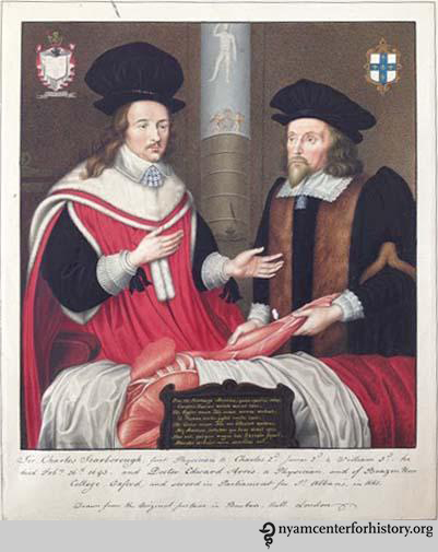
English Physicians Charles Scarborough and Edward Arris performing an anatomical dissection in 1651. After an original watercolor by G.P. Harding. Click to enlarge.
Accepted as a gift by The New York Academy of Medicine in 1975, the Ladd Collection comprises 671 prints dating from the early 17th century to the first half of the 19th century. The prints, which demonstrate a variety of printing processes including etching, engraving, mezzotint, stippling, lithography, and hand coloring, primarily depict people who have made historically significant contributions to the fields of science and medicine, as well as some medical institutions, procedures, and other health-related topics. William S. Ladd, a former dean of Cornell University Medical College, accumulated the collection during the first half of the 20th century, purchasing many of the prints as deaccessioned duplicates from the Ashmolean Museum at Oxford University.

Georg Faber von Rottenman. Engraving by Bernard Strauss. Von Rottenman was a maker of pills in Ratisbon ca. 1648. Click to enlarge.
Erich Meyerhoff, librarian of Cornell’s Medical Library from 1970 to 1986, recognized the research value of the collection and suggested it be given to the NYAM Library because, as he stated in his correspondence to NYAM librarian Alfred Brandon in 1975, “[NYAM] has the most important collection in the history of medicine in our region, which includes an extensive collection of portraits listed in its ‘Portrait Catalog.’”
The Ladd Collection was previously housed in a basement storage room in 27 flat-file drawers, which were overstuffed, dirty, and causing damage to the portrait mats. Our goals for the project—which began in January 2014 and finished in August 2014—were to clean the portraits, rehouse them to prevent further deterioration, and increase access to the collection by creating a digital inventory and location guide.
Click an image to view the gallery:


To begin, all of the portraits were dry cleaned using a smoke sponge.

Cleaning with a smoke sponge.
Many of the portraits also needed new mats (because the originals were either damaged or unacceptably acidic), as well as new interleaving tissue to replace tissue that had become stained and torn.
Portrait in need of a new mat and interleaving tissue.
We created new window mats for the portraits and hinged them to archival mat board supports using Japanese tissue and wheat starch paste. Because the prints themselves are in good condition, very few needed extensive repairs.

Cutting new mats.
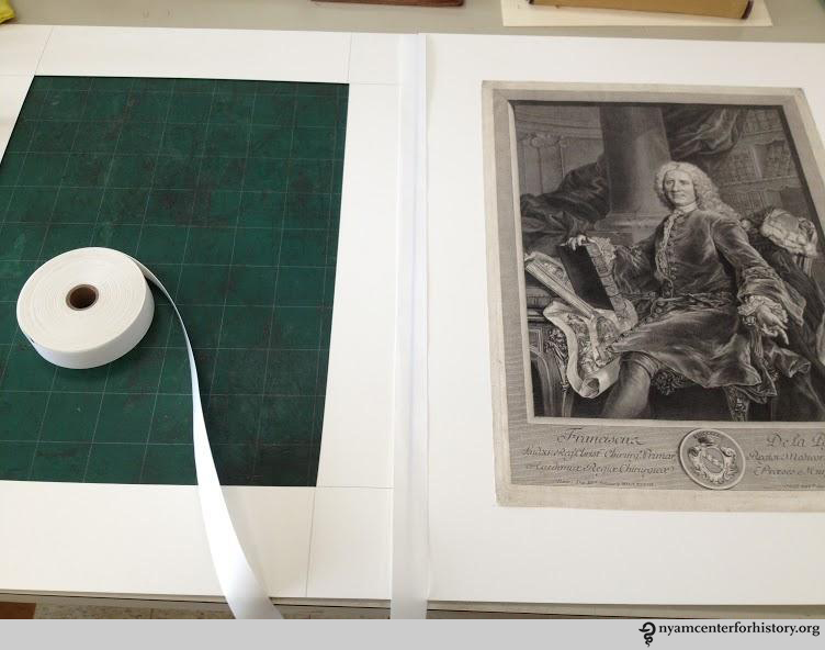
New window mat hinged to archival mat board supports.
That said, about 10 of the portraits needed washing in order to remove thickly applied, brittle adhesive residue that was causing damage to the edges of the prints. First, we tested the inks for solubility to determine whether an aqueous treatment was appropriate. Once we determined that the inks were stable, we washed the prints in a slightly alkaline bath.
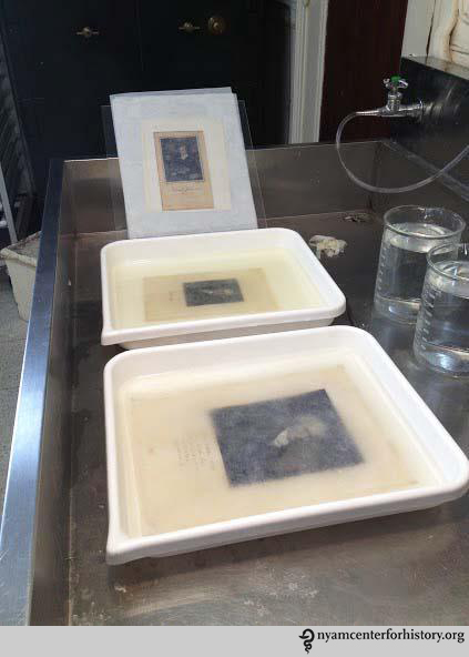
Prints in a slightly alkaline bath.
Rather than returning the collection to flat-file drawers, the conservation team made the decision to rehouse the matted prints (alphabetically and according to size) in acid and lignin-free, custom-ordered drop-front boxes from Talas that will be stored in climate-controlled conditions in NYAM’s recently renovated rare book storage stacks.

Prints rehoused in drop-front boxes.
Although the collection had been described and cataloged at the time of its acquisition in 1975, it had no online presence and was virtually undiscoverable to the average user. Thus, over the course of the project, staff completed a digital inventory and location guide with the aim of increasing accessibility. This will be made available online soon.

The end result.
These prints have importance not only because of their subject matter but also because of their aesthetic and art historical value. As a result of this project, scholars of the history of medicine, art, and printing can now use these prints as primary resources in their studies.
To view the collection or to access the collection guide, contact history@nyam.org or call 212-822-7313.


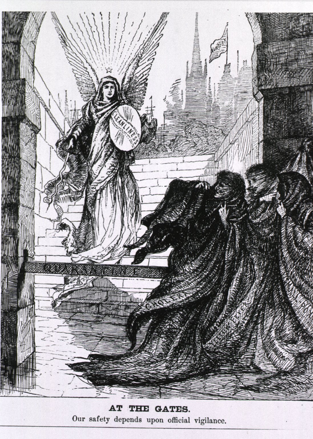
![Poultney Bigelow, The Pest at Our Gates, ([New York] : Merchants’ Association of New York, [1908])](https://nyamcenterforhistory.org/wp-content/uploads/2014/08/pestatourgates1908_watermark.jpg)