Spoiled by a Certain Englishman? The Copying of Andreas Vesalius in Thomas Geminus’ Compendiosa

Laura Robson, the author of today’s guest post, is our 2014–2015 Helfand Research Fellow. She completed her PhD in Classics at the University of Reading, UK.

“I wish the Epitome had not been spoiled so disgracefully by a certain Englishman (who I think lived with my brother for a time). He took what had been written with great care succinctly as a list in the Epitome and expanded it with excerpts taken from the books of the Fabrica… He utterly corrupted what had made it most praiseworthy and so roughly and absurdly copied what had been set forth with elegant drawing and engraving that he preserved no appearance of Oporinus’ majestic edition.”1

These were the concerns of Franciscus Vesalius, brother of the famous anatomist Andreas Vesalius, published in the preface to The China Root Epistle in 1546. Within only three years of the original publication of Andreas Vesalius’ De Humani Corporis Fabrica and its companion work, the Epitome, a number of medical authors had copied and reproduced the beautiful illustrations that had made Andreas Vesalius and his work so famous.2

Andreas Vesalius (1514-1564). De humani corporis fabrica libri septum. Basel: Johannes Oporinus, 1543. The most famous illustrations are the series of fourteen muscle men, progressively dissected. Some figures, such as this one, are flayed. Hanging the muscles and tendons from the body afforded greater detail, not only showing the parts, but how they fit together.

Andreas Vesalius (1514-1564). De humani corporis fabrica libri septum. Basel: Johannes Oporinus, 1543. Click to enlarge.

The Fabrica was one of the first anatomical treatises of the 16th century to present illustrations of the anatomised body in a naturalistic way. Vesalius promoted the dissection of the human body as the best way to learn about anatomy. By performing human dissections, he uncovered errors in the work of the ancient anatomist Galen, whose use of animals as dissection material to substitute for the lack of human cadavers had dominated the understanding of the body for centuries. Vesalius was caught in a conflict: how to show the anatomical errors in Galen’s treatises without going against such an important medical authority and potentially damaging his own medical career. He used the Fabrica to present his findings and to build on Galen’s important work.

The Fabrica contained 700 folio pages of Latin text and beautiful woodcut illustrations depicting the anatomical body in different poses. Readers were signposted to turn back and forth several times between image and text. This, as well as each image being accompanied by a letter key, encouraged a very active reading of Vesalius’ treatise.3 The work proved popular, with people taking a particular interest in the figures, although people copied, adapted, and reused both the images and text of Vesalius’ work in many different medical treatises. Due to the size and high cost of the Fabrica, cheaper copies were often more accessible than the original, even though the pirating enraged Vesalius and his close circle. Book piracy was common at this time. There was not the modern sense of intellectual property or copyright legislations. Licenses allowed particular printers to print works first, but the Venetian and imperial privileges obtained by the authors to try and protect their books from piracy did little to stop others from copying them.4

In fact, Franciscus Vesalius accused the wrong man of copying his brother’s work (suggesting, perhaps, that he had not seen a copy of the offending book). The only Englishman known to live with Vesalius was John Caius, when they lodged together in Padua during their studies. Caius went on to be physician to King Edward VI. There is no evidence that he pirated any version Vesalius’ work.5

The work Franciscus refers to is in fact Thomas Geminus’ Compendiosa.6 The first edition is predominantly made up of the Latin text of the Epitome and its illustrations, with the addition of many Fabrica figures. It is believed to be one of the first books to use copperplate illustrations.7 Geminus stated in his dedication that he followed Vesalius, but shortened his book to make it more useful to readers, in particular students.8 Reproducing the images of the Fabrica with the text of the Epitome meant that there was little interaction and connection between the two. However, the publication of the Compendiosa did bring Vesalius’ illustrations to a wider audience as the book was shorter and therefore cheaper than the original.

In order to make the work more accessible to those who could not read Latin, Geminus published an English edition of the Compendiosa in 1553. Nicholas Udall translated the short captions from the Fabrica figures into English. However, the main text of the Epitome was not translated. Instead the illustrations were placed after the text of Thomas Vicary’s The anatomie of mans body, first published in 1548.9 Geminus rearranged this text to follow the order of dissection for the parts of the body that decayed the fastest—the abdomen, the thorax, and the head. Although the images were not rearranged to fit this order, they connected more strongly to the text than in the Latin edition, as readers were signposted to particular figures discussing different parts of the body.

In his preface to the English Compendiosa, Nicholas Udall puts forward some interesting points about the uses of images and texts in medical manuals of his time. He said he did not know whether images or texts were more important when presenting anatomical information. He argued that information is set forth in writing for “high learning” and in pictures for the unlearned. He also explained that surgeons often performed duties like resetting bones by looking at figures alone.10 Surgeons had a low status in the medical profession at this time. They were not university educated like physicians and they were accused of having little knowledge about the science of medicine and healing. This suggests that readers who could not understand Latin, like surgeons, used anatomical figures and not written texts. I believe the English version of the Compendiosa was an attempt at encouraging these readers to read the text as well, by providing it in the vernacular language.

The coat of arms, left, and title page, right, of the Academy's copy of the 1559 English edition of Geminus’ Compendiosa.

The coat of arms, left, and title page, right, of the Academy’s copy of the 1559 English edition of Geminus’ Compendiosa. Click to enlarge.

In 1559 the English edition of Geminus’ Compendiosa was reissued.11 The annotated copy of this edition in the collection at the New York Academy of Medicine reveals hints as to how this anatomical text was used by readers at the time. The coloured and illuminated title page includes a portrait of Queen Elizabeth I. Slithers of gem stones have been attached to her necklace and the coat of arms opposite her on the adjacent page. The nude figures known as Adam and Eve are also coloured, and a reader inscribed the verse, “The Eyes of Them Both were opened, and They knew that They were naked: Genesis Chapter 3 Verse 7.”

Adam and Eve in the Academy's copy of the 1559 English edition of Geminus’  Compendiosa. Click to enlarge.

Adam and Eve in the Academy’s copy of the 1559 English edition of Geminus’ Compendiosa. Click to enlarge.

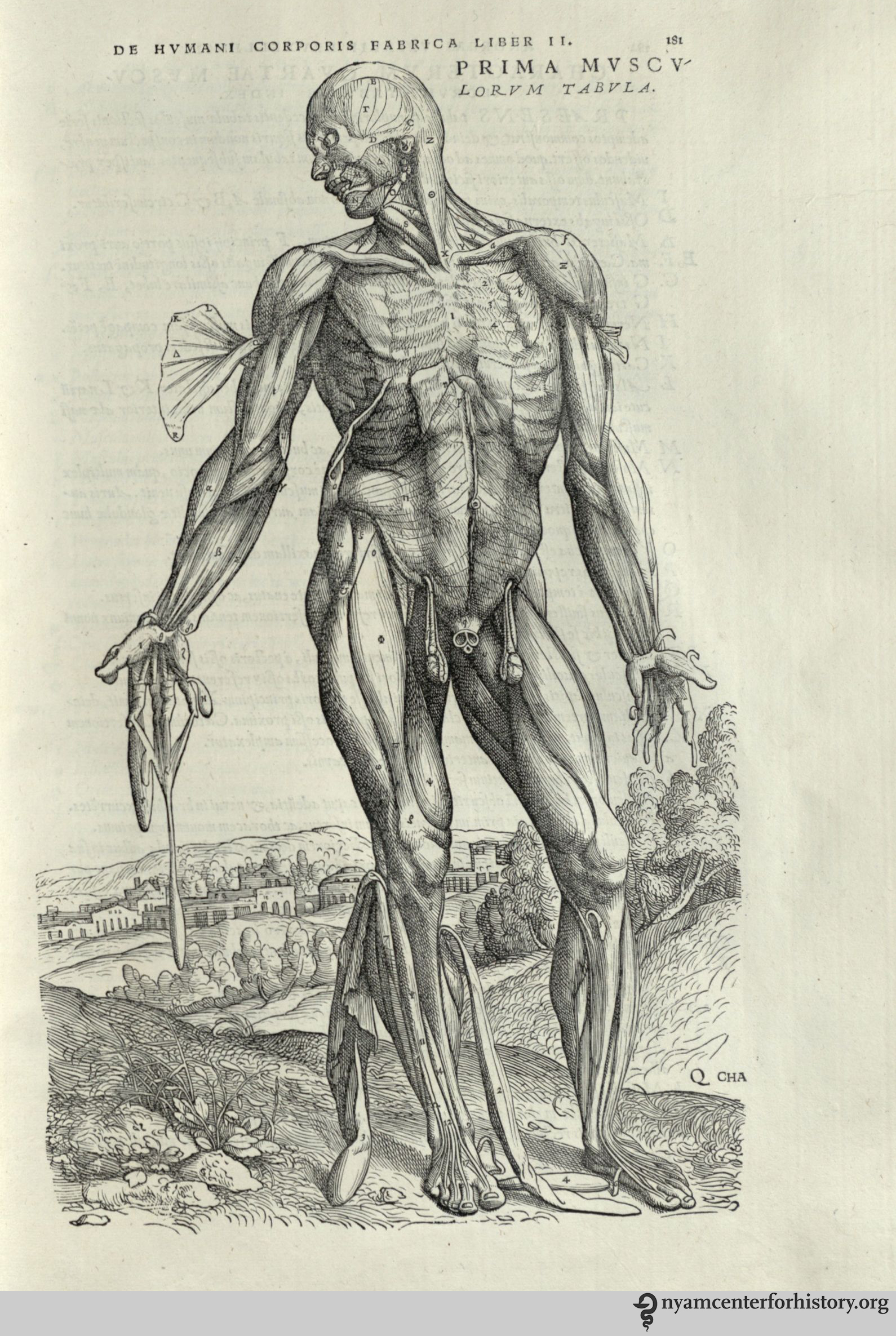
Two readers annotated this copy, both with different handwriting from the owner who in 1769 wrote his name—“G. Molesworth”—on the title page. One reader underlined key words and sections of the text, marking these with almost illegible notes in the margins. The other reader, though, focused on the illustrations. This second reader annotated the first three Vesalian musclemen images in the copy, adding the letters of the key, along with the Latin names for the body parts they represent.12 This English edition of the Compendiosa seldom uses the Latin names for parts of the body. So our reader did not get the information for his notes from this edition of the text. He must have consulted another text, such as the Latin edition of 1545, or even the original work of Vesalius’ Fabrica, in order to make his annotations.

Image of annotated muscleman figure  in the Academy's copy of the 1559 English edition of Geminus’  Compendiosa. Click to enlarge.

The annotated muscleman figure in the Academy’s copy of the 1559 English edition of Geminus’ Compendiosa. Click to enlarge.

This demonstrates the culture of active reading in the early modern period. This reader engaged with more than one treatise, perhaps even several works, when learning about the dissection of the body. He was familiar with the Latin language, and was therefore not one of the unlearned readers mentioned by Nicholas Udall in his preface to the work.

While Andreas and Franciscus Vesalius opposed the reproduction of the Fabrica and Epitome, the works that copied, adapted, and reused material from these texts allowed for the transmission of Vesalius’ knowledge of the body to a wider audience than the original works could reach. And this new audience interacted with the material, coming to a greater understanding of the dissected human body in the early modern period.

References

1. Andreas Vesalius (1546), Vesalius: The China Root Epistle, translated by Daniel H. Garrison (2015), p. 6.

2. Andreas Vesalius (1543a), De Humani Corporis Fabrica Libri Septem, Basel and Andreas Vesalius, (1543b), Andreae Vesalii Suorum de Humani Corporis Fabrica Librorum Epitome, Basel.

3. On active reading in Vesalius’ anatomical texts see, Nancy Siraisi (1994), “Vesalius and Human Diversity in De humani corporis fabrica”, in Journal of the Warburg and Courtauld Institutes, Vol. 57 p.64 and Sachiko Kusukawa (2012), Picturing the Book of Nature: Image, Text, and Argument in Sixteenth-Century Human Anatomy and Medical Botany, Chicago and London, p. 24.

4. On history of copyright and pirating see Christopher L. C. E. Witcombe (2004), Copyright in the Renaissance: Prints and Privilegio in Sixteenth-Century Venice and Rome, Leiden.

5. See Charles O’Malley (1955), “The Relations of John Caius With Andreas Vesalius and Some Incidental Remarks on the Guinta Galen and on Thomas Geminus,” in Journal of the History of Medicine and Allied Sciences Vol. 10.2 pp.147-172.

6. Thomas Geminus (1545), Compendiosa totius anatomie delineatio, aere exarata: Thomam Geminum, London.

7. Leroy Crummer (1926), “The Copper Plates of Raynalde and Geminus”, in Proceedings of the Royal Society of Medicine Vol 20.1 p. 53.

8. Thomas Geminus (1545), Compendiosa totius anatomie delineatio, aere exarata: Thomam Geminum, London, p. 1.

9. Thomas Vicary (1577 [1548]), A Profitable Treatise of the Anatomie of mans body: compyled by that excellent chirurgion, M. Thomas Vicary esquire, seriaunt chirurgion to king Henry the eyght, to king Edward the. vj. to Queene Mary, and to our most gracious Soueraigne Lady Queene Elizabeth, and also cheefe chirurgion of S. Bartholomewes Hospital. Which work is newly reuyued, corrected, and published by the chirurgions of the same hospital now beeing, London.

10. Thomas Geminus (1553), Compendiosa totius anatomie delineatio, aere exarata: Thomam Geminum, London, p.1.

11. Thomas Geminus (1559), Compendiosa totius anatomie delineatio, aere exarata: Thomam Geminum, London.

12. Ibid. p. Cii.

Making Collections Accessible: The New York Academy of Medicine Library’s Health Pamphlet Collection

By Katarzyna Bator, Collections Care Assistant, Gladys Brooks Book & Paper Conservation Laboratory

Every library is likely to find parts of its collection in need of protective enclosures and unique storage solutions. As part of our responsibility for the physical care of the collections at The New York Academy of Medicine, staff in the Gladys Brooks Book & Paper Conservation Laboratory routinely engages in large-scale rehousing projects. One such project currently underway is rehousing the library’s Health Pamphlet Collection. We estimate the project will take up to three years to complete.

The Health Pamphlet Collection consists of 19th– and early 20th-century health-related publications in many languages. It covers a wide range of topics, such as nutrition, proper hygiene, exercise, as well as medical innovations and research.

A damaged document box housing health pamphlets.

A damaged document box housing health pamphlets.

Approximately 50,000 health pamphlets are currently housed in acidic envelopes or plastic bags. These are in oversized boxes too big for the compact shelving unit on which they are stored. This limits accessibility, as the compact shelving cannot move properly, leaving little room for a librarian to retrieve each box from the shelf. In addition, each box is heavy and overstuffed with materials. This puts the pamphlets at risk of damage during storage and retrieval, and is problematic for staff who have to move and transport heavy boxes for patron use.

Previous storage space, with overstuffed document boxes.

Previous storage space, with overstuffed document boxes.

As part of the rehousing efforts for this collection, staff members place each pamphlet in an archival envelope with a 10-point folder stock insert for additional support, and then into a custom-made enclosure. The design of the enclosure is borrowed from the New-York Historical Society Library’s conservation laboratory. It is economic, sturdy, easy to make, and most importantly allows for safe and easy access to the collection.

Storage space with rehoused pamphlets

Storage space with rehoused pamphlets

The process of rehousing a collection involves more than simply making new enclosures and moving items to a new space. In order to make the Health Pamphlet Collection more accessible, staff members also dry clean each item with soot sponges—absorbent vulcanized rubber dirt erasers— and assess them for other conservation treatment needs, which they record in a spreadsheet to address as needed over time. In addition, a volunteer is creating an accurate inventory of all of the pamphlets to aid in future cataloging updates.

The Health Pamphlet Collection contains a wealth of information for researchers; through this project, conservation staff hopes to guarantee its accessibility to patrons both today and for generations to come.

Visiting Ellis Island’s Hospital Complex

On June 7, our Friends of the Rare Book Room and ARCHIVE Global: Architecture for Health enjoyed a private visit to the hospital zone on Ellis Island. The private support group Save Ellis Island offers hardhat tours of the hospital complex, which is adjacent to the main reception center operated by the National Park Service. More than 30 people took the ferry from Battery Park across New York Harbor to Ellis Island to learn more about the site and its importance to the history of public health in New York City. Some stayed on for lunch afterward at historic Fraunces Tavern.

Our Save Ellis Island tour guide gives safety instructions before the group enters the hospital zone.

Our Save Ellis Island tour guide gives safety instructions before the group enters the hospital zone.

On the south side of the island, and out of use since 1954, the hospital complex housed would-be immigrants who were not permitted to immediately enter the country. All steerage passengers were inspected—usually for only a few seconds, given their great numbers—and some 1 to 2% were detained for health reasons. Completed in 1909, the 750-bed hospital included wards for infectious diseases, kitchens, massive laundry facilities, an autopsy room, and recreation spaces for patients and staff alike.

The autopsy room.

The autopsy room.

Even in its semi-derelict condition, the complex is one of the few remaining “pavilion” style hospitals in the country. Pavilion hospitals were first built in France in the 18th century, and were enthusiastically endorsed by reformers such as Florence Nightingale in the 19th century. The design emphasized the need for ventilation, with wards built to promote sanitary conditions, provide light, and maximize the circulation of air. Pavilion hospital design fell out of use in the 20th century.

Caged verandas allowed patients access to fresh air while controlling their movement around the complex.

Caged verandas allowed patients access to fresh air while controlling their movement around the complex.

The Ellis Island site, already abandoned and crumbling, was further damaged during hurricane Sandy, and Save Ellis Island is working to stabilize the buildings, while preserving the sometimes eerie atmosphere of the site, now partially overgrown with vegetation. For more information about the complex, and the Save Ellis Island project to bring it back into public view, see: http://www.saveellisisland.org/history/hospital-complex

20150607_122018

Wall with a tide mark showing the level of water during Hurricane Sandy.

We are increasingly offering our Friends group exclusive events such as this visit. If you are interested in becoming a Friend, find out more here. Friends who missed out on this sold-out event should e-mail culturalevents@nyam.org to express their interest in another tour at a later date.

Apply for our 2016 Research Fellowships

Are you working on a history of medicine project that would be enhanced by spending a month mining our collections?

NYAM Library, Rare Book Room photos by Amy Hart © 2012We are now accepting applications for the Paul Klemperer Fellowship in the History of Medicine and the Audrey and William H. Helfand Fellowship in the History of Medicine and Public Health. Each fellow receives a stipend of $5,000 to support travel, lodging, and incidental expenses for a flexible period between January 1, 2016 and December 31, 2016. Fellows are expected to spend at least four weeks in New York City, working at The New York Academy of Medicine. Besides completing a research project, each fellow will be expected to make a public presentation at the Academy and submit a final report.

Both fellowships are designed to support researchers who can demonstrate how an opportunity to immerse themselves in our rich holdings will enhance their work. Preference in the application process is given to early career scholars, although the fellowships are open to anyone who wishes to apply, regardless of academic status, discipline, or citizenship. While both fellowships are for researchers engaged in history of medicine projects, the Helfand Fellowship emphasizes the role of visual materials in understanding that history.

The application deadline is Monday, August 17, 2015. Letters of recommendation must be received by Friday, August 21, 2015. Fellowship recipients will be notified by Thursday, October 1, 2015.

Perspective applicants are encouraged to contact Arlene Shaner, Reference Librarian for Historical Collections, at 212-822-7313 or history@nyam.org with questions or for  assistance identifying useful materials in the library collections.

Dr. Evelyn Hooker and the Acceptance of Homosexuality

By Paul Theerman, Associate Director, Center for the History of Medicine and Public Health

In the late 19th century, new legal, criminal, and scientific frameworks emerged seeking to understand, define, and in some cases control, human sexuality. In particular, homosexual activity between men became illegal in many countries, which opened up discussion about what counted as “normal” or “deviant” sexual expression. A significant body of research work began to be generated, such as Richard von Krafft-Ebing’s Psychopathia Sexualis (1886), seeking to understand the range of human sexuality and arguing that “deviancy” should be treated as a medical rather than criminal issue.1

Evelyn Hooker. Courtesy of UC Davis.

Evelyn Hooker. Courtesy of UC Davis.

By the 20th century, pioneering researchers like Evelyn Hooker (1907–1996) had begun to question whether homosexuality should be considered in medical terms. Hooker administered standard psychological tests to carefully selected groups of gay and straight men, who performed virtually identically. Her work was one in a series of investigations that eventually led to the removal of homosexuality from the list of mental disorders in the major official categorization of mental illness in the United States, the Diagnostic and Statistical Manual of Mental Disorders.

Born in Nebraska in 1907, Hooker went to the University of Colorado for her bachelor’s and master’s degrees in psychology. She undertook her Ph.D. work at Johns Hopkins, graduating in 1932. For the next eight years, she worked in a number of colleges, including Whittier College, was laid up with tuberculosis for two years, and had a fellowship year in Berlin. In 1940, she took up a research associate position at UCLA, where she remained for the next 30 years.2

Teaching was part of her purview. As the story goes, a friendship with a student who was gay, struck up in the mid-1940s, led to the student’s request that she research the gay community in Los Angeles. By 1953 she felt ready to do a controlled study, aided by a grant from the National Institute of Mental Health (NIMH). She assembled a group of 60 men, equal numbers of gay and straight, and matched for age, IQ, and education. In addition, the subjects had to be otherwise mentally healthy, that is, not in therapy nor showing any obvious mental disturbance. Finally, all were supposed to be “pure” in their orientation: purely heterosexual or purely homosexual. To this group, Hooker administered the Rorschach test, the Thematic Apperception Test (TAT), and the Make-A-Picture Story (MAPS) test, designed to measure personality, emotional stability, and coherence of thought. Recognized psychological experts evaluated the tests. After reviewing the results, Hooker found that they could not distinguish the tests completed by gay men from those by straight men. Any mental illness in this group was as likely to be found among heterosexual men as among homosexual ones.3

"Table II—Ratings on Overall Adjustment—Rorschach." In Hooker, “The Adjustment of the Male Overt Homosexual,” Journal of Projective Techniques 21 (1958): 18-31.

“Table II—Ratings on Overall Adjustment—Rorschach.” In Hooker, “The Adjustment of the Male Overt Homosexual,” Journal of Projective Techniques 21 (1958): 18-31. Click to enlarge.

Hooker presented her results at the 1956 meeting of the American Psychological Association. The editors of the Journal of Projective Techniques persuaded her to publish the results despite her wish to continue work until they were “incontrovertible.”4 In the following years, she continued to work and publish on the topic of gay men’s mental health—women were very little studied, the researchers themselves noted—with continued support from the NIMH. In 1967, the director of the NIMH, Dr. Stanley F. Yolles, appointed her the chair of the Institute’s Task Force on Homosexuality. Two years later, the task force finished its work. Its report concluded that “Homosexuality represents a major problem for our society largely because of the amount of injustice and suffering entailed in it, not only for the homosexual but also for those concerned about him.”5 It recommended establishing a Center for Study of Sexual Behavior within NIMH, to support research and training especially for mental health professionals, law enforcement personnel, and guidance and caretaking personnel.

 Hooker, “The Adjustment of the Male Overt Homosexual,” Journal of Projective Techniques 21 (1958): 18.

Hooker, “The Adjustment of the Male Overt Homosexual,” Journal of Projective Techniques 21 (1958): 18.

And yet . . . if gay people didn’t track differently than straight people on a whole range of mental disorders, they definitely did in one instance, according to the diagnostic standards of the times. Homosexuality itself was considered to be a mental disorder. And as jarring as it is to see, the same task force report from 1969 included as its final working paper “Treatment of Homosexuals,” detailing psychoanalytic, group, and drug- and electric shock–based aversion therapies, all intended to redirect sexual orientation.6 At the same time, though, countervailing political and cultural forces pushed towards acceptance of homosexuality, its normalization and de-medicalization. A recent New York Times article captures some of that flavor, expressed in the pre-Stonewall 1960s. As is well known, the Diagnostic and Statistical Manual included homosexuality as a mental disorder as late as its second edition in 1968; the American Psychiatric Association removed this designation in 1973, and the third edition of the DSM, published in 1980, included only the disorder “ego-dystonic homosexuality,” for those gay people uncomfortable with their orientation.7 By the 1987 revision of DSM, this condition was further downgraded to a “disorder not otherwise specified.”8

“Other Psychosexual Disorders. 302.00 Ego-dystonic Homosexuality,” American Psychiatric Association, Diagnostic and Statistical Manual of Mental Disorders (Third Edition) (Washington, DC: American Psychiatric Association, 1980), p. 281.

“Other Psychosexual Disorders. 302.00 Ego-dystonic Homosexuality,” American Psychiatric Association, Diagnostic and Statistical Manual of Mental Disorders (Third Edition) (Washington, DC: American Psychiatric Association, 1980), p. 281.

Evelyn Hooker went on to be a beloved mentor, especially for psychiatrists and psychologists interested in gay studies.9 She was the subject of a 1991 documentary, Changing our Minds: The Story of Dr. Evelyn Hooker,10 and the recipient of many awards, including the Distinguished Contribution in the Public Interest Award of the American Psychological Association. She passed away in 1996.

References

1. A brief introduction to the history of sexology can be found at The Kinsey Institute, which continues to explore sexual health and knowledge worldwide: www.kinseyinstitute.org/resources/sexology.html. See also APA Task Force on Appropriate Therapeutic Responses to Sexual Orientation, Report of the Task Force on Appropriate Therapeutic Responses to Sexual Orientation (Washington, DC: American Psychological Association, 2009), especially chapter 2, “A Brief History of Sexual Orientation Change Efforts,” pp. 21–25; Jonathan Katz’s Gay American History: Lesbians and Gay Men in the U.S.A. (1976; reprint ed., New York: New American Library, 1992) and The Invention of Heterosexuality (New York: Dutton, 1995); and Jennifer Terry, An American Obsession: Science, Medicine, and Homosexuality in Modern Society (Chicago: The University of Chicago Press, 1999).

2. Biographical material on Evelyn Hooker, here and below, comes from “Psychology’s Feminist Voices: Evelyn Gentry Hooker,” http://www.feministvoices.com/evelyn-gentry-hooker/, accessed June 10, 2015.

3. Evelyn Hooker, “The Adjustment of the Male Overt Homosexual,” Journal of Projective Techniques 21 (1958): 18-31.

4. Ibid., quotation from footnote on page 18.

5. National Institute of Mental Health Task Force on Homosexuality, Final Report and Background Papers, edited by John M. Livingood (Rockville, MD: U.S. Dept. of Health, Education, and Welfare, Public Health Service, Alcohol, Drug Abuse, and Mental Health Administration, National Institute of Mental Health, 1972), quotation from page 2.

6. NIMH Task Force on Homosexuality, Final Report: Task Force Working Papers, “Treatment of Homosexuals,” by Jerome D. Frank, pp. 63–68.

7. APA Task Force, Report, p., 23; American Psychiatric Association, Diagnostic and Statistical Manual of Mental Disorders (Third Edition) (Washington, DC: American Psychiatric Association, 1980), pp. 281–83.

8. American Psychiatric Association, Diagnostic and Statistical Manual of Mental Disorders (Third Edition–Revised) (Washington, DC: American Psychiatric Association, 1987), p. 296.

9. See in particular, Linda D. Garnets and Douglas C. Kimmel, eds., Psychological Perspectives on Lesbian, Gay, and Bisexual Experiences, 2nd ed. (New York: Columbia University Press, 2003), especially the chapter “What a Light It Shed: The Life of Evelyn Hooker,” by Garnets and Kimmel.

10. Changing Our Minds: The Story of Dr. Evelyn Hooker, directed by Richard Schmiechen, DVD, 75 mins. (San Francisco: Frameline, 1991).

Garbage and the City

By Lisa O’Sullivan, Director, Center for the History of Medicine and Public Health

This summer we are proud to present a new collaborative series, “Garbage and the City: Two Centuries of Dirt, Debris and Disposal.”

Together with our partners the Museum of the City of New York and ARCHIVE Global: Architecture for Health, “Garbage and the City” presents three moments in the city’s battle with sanitation and waste disposal challenges in a rapidly growing urban environment. Catherine McNeur will set the scene with “Hog Wash, Swill Milk, & the Politics of Waste Recycling in Antebellum Manhattan” on July 1. Julie Sze will discuss “Noxious New York: Race, Class and Garbage” on August 3, and finally, Robin Nagle, anthropologist-in-residence for New York City’s Department of Sanitation, will consider the daily practice of garbage collection and management in the city today with “Life Along the Curb: Inside the Department of Sanitation of New York” on August 17. All three events are free with advance registration.

New York City garbage truck, circa 1929. Photo from The New York Academy of Medicine Committee on Public Health Archive.

New York City garbage truck, circa 1929. Photo from The New York Academy of Medicine Committee on Public Health Archive.

The Academy has a long history tackling questions related to New York City’s sanitation infrastructure. Waste management and disposal was an ongoing concern as the city grew. Despite the creation of the Department of Street Cleaning in 1881, street cleaning and garbage removal contracts, like many other services enmeshed in the politics of city, included the trading of political favors, jobs for constituents, and the creation of slush funds. The threat or occurrence of epidemic disease triggered attempts to improve the situation, but at the turn of the 20th century, sanitation and waste disposal efforts remained haphazard and slow to change.

Many sanitation advocates of the late 19th century blamed disease on filth and refuse and the foul-smelling miasmas they produced. The emergence of new bacterial theories and techniques linked disease to the presence of specific pathogens. Whichever approach to disease was taken, the reality was clear: keeping the city clean from refuse was critical to minimizing the spread of infectious diseases such as cholera, making dealing with garbage a critical issue for the health of the city.

An open letter to mothers from the Committee of Twenty.

An open letter to mothers from the Committee of Twenty. Click to enlarge.

The Academy’s Committee for Public Health proposed new street cleaning methods periodically in the early 1900s. At this time, most of New York City’s garbage was carried out to sea in barges and dumped into the ocean. Collaborating with municipal officials and around a dozen civic organizations, the Academy appointed a Committee of Twenty on Street and Outdoor Cleanliness (a subcommittee of its Committee on Public Health). Its goal was public education, and included signage urging people to clean the sidewalks and curb their dogs, and a competition to design a more effective trash basket. The Committee reported on topics as varied as the effective design of dump trucks; conditions at the city’s open air markets and suggestions for their improvement; education campaigns instructing “every mother in this neighborhood” to teach their children to “refrain from this obnoxious practice” of throwing litter in street; and air pollution from fires on Rikers Island.1

Pamphlets reflecting the work of George Soper and the Committee of Twenty.

Pamphlets reflecting the work of George Soper and the Committee of Twenty.

In the 1930s, George Soper, the sanitation engineer best known for identifying Mary Mallon (“Typhoid Mary”) as a carrier of typhoid,2 was sent by the Committee of Twenty to take a trash tour of Europe. He attended the 1931 International Conference on Public Cleansing in London; measured the plowing capacity of German snow trucks; visited 14 incineration plants; and documented varied street sweeping methods during his extensive travels. The evidence he brought back all pointed in the same direction: whatever its successes, New York City was behind the times when it came to dealing with trash. The Academy used Soper’s reports to urge significant changes in the infrastructure of New York City’s garbage collection and disposal.

By 1933, politics struck again. The Committee chairman’s report stated that “the activities of the Committee of Twenty were considerably curtailed by the unexpected changes in City administration.”3 The Committee bemoaned the fact that despite better cooperation between the Police and Sanitation Departments, new ordinances and regulations were not systematically followed, and the “streets of New York City remain an untidy, if not disgraceful, condition.”4 Despite their concerns, the Committee concluded that the combination of political change and worsening economic conditions meant their attention would be better directed towards other efforts at a national level.

On a more positive note, the 1930s saw considerable resources expended, partly through New Deal projects, building new sanitation infrastructure, particularly sewage treatment.5 A 1934 law curtailed the dumping of municipal waste at sea, beginning a new era of sanitary landfills.6 Throughout the decade the Department of Sanitation (renamed from the Department of Street Cleaning in 1929) introduced new mass-produced garbage truck able to better compact and transport garbage. The winning entrant of the Committee’s competition for a more effective trash basket however, has sadly been lost to time.

New York City garbage truck circa 1930.

New York City garbage truck, circa 1930. Photo from the New York Academy of Medicine Committee on Public Health Archive.

The “Garbage and the City” series is presented in collaboration with the Museum of the City of New York and ARCHIVE GLOBAL and is supported by a grant from the New York Council for the Humanities. Any views, findings, conclusions or recommendations expressed in this program do not necessarily represent those of the National Endowment for the Humanities.

References

1. Committee of Twenty on Street and Outdoor Cleanliness, Committee on Public Health Archive, New York Academy of Medicine.

2. George A. Soper, “The Curious Career of Typhoid Mary,Bulletin of The New York Academy of Medicine, 1939 Oct; 15(10): 698–712.

3. Presumably a reference to Mayor John O’Brien, who served a one year term in 1933 before being defeated by Fiorello LaGuardia. O’Brien is now regarded as the last of the “Tammany Hall” mayors, criticized for his lackluster response to the impact of the Depression on the New York population. See: “Mayor John O’Brien: His Heart Is As Black As Yours!” Bowery Boys blog, February 25, 2010.

4. Report of the Chairman at the meeting of March 23, 1933, Committee of 20 on Street and Outdoor Cleanliness, New York Academy of Medicine Archives.

5. John Duffy, A History of Public Health in New York City 1866-1966 (Russell Sage Foundation: New York, 1968), 521.

6. George S. Soper, “Disposal of waste an urgent problem: Supreme Court order against dumping at sea points the need for incinerators,” The New York Times, March 18, 1934.

The Legacy of Aloysius “Alois” Alzheimer

By Danielle Aloia, Special Projects Librarian

Alois Alzheimer in 1884. In Maurer, Maurer, and Levi, Alzheimer: The Life of a Physician and the Career of a Disease, 2003.

Alois Alzheimer in 1884. In Maurer, Maurer, and Levi, Alzheimer: The Life of a Physician and the Career of a Disease, 2003.

Eminent German scientist Aloysius “Alois” Alzheimer was born on June 14, 1864. He considered himself a psychiatrist, because he “not only introduced the art of microscopy into psychiatry, but also contributed to psychiatry’s greatest interest in talking with patients.”1 His early work was at the Asylum for the Insane and Epileptic or “The Castle of the Insane,” where he spent hours listening to and examining patients, documenting each case.2 He also became a court-appointed forensic physician, treated private patients, and performed histological research.

While at the Asylum for the Insane and Epileptic, he met Auguste D., a 51-year-old wife and mother with symptoms of forgetfulness and jealously, whose husband could not take care of her anymore. Her case was curious because she was so young and had led a relatively healthy life. Senile dementia had been documented in patients in their 70s and 80s but none declined so rapidly as in Auguste D. in her last 4 years.

Emil Kraepelin circa 1910. In Kraepelin, Memoirs, 1987.

Emil Kraepelin circa 1910. In Kraepelin, Memoirs, 1987.

Alzheimer resigned from the asylum in 1903 and began work at the Psychiatric Clinic on Nussbaumstrasse in Munich under pioneering psychiatrist Emil Kraepelin. Much of Alzheimer’s early work focused on dementia caused by syphilis. Working with Kraepelin, he delved much deeper into the presenile dementia he first noted with Auguste D. at the asylum, what was to become the pinnacle of his career.

Together, Kraepelin and Alzheimer wanted to prove that psychiatric symptoms could be traced back to physical causes in the central nervous system.3 But when Kraepelin presented this hypothesis at the annual meeting of German Association for Psychiatry in 1906, they were not taken seriously. In April 1906, Auguste D. died and Alzheimer acquired her brain and clinical records. He began to compile his evidence of their “anatomical doctrine.”4

Left to right: Alzheimer and Kraepelin with psychiatrist Robert Gaupp and neuropathologist Franz Nissl, circa 1908.  In Kraepelin, Memoirs, 1987.

Left to right: Alzheimer and Kraepelin with psychiatrist Robert Gaupp and neuropathologist Franz Nissl, circa 1908. In Kraepelin, Memoirs, 1987.

With this doctrine, Alzheimer became the first person to describe the plaques and neurofibrillary tangles now known as indicators of Alzheimer’s disease.5 But the first article, published in 1906 in Zeitschrift fuer Psychiatrie und Psychisch-Gerichtliche Medizin, “Ueber eine eigenartige Erkrankung der Hirnrinde” [About a peculiar disease of the cerebral cortex], was not well received and garnered little attention. A second longer article published in 1911 in Zeitschrift fuer die gesamte Neurologie und Psychiatrie, “Ueber eigenartige Krankheitsfaelle des spaeteren Alters” [On peculiar cases of disease at higher age], drew much more attention. It included histological drawings of the tangles of the disease progression. Soon, other reports of similar cases started to appear in the literature.6

Drawings of histological preparations of Auguste D’s material, stained by Bielschowsky’s technique to demonstrate tangles, and their stages. Beginning of the disease. In Alzheimer, Ueber eigenartige Krankheitsfaelle des spaeteren Alters [On peculiar cases of disease at higher age]. Zeitschrift fuer die gesamte Neurologie und Psychiatrie 1911;4:356-385.

Drawings of histological preparations of Auguste D’s material, stained by Bielschowsky’s technique to demonstrate tangles, and their stages. Beginning of the disease. In Alzheimer, Ueber eigenartige Krankheitsfaelle des spaeteren Alters [On peculiar cases of disease at higher age]. Zeitschrift fuer die gesamte Neurologie und Psychiatrie 1911;4:356-385.

Drawings of histological preparations of Auguste D’s material, stained by Bielschowsky’s technique to demonstrate tangles, and their stages. 8. Advanced stage; and 9. Terminal state of the disease.. In Alzheimer, Ueber eigenartige Krankheitsfaelle des spaeteren Alters [On peculiar cases of disease at higher age]. Zeitschrift fuer die gesamte Neurologie und Psychiatrie 1911;4:356-385.

Drawings of histological preparations of Auguste D’s material, stained by Bielschowsky’s technique to demonstrate tangles, and their stages. 8. Advanced stage; and 9. Terminal state of the disease. In Alzheimer, Ueber eigenartige Krankheitsfaelle des spaeteren Alters [On peculiar cases of disease at higher age]. Zeitschrift fuer die gesamte Neurologie und Psychiatrie 1911;4:356-385. Click to enlarge.

Kraepelin named this new dementia after Alzheimer in the 1910 edition of his psychiatry textbook, Psychiatrie: Ein Lehrbuck für Studi[e]rende und Aerzte. They worked together from 1903 to 1912, when Alzheimer left to become chair of psychiatry at the Psychiatric Clinic in Breslau. Alzheimer died in 1915, at the age of 51, after a serious illness. During the course of his career Alzheimer made strides in the understanding of other diseases, such as epilepsy, and his work serves as the foundation for the continued development of a cure for the disease.7

References

1. Maurer K, Maurer U, Levi N (Trans.). Alzheimer: The Life of a Physician and the Career of a Disease. New York: Columbia University Press; 2003.

2. Ibid.

3. Ibid.

4. Ibid.

5. Hippius H, Neundörfer G. The discovery of Alzheimer’s disease. Dialogues in Clinical Neuroscience. 2003;5(1):101-108.

6. Zilka N, Novak M. The tangled story of Alois Alzheimer. Bratisl Lek Listy 2006;107(9-10):343-345.

7. Zilka N, Novak M. The tangled story of Alois Alzheimer. Bratisl Lek Listy 2006;107(9-10):343-345.

An Eye for Conservation: William Clift, Fenwick Beekman, and John Hunter

By William Buie, MA (History), Rutgers University-Camden, Spring Intern

“After [John] Hunter’s death, his great rambling mansion, three blocks thrown into one, passed through many hands. Till 1806 the Museum was still filled with his collection. … And there is a tradition that Stevenson drew from them his picture of the house and museum of Dr. Jekyll.” (Paget 1897, 155)1

Author Robert Louis Stevenson’s description of Dr. Jekyll’s fictional residence caused some of his contemporaries to suspect that he had used the real home and museum of the pioneering 18th-century Scottish surgeon John Hunter as a model; whether or not this was really the case is up for debate.

Dr. Jekyll and Mr. Hyde. Color lithograph by National Printing & Engraving Company, 188?. Courtesy of the Library of Congress Prints and Photographs Division.

Dr. Jekyll and Mr. Hyde. Color lithograph by National Printing & Engraving Company, 188?. Courtesy of the Library of Congress Prints and Photographs Division.

Stevenson did not mention Hunter in any of his notes. Yet the rumor became so widespread that Stephen Paget casually referred to the “tradition” in his 1897 biography of John Hunter.2 Speculation continues well into the 21st century.3 We may never know for sure if Dr. Jekyll and Mr. Hunter both lived at No. 28, Leicester Square, but we can get a sense of what the residence looked like thanks to one of the few surviving design plans, in the library’s collection.

Pencil copy of William Clift's drawing of the "Ground plan of Mr. Hunter's Premises level with Street, or Parlour-Floor level."

Pencil copy of William Clift’s drawing of the “Ground plan of Mr. Hunter’s Premises level with Street, or Parlour-Floor level.” Click to enlarge.

Much of what we know about Hunter’s Leicester Square residence comes from a ground floor plan reproduced from memory by William Clift, Hunter’s assistant and the first conservator of the Hunter Museum. Clift was born 1775 in Bodmin, a town in Cornwall, England. He had a difficult upbringing. He lost his father, Robert Clift, at a very young age. According to Clift, his mother Joanna Courts occasionally “starved herself to save threepence a week” in order educate her son. She died when Clift was eight, leaving him “cast adrift on the wide wide world.”4 He eventually found work under a nurseryman named George King, a man given to occasional brandy-fueled outbursts. Out of the blue one day, an inebriated King chased Clift through the nursery. Clift managed to escape despite that fact that his pursuer was riding a horse. Clift drew a caricature of the incident sometime later and used it to entertain his coworkers. King became aware of the drawing and fired Clift.

Luckily, Nancy Gilbert of Priory, Bodmin became aware of Clift’s circumstance. Gilbert was a childhood friend of Anne Home, whom Hunter had married in 1771. Gilbert knew that the surgeon needed a new assistant and recommended Clift to Hunter, who gave him the job. On February 14, 1792, his and Hunter’s birthday, Clift arrived at the Leicester Square residence. In addition to the clothes on his back, he had only “four changes of shirts and neckcloths” to his name.4

Hunter was an accomplished anatomist, surgeon, lecturer, and a pioneer of evidence-based medicine.  During his lifetime, Hunter developed new methods for treating gunshot wounds and venereal disease. He stressed experimental research and encouraged his students to pay close attention to the way that the human body responded damage. Because of that approach, Hunter is now known as the pioneer of “scientific surgery.” He was also an avid collector of animal and plant specimens. His museum contained approximately 14,000 preparations of more than 500 different species.5

John Hunter, "engraved by W. O. Geller from the original picture by Sir Joshua Reynolds in the Royal College of Surgeons," 1836.

John Hunter, “engraved by W. O. Geller from the original picture by Sir Joshua Reynolds in the Royal College of Surgeons,” 1836.

Clift worked for Hunter for less than two years, but the days were full. Each morning, Clift assisted with dissection and the preparation of specimens for display. He put his  penmanship to use each evening when Hunter required he take dictation, copy his employer’s nearly illegible notes, and answer personal correspondence. The sudden death of Hunter on October 16, 1793 threatened to cast Clift adrift once more. Although Hunter’s income was steady and substantial, he borrowed from creditors to keep a ready supply of cash to cover household expenditures. Following his death, Mrs. Hunter left the house and rented out her room. She allowed only Clift and Elizabeth Adams, who worked as the Hunter’s housekeeper, to remain on staff. Clift was to look after the museum for a salary of £21 a year.6

Recognizing the intellectual value of Hunter’s manuscripts and collection of specimens, Clift set himself to the task of preserving them. He copied by hand nearly half of Hunter’s manuscripts. He also cared for the specimens in Hunter’s museum. Clift dutifully and quietly carried on for six years. His efforts were rewarded when the British government bought the collection in 1799 and transferred its care to the Royal College of Surgeons. A board of curators assembled by the RCS appointed Clift the official conservator of the museum. During his time as conservator, Clift cared for the physical condition of the collection and maintained its original order.7

Clift oversaw the Hunter Collection for the next 42 years, during which time the RCS transferred the materials from Leicester Square to a new building near Lincoln’s Inn Fields. Late one night after the move, as his time as conservator was coming to an end, Clift drafted the ground floor plan that we have today.8 Simon Chaplin, director of culture & society of the Wellcome Trust, has conducted extensive research into Hunter and the Leicester Street house and considers the drawing to be fairly accurate.9 Given that no complete plan exists for the Leicester Square property for the time when the Hunters lived there, Clift’s plan remains one of the best representations that we have today.

"Ground Plan of Mr. Hunter's Premisis, level with Street or Parlour-Floor level." Copied from original in Royal College of Surgeons.  Click to enlarge.

“Ground Plan of Mr. Hunter’s Premisis, level with Street or Parlour-Floor level.” Copied from original in Royal College of Surgeons. Click to enlarge.

Scholars typically describe the Leicester Square property as made up of two separate buildings that were originally separate structures. However, it may be helpful to think of the property as composed of four structures. There were two main buildings. Twenty-eight Leicester Square faced west. Thirteen Castle Street faced east. Situated between the two main buildings were two smaller structures. One building contained Hunter’s picture gallery. The other contained his “conversatione room,” lecture theater, and museum. The second and third floors of the Leicester Square building contained the Hunters’ private rooms. Mr. and Mrs. Hunter conducted many of their public affairs on the ground floor. It was there that Anne Home, a well-educated poet with connections in London’s literary and artistic scene, entertained guests. The Castle Street (now Charing Cross Road) building contained Hunter’s dissecting room, preparations room, a dining room for students, and a room for housekeeper Elizabeth Adams.10

Portrait of Dr. Fenwick Beekman. In Annan, G. L. (1961) "The Fenwick Beekman Collection." Bulletin of the New York Academy of Medicine 37(4):  277–280.

Portrait of Dr. Fenwick Beekman. In Annan, G. L. (1961) “The Fenwick Beekman Collection.” Bulletin of the New York Academy of Medicine 37(4): 277–280.

A guide to the Fenwick Beekman Collection of images is now available online here. Before I discovered who Beekman was, the images collected appeared to have been randomly assembled. Biographical research revealed that Beekman was the foremost private collector of material related to 18th-century Scottish physician John Hunter. Beekman spent years researching and writing about him. When Beekman donated his Hunterian collection in 1960 it was considered the best in private hands. Only the Royal College of Surgeons could boast of a superior collection of Hunter-related materials. It is thanks to Beekman that the New York Academy of Medicine came to own a copy of Clift’s ground plan, along with the other items related to Hunter. After researching Beekman, I began looking into the history of each item in the collection. Thematically, the images reflect the diverse interests of Beekman and Hunter. In addition to the various images, there are several handwritten letters by Hunter that shed light on the early days of his career. I came away from the collection informed and entertained. I have no doubt others will as well.

References

1. Stephen Paget, John Hunter, Man of Science and Surgeon, (London: T. Fisher Unwin, 1897), 155.

2. Simon David John Chaplin, “John Hunter and the ‘Museum Oeconomy’, 1750-1800” (PhD diss., University of London, 209).

3. Lloyd Axelrod, “Strange Case of Dr. Jekyll and Mr Hyde – and John Hunter,” The American Journal of Medicine 125 (2012): 618.

4. Arthur Keith, “The Dicary Lecture on the Life and Times of William Clift, First Conservator of the Museum of the Royal College of Surgeons of England. Given in the Theatre of the Royal College of Surgeons, Friday, December 7th, 1923,”The British Medical Journal 2 (1923): 1127, 1128.

5. “John Hunter,” The Royal College of Surgeons, accessed April 23, 2015, https://www.rcseng.ac.uk/museums/hunterian/history/johnhunter.html.

6. Keith, “Life and Times of William Clift,” 1127-29.

7. Ibid, 1129; Jessie Dobson, “William Clift, F.R.S., First Conservator of the Hunterian Museum,” Proceedings of the Royal Society of Medicine 48 (1955): 324-325.

8. Keith, “Life and Times of William Clift,” 1129.

9 Chaplin, “John Hunter.”

10. Ibid.

Did Corsets Harm Women’s Health?

By Johanna Goldberg, Information Services Librarian

“It is difficult to imagine a slavery more senseless, cruel, or far-reaching in its injurious consequences than that imposed by fashion on civilized womanhood during the past generation. Her health has been sacrificed, and in countless instances her life has paid the penalty; while posterity has been dwarfed, maimed, and enervated, and in body, mind, and soul deformed at its behests. … [T]he tight lacing required by the wasp waists has produced generations of invalids and bequeathed to posterity suffering that will not vanish for many decades. By it, as has been pointed out by the authorities cited, every vital organ in the body has been seriously affected.”1

The title page of

The title page of “Fashion’s Slaves,” 1892. Click to enlarge.

So writes Benjamin Orange Flower in “Fashion’s Slaves,” a 32-page pamphlet published in 1892 as an appeal for women’s dress reform. One of the many causes Flower takes up is the corset, expressing his concern that the undergarment causes damage to internal organs. He continues, “If women will continue this destructive habit, the race must inevitably deteriorate.”1

Certainly, many women felt fettered by their restrictive clothing or there would never have been a dress reform movement. But just how damaging were corsets?

Not all corsets were alike. Tight lacing—cinching a corset to achieve a very small, or wasp, waist—began in the 1820s and 1830s after the advent of corsets made with metal eyelets. Medical professionals came out strongly against the practice.2 As shown in dramatic X-ray images in Ludovic O’Followell’s Le Corset, tightly laced corsets could change the shape of the rib cage,3 but there is no evidence that women had lower ribs removed to decrease their waists.4

Click on an image to view the gallery from Le Corset.

By measuring 19th-century corsets and dresses, historians have determined that women probably did not cinch their waists below 20 inches.4 (By comparison, today many U.S. stores list their XXS waist size at 23.5 inches.5,6) While many waists were still quite small, they may never have gone to the 14-inch extremes reported in women’s magazines, regardless of what fashion drawings depicted.4

A tightly laced corset could reduce lung capacity, irritate skin, and weaken back and chest muscles used to being supported.2 Whether tight lacing caused long-term health issues, like reduced pelvis size, constipation and digestive issues, and reproductive problems ranging from miscarriage to uterine prolapse, is more difficult to assess and remains unclear.2,4,7

Dr. Warner trade card, inside and out. An 1883 article from Godey’s Lady’s Book and Magazine hailed Dr. Warner’s Coraline Corset as a model of comfort, superior to whalebone and horn corsets, and endorsed the model as a substitute for tight-laced models: “They have demonstrated that tight lacing is not essential to grace or beauty of form; and while impractical dress reformers have been preaching reforms which no one would adopt, Warner Brothers, by introducing properly fitting corsets, have given practical aid to the health and comfort of several million ladies.”8

Dr. Warner trade card, inside and out. An 1883 article from Godey’s Lady’s Book and Magazine hailed Dr. Warner’s Coraline Corset as a model of comfort, superior to whalebone and horn corsets, and endorsed the model as a substitute for tight-laced models: “They have demonstrated that tight lacing is not essential to grace or beauty of form; and while impractical dress reformers have been preaching reforms which no one would adopt, Warner Brothers, by introducing properly fitting corsets, have given practical aid to the health and comfort of several million ladies.”8 Click to enlarge.

Many health problems once blamed on the corset are now clearly not the fault of the undergarment. Death caused by postpartum infections, or childbed fever, became relatively rare with the advent and spread of antiseptic techniques. With the discovery of the tubercle bacillus in 1882, it became clear that corsets did not cause the disease. Incidence of breast cancer did not decrease after corsets that did not compress the breasts came into vogue. As Gerhart S. Schwartz wrote in a 1979 Bulletin of the New York Academy of Medicine article, “one disease after another found an explanation which was unrelated to the corset.”9

Many of the doctors against tight lacing, including O’Followell, did not condemn corsets as a whole. Instead, they championed designs less tightly laced. Several pamphlets in our collection feature what they claim to be medically sound corsets.

In one, “La Grecque Corset as an Aid to the Physician and Surgeon,” printed circa 1911, the van Orden Corset Company advertises corsets that pull in the abdomen while reducing strain on abdominal muscles.10

Incorrect and correct pressure applied by corsets. In

Incorrect and correct pressure applied by corsets. In van Orden Corset Company, “La Grecque Corset as an Aid to the Physician and Surgeon,” circa 1911. Click to enlarge.

The pamphlet also features a maternity corset, “designed for a natural change of figure.”10 Corsets for maternity came on the market in the 1830s, and were often tightly laced.11 Yet the medical literature of the period does not discuss dangers of maternity corsets to the fetus or the mother,11 either due to taboos of the time or because negative impacts were rare or unreported. The maternity corset advertised by the van Orden Corset Company, from the early 1900s, was not tightly laced, taking advantage of new elasticized fabrics to expand as needed.10

La Grecque Maternity Corset. In

La Grecque Maternity Corset. In van Orden Corset Company, “La Grecque Corset as an Aid to the Physician and Surgeon,” circa 1911. Click to enlarge.

Corsets from a Surgical Standpoint,” from H. W. Gossard and Company (still in business today as a lingerie company), describes to physicians the benefits of prescribing their pliable front-laced corsets. These corsets, they claimed, improved posture and “preserve[d] the lines demanded by fashion, but without discomfort or injury.”12

Figures 5 and 6 in

Figures 5 and 6 in H. W. Gossard and Company, “Corsets from a Surgical Standpoint,” 1909. Click to enlarge.

Both of these pamphlets were published at the end of an era. The advent of elasticized fabric paved the way for the creation of an early bra, displayed by Herminie Cadolle at the Exposition Universelle in 1889.13 The corset’s final death knell was World War I. Women could not work in factories or the field while wearing restrictive clothing. Once household staff went to work for the war effort, upper-class women had no one to help them dress. Girdles and bras took over the corset’s supporting role, about 20 years after Flower’s calls for the end of the “destructive habit” of corsetry.1,13

References

1. Flower BO. Fashion’s slaves. Boston: Arena Pub. Co.; 1892.

2. Starr M. Vintage X-rays reveal the hidden effects of corsets. CNET. 2015. Available at: http://www.cnet.com/au/news/vintage-x-rays-reveal-the-hidden-effects-of-corsets. Accessed May 14, 2015.

3. O’Followell L. Le corset; histoire, médecine, hygiène. volume 2. Paris: Maloine; 1908.

4. Davis L. No, corsets did not destroy the health of Victorian women. io9. 2014. Available at: http://io9.com/no-corsets-did-not-destroy-the-health-of-victorian-wom-1545644060. Accessed May 14, 2015.

5. LOFT: Size Chart. Available at: http://www.anntaylor.com/catalog/sizeChartPopup.jsp. Accessed May 18, 2015.

6. Gap – women’s size chart. Available at: http://www.gap.com/browse/sizeChart.do?cid=2081. Accessed May 18, 2015.

7. Klingerman KM. Binding femininity: An examination of the effects of tightlacing on the female pelvis. 2006. Available at: http://etd.lsu.edu/docs/available/etd-04072006-115441/unrestricted/Klingerman_thesis.pdf. Accessed May 14, 2015.

8. Coraline: Its discovery and use in the manufacture of corsets. Godey’s Lady’s B Mag. 1883:468–469. Available at: https://books.google.com/books?id=nXA-AQAAMAAJ&pgis=1. Accessed May 18, 2015.

9. Schwarz GS. Society, physicians, and the corset. Bull N Y Acad Med. 1979;55(6):551–90. Available at: http://www.pubmedcentral.nih.gov/articlerender.fcgi?artid=1807654. Accessed May 14, 2015.

10. La Grecque corset as an aid to the physician and surgeon. New York: van Orden Corset Co.; 1911?

11. Summers L. Bound to please: A history of the Victorian corset. Oxford, New York: Berg; 2001.

12. Corsets from a surgical viewpoint. Chicago: Gossard Co.; 1909.

13. Fontanel B. Support and seduction: The history of corsets and bras. New York: Abrams; 1997.

Aging Through Time

By Danielle Aloia, Special Projects Librarian

“Ah, but I was so much older then
I’m younger than that now”
– Bob Dylan, My Back Pages

May celebrates Older Americans Month, which this year focuses on promoting health and community engagement of seniors across the nation. Today, 14.1% of the U.S. population is aged 65 and older1 and by 2030, 20% of the population will be over 65.2 We are living longer, healthier, and more productive lives than ever before.

Over the years there has been investigation into aging and the life course, reflecting beliefs informed by the average lifespans of the time. The Art of Invigorating and Prolonging Life by William Kitchiner, published in 1821 in the U.K. and two years later in the U.S., suggested that life is divided into three stages, each stage requiring a different regimen of “food – clothes – fire – air – exercise – sleep – wine – &c.”3 Kitchiner cautions that people only realize the importance of these elements after they become enslaved to other, detrimental, habits. They may need to proceed gradually with his recommendations in order to correct the bad habits and form new ones.

The first stage of life is a period of preparation, from birth to 21. In this stage, people should take in as much healthful food as can be digested for the body to convert into Chyle (bodily fluids). The second stage, the period of active usefulness (ages 21-42), should include plenty of “hard exercise in the open air” to restore the body’s constitution. In the third stage, the period of decline, the rate of decline is based on the strength of the constitution built during the active stage of life. Without “due attention to Diet &c., the Third period of Life is little better than a Chronic Disease.”4

Pages 34-35 of Kitchiner, The Art of Invigorating and Prolonging Life, 1823 edition.

Pages 34-35 of Kitchiner, The Art of Invigorating and Prolonging Life, 1823 edition. Click to enlarge.

According to Kitchiner, by 42 years of age humans are on the decline; they are ancient by 63. Put in context, life expectancy in the 1900s for men was 46.3 in the U.S.and 44 in the UK.5,6 But still, Kitchiner believed it was never too late to make up for lost time.

Kitchiner incorporated William Jones’s andrometer,7 a chart intended as a tool for people to gauge their progress through life.8

William Jones' Andrometer, on pages 36-37 of Kitchiner, The Art of Invigorating and Prolonging Life, 1823 edition.

William Jones’ Andrometer, on pages 36-37 of Kitchiner, The Art of Invigorating and Prolonging Life, 1823 edition. Click to enlarge.

Unfortunately, Jones passed away in his late 40s and Kitchiner in his 50s. Sadly, neither got to experience “a glorious retirement” or the “universal respect” due after the age of 60.

More than 100 years later, in 1974, a new benchmark showed similarities to Kitchiner’s book while offering a 20th-century outlook. D. D. Stonecypher published Getting Older and Staying Young: A Doctor’s Prescription for Continuing Vitality in Later Life to give readers reliable and practical advice about aging, because “the quality of one’s later years grows out of the choices the individual makes.”9 In 1974 life expectancy in the United States was 68.2 for men and 75.9 for women.10

Stonecypher had specific audiences in mind for his work: middle-aged readers wondering about their aging bodies who may be modifying activities in order to preserve their vigor; older readers looking to gain insights into preserving mental and physical vitality; and younger readers who wished to assist the elderly and gain insight and perspective on their own aging process. He also notes another type of reader, the policy maker or community worker who “holds the key to the mounting social problems of aging.”

Stonecypher offered the following questionnaire as a way for readers to assess the probability of living a long life, but goes on to explain that medical science was advancing so rapidly it could be possible to double the life span to over 100 years. Citing that in classical Greece and Rome average life expectancy was 18 years, he writes that by the 1800s it had doubled to 35 years and between 1800 and 1970 it doubled again to 72 years.

In Stonecypher, Getting Older and Staying Young, 1974.

In Stonecypher, Getting Older and Staying Young, 1974.

In 2013, life expectancy in the United States was 76.4 for men and 81.2 for women,11 a substantial increase even from 1974. The longer one lives the more productive one may need to be: “a glorious retirement” may not be the answer to a healthy old age. Stonecypher tries to persuade his audience: “It is prejudice that has justified the compulsory retirement, inadequate pensions, the ostracism, and the other stresses which have come to seem a normal part of life after 65.”

This year, the Medicare, Medicaid and the Older American Act celebrates its 50th anniversary.12 The Act led to programs that have ensured access to health care, community services, and protections of the rights of elders. We have come a long way, but have even more work to do to support health and productivity of seniors as the population ages.

References

1. U.S. Census Bureau. State and County QuickFacts. http://quickfacts.census.gov/qfd/states/00000.html. Accessed May 21, 2015.

2. U.S. Census Bureau. An Aging Nation: the Older Population in the United States. Washington, D.C.: U.S. Census Bureau; 2014. http://www.cdc.gov/nchs/data/nvsr/nvsr62/nvsr62_07.pdf. Accessed May 21, 2015.

3. Kitchiner William. The Art of Invigorating and Prolonging Life. Philadelphia : H. C. Carey & I. Lea; 1823.

4. Ibid.

5. U.S. Census Bureau. United States Life Tables, 2009.. Washington, D.C.: U.S. Census Bureau; 2014. http://www.cdc.gov/nchs/data/nvsr/nvsr62/nvsr62_07.pdf. Accessed May 21, 2015.

6. England. Office for National Statistics. Mortality in England and Wales: Average Life Span, 2010. England: Office for National Statsitcs; 2012. http://www.ons.gov.uk/ons/dcp171776_292196.pdf. Accessed May 22, 2015.

7. Mental Floss. The Andrometer: an 18th-Century Measuring Stick for Success in Life. http://mentalfloss.com/article/58057/andrometer-18th-century-measuring-stick-success-life. Accessed May 21, 2015.

8. Jones, William. The works of Sir William Jones, Volumes 1-2, 1807. http://books.google.com/books?id=PW5KAAAAYAAJ&printsec=frontcover&source=gbs_ge_summary_r&cad=0#v=onepage&q&f=false. Accessed May 21, 2015.

9. Stonecypher D. D. Getting Older and Staying Young. [1st ed.]. New York : Norton; 1973.

10. U.S. Census Bureau. United States Life Tables, 2009.. Washington, D.C.: U.S. Census Bureau; 2014. http://www.cdc.gov/nchs/data/nvsr/nvsr62/nvsr62_07.pdf. Accessed May 21, 2015.

11. U.S. Census Bureau. Health, United States, 2014. Washington, D.C.: U.S. Census Bureau; 2014. http://www.cdc.gov/nchs/data/hus/hus14.pdf. Accessed May 21, 2015.

12. 2015 White House Conference on Aging. http://www.whitehouseconferenceonaging.gov/about/index.html. Accessed May 21, 2015.